ABSTRACT
CHEN, TIANCHI. Free Energy and Structural Characteristics of Proline-rich Peptides with Distributed Multiple Guests. (Under the direction of Christopher Roland and Celeste Sagui.)
Folded polyproline can exist as either a left-(PPII) or right-handed (PPI) helices,
depending on their environment. We have characterized the free energy and structural
characteristics of proline-rich peptides with distributed multiple guests in an implicit
solvent environment. Specifically, the peptides are based on a motif of repeated (P ro− P ro−X) and (P ro−X −X) (X a natural amino acid) units. In addition, selected peptides involving hydroxyl prolines, as found in collagens, have also been considered.
These investigations are based on classical molecular dynamics simulations combined
with replica-exchange methods. Previous results for pure polyproline peptides have shown
that the space of peptide structures is characterized not just by the pure PPII and PPI
structures, but rather a broad distribution of stable minima with similar free energy.
Introducing distributed guests into the system changes this picture: while there still exists
a large number of possible states, the distribution of probable states tends to be strongly
skewed in favor of a small number of PPII-like structures. Moreover, the addition of
hydroxyl proline over proline enhances this trend, suggesting that (hydroxyl) proline-rich peptides with distributed guest might make good molecular rulers in structural biology.
We have also characterized the secondary structure characteristics of these peptides based
on a Ramachandran analysis, the cis/trans isomerization of the prolyl bonds, and an
odds-ratio analysis. Qualitatively, the obtained results resemble those of previously investigated
©Copyright 2014 by Tianchi Chen
Free Energy and Structural Characteristics of Proline-rich Peptides with Distributed Multiple Guests
by Tianchi Chen
A thesis submitted to the Graduate Faculty of North Carolina State University
in partial fulfillment of the requirements for the Degree of
Master of Science
Physics
Raleigh, North Carolina
2014
APPROVED BY:
Christopher Roland Co-chair of Advisory Committee
Celeste Sagui
Co-chair of Advisory Committee
DEDICATION
BIOGRAPHY
The author was born in Dalian, China. He came to NCSU in 2011 and finished his M.S.
ACKNOWLEDGEMENTS
This research was supported by NSF grant 1148144.We thank the NC State HPC Center
for extensive computational support.
I would like to thank my advisers Dr. Christopher Roland and Dr. Celeste Sagui for
providing me guidance and assistance on this project.
I also would like to thank my friends Dr. Mahmoud, Yuan Zhang and Feng Pan for
their kind help.
TABLE OF CONTENTS
LIST OF TABLES . . . vi
LIST OF FIGURES . . . viii
Chapter 1 INTRODUCTION . . . 1
Chapter 2 Methodology and Simulation Details . . . 6
2.1 Free energy and collective variabiles . . . 6
2.2 Sampling protocol . . . 8
2.3 Simulation Details . . . 10
2.4 Odds ratio construction . . . 12
2.5 Secondary structure and PPII content analysis . . . 13
Chapter 3 Results . . . 15
3.1 Free energy and structure of polyproline-rich peptides . . . 15
3.2 Secondary structural characteristics and Ramachandran plots . . . 17
Chapter 4 Discussion . . . 20
Chapter 5 Summary . . . 23
References . . . 24
Appendices . . . 31
Appendix A Tables . . . 32
LIST OF TABLES
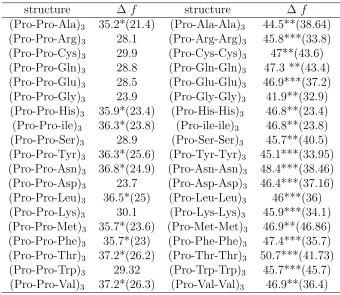
Table A.1 Comparison of free energy difference f(P P I)−f(P P II) (kcal/mol) in implicit water for short polyproline and hydroxyl proline peptides from (Ω,Λ) plots. Data for the pure proline is taken from Ref. [48]. . . 32 Table A.2 Estimated free energy difference ∆f =f(P P I)−f(P P II) (Kcal/mol)
for (P ro−P ro−X)3 and (P ro−X−X)3peptides in implicit water. We
emphasize that these numbers represent estimates only, since many of free energy landscapes do not have an unambiguous minima associated with a pure PPI state. For (P ro−P ro−X)3, such peptides are marked
with an *; the given number then represents the free energy difference between the estimated zero level and the PPII minima, while the num-ber in brackets corresponds to the difference between the lowest Ω = 7 minima and the PPII minima. Similarly for the (P ro−X−X)3
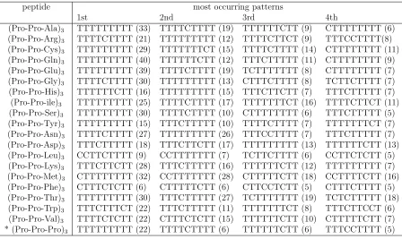
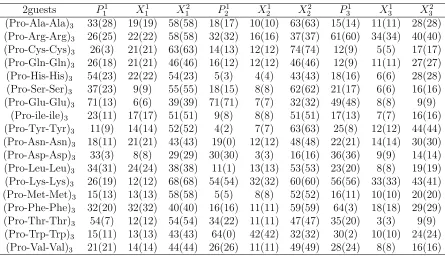
pep-tides, except that in this case, ** (***) marks the difference between the Ω = 7 (Ω = 5) states. . . 33 Table A.3 The four most commonly structures for (P ro−P ro−X)3 peptides
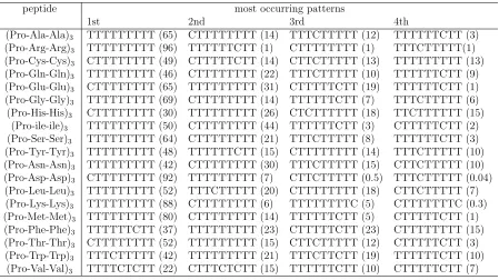
in implicit water along with a percentage of their occurrence (brack-eted numbers), as estimated by HT-REMD method. Data for proline (marked with an *) is taken from Ref. [48]. Only prolyl bonds are con-sidered. . . 34 Table A.4 The four most commonly structures for (P ro−X−X)3 peptides in
implicit water along with a percentage of their occurrence (bracketed numbers), as estimated by HT-REMD method. . . 35 Table A.5 The F and PPII (bracketed number) content as a percentage for (P ro−
P ro−X)3 peptides in implicit water based on a cluster analysis. . . 36
Table A.6 The F and PPII (bracketed number) content as a percentage forP ro− X−X)3 peptides in implicit water based on a cluster analysis. . . . 37
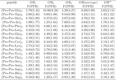
Table A.7 Odds ratio (OR) based on F and PPII (bracketed) content for (P ro− P ro−X)3 peptides with distributed guests. Here, the subscript on the
ORs (i.e.,OR1,2,3) indicates the location of the Pro-Pro-X triplet within
the peptide that is being considered. Thus, OR1 is associated with the
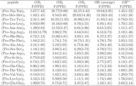
Table A.8 Odds ratio (OR) based on F and PPII (bracketed) content for (P ro− X −X)3 peptides with distributed guests. Here, the subscript on the
ORs (i.e.,OR1,2,3) indicates the location of the Pro-Pro-X triplet within
the peptide that is being considered. Thus, OR1 is associated with the
P2
1 −X11 residues, etc. Data marked OR
∗∗ is taken from Ref. [50] for
single-isolated amino acid guests, and is shown for comparison purposes only. . . 39 Table A.9 Grouping of amino acid guests according to their nature and the strength
LIST OF FIGURES
Figure B.1 Schematic illustrating the the backbone dihedral anglesψ,φ andωfor proline and hydroxyl proline. . . 41 Figure B.2 The (Ω,Λ) free energy landscapes in kcal/mol for pure hydroxyl
poly-roline peptides of different lengths: (a) 4-mer; (b) 5-mer; (c) 6-mer; (d) 7-mer; (e) 8-mer and (f) 9-mer. . . 42 Figure B.3 The (Ω,Λ) free energy landscapes (kcal/mol) for selected peptides
(P ro− P ro−X)3 (left panel) and (P ro −X −X)3 (right panel).
From top to bottom, here guest X is: (a) Arg; (b) Gln; (c) Gly and (d) Ser. . . 43 Figure B.4 This shows the Ramachandran plot for the (Pro-Pro-Arg)3 peptides.
The regions marking the different secondary structure for each residue are marked and color coded with black representing F-region data, blue αR data and pink the β-region data. . . 44
Figure B.5 This shows the Ramachandran plot for the (P ro−Arg−Arg)3
Chapter 1
INTRODUCTION
Many biomolecules exist as chiral images of each other, forming enantiomeric structures
which depend on the chirality of the molecular building blocks. For instance, most
nat-ural proteins formed from L-amino acids give rise to right-handed helices, while those
formed from D-amino acids favor left-handed structures. Considerably less common are
biomolecules that form left- and right-handed structures from the same chiral unit. A par-ticularly important and prominent example of this is provided by polyproline, which forms
helical structures with two well-characterized conformations. A left-handed polyproline
helix (PPII) is formed when all the sequential residues adopt the dihedral backbone
an-gles (φ, ψ) = (−75o,146o) with the prolyl bonds in the trans-isomer conformation (i.e.,
the backbone dihedral angleω= 180o with three residues per turn), and a more compact
right-handed polyproline helix (PPI) forms when the dihedral residues adopt dihedral
angles of roughly (−75o,160o) and all the prolyl bonds assume a cis-isomer conforma-tion (i.e., ω = 0o with 3.3 residues per turn). See Fig. 1 for a schematic of a proline dimer illustrating the different angles. Of the twenty natural amino acids, only proline
is comfortable in the cis-isomer conformation and with the effective stabilization of the left-handed helices.
In terms of the biological activity of proline peptides, much of it is associated with
the left-handed PPII structure. Thus, PPII plays an important role in cell processes such
as transcription, signal transduction, and cell motility. Proline’s propensity to form
left-handed helices is also crucial for cellular structural integrity, in particular for plant cell
wall proteins and collagen. PPII helices also commonly occur in globular proteins [1],
of the protein domains such as WW and EVH1. The SH3 (Src homology 3) domains have
been shown to mediate protein-protein interactions by binding to Pro-rich sequences of
10 amino acids in a PPII conformation [16,57]. The requirement that the Pro-rich ligands
be in a PPII conformation is of interest because such conformations can have therapeutic
applications, since SH3 domains are found in kinases, adapter proteins, lipases, GTPases,
etc. Perhaps because of their important role, prolines are consistently conserved at a level
of 100-80% in proteins with a sequence identity above 20% [2].
Proline oligomers also have considerable interest in their own right. Traditionally,
the relatively rigid structure of PPII has been used as a “molecular ruler” in structural
biology [18, 28, 32, 54], especially for the validation of spectroscopic rulers in F¨oster reso-nance energy transfer (FRET) experiments [66]. However, both FRET and
photoinduced-electron transfer [21] (PET) studies show deviations of experimentally observed
end-to-end distances of polyproline from theoretical predictions. These experimental studies, in
agreement with new theoretical work [47,48,51], conclude that polyproline exhibits static
structural heterogeneity with subpopulation of distinct end-to-end distances that do not
interconvert on the scales from nano- to milliseconds. This heterogeneity is attributed to
interspersed cisisomers that disrupt the otherwise ideal all trans PPII structure. PPII type of helices are also believed to play an important role in protein denatured
states, even in molecules that do not contain a single proline, such as diverse Ala-based peptides. Experimentally, considerable similarities were noted between the ultraviolet
circular dichroism (CD) spectra of denatured proteins and that of PPII [68]. This led to
the proposal that the PPII structure represents an alternate to the random coil model
for disordered peptides and unfolded proteins [67]. Although dating back all the way
to 1968, this proposition was revived in the last decade, with a common consensus that
PPII conformations are indeed part of the denatured states, but with considerable dissent
with regards to the proportion of these PPII states versus other states. Thus, for
Ala-rich peptides, the two contrasting views are that Ala has a very high PPII propensity
(between 80% and 100%) [7, 27, 33, 44, 46, 61–63]; or that the PPII conformation is just one of many local conformational states, and not the overall conformation of the unfolded
peptides [8, 22, 24, 35, 41–43, 53, 69, 70, 73].
Experimentally, there has been considerable emphasis on deriving an intrinsic PPII
propensity scale for individual amino acids in a proline-rich host-guest environment [13–
tool for probing the overall structural characteristics of peptides, such as their α-helix [12,38] andβ-sheets [45,65] content. In the proline-based experiments, a number of guest amino acids (usually of the same kind) are inserted in the middle of a short polyproline
peptide, usually consisting of three prolines on either side of the guest. Since a proline
“guest” is expected to form a stable PPII helix in aqueous solution, deviations from
this conformation induced by non-proline guests are expected has provide a measure of
their PPII propensity. In particular, the CD spectrum of pure proline has a maximum at
228 nm [34], which is assumed to be proportional to the PPII content because the PPII
helix is the only secondary structure known to have a positive band in this wavelength
range [59]. In a single-guest setting, pure proline was estimated to be on average 67% PPII helical at 5◦C, and other amino acids such as Gln, Ala and Gly were deemed to have
high PPII propensity in this context [14, 34, 60]. In contrast, amino acids such as Val and
Ile have disfavor for PPII conformation. Although the earlier works tried to quantify the
PPII content by comparing the CD spectra of Xn’s to some reference peptides, the more
recent works [13–15, 72] make the comparison in a more qualitative manner. In addition
to single-guest systems, peptides with 2, 3, 5, and 7 guest alanines (A=Ala) have been
also investigated [13, 14, 34, 60, 72], often by comparing their CD spectra to the pure
proline peptides. The studies concluded that multiple-guest Ala’s also possesses a high
PPII content, and that a short sequence of Ala’s can adopt the PPII conformation [14]. We recently examined this problem theoretically using new free energy methods in
combination with classical molecular dynamics with state-of-the-art force fields,
investi-gating the structural characteristics of proline-based oligopeptides with single and
mul-tiple guest amino acids [50, 52]. We extended the list of guests to cover all twenty natural
amino acids, and also considered the case of the experimentally examined multiple guests.
In terms of the amino acid guests in proline-rich peptides, we found that the average PPII
content of these peptides to be in qualitative agreement with their experimental values.
These peptides also share similar structural features, and that a population analysis of
secondary structure motifs (residue by residue) is in agreement with the results of Vilathe guest residues (other than Pro) do not favor the PPII region. We found that there is no
need to invoke an “intrinsic PPII propensity” in order to explain the experimental results.
Rather, these may be understood in terms of the following. (i) As it is well-known [17,39],
proline peptides are conformationally restricted by their pyrrolidine rings, and by steric
pre-ceding residue to 50◦ < ψ < 180◦ (except for Gly), forcing the preceding residue to be in either a β or F region, according to the value of φ. In the present host-guest setup, there is a statistical, Boltzmann-weighted distribution of conformations, with the highest
percentage of the guest population to be found in theβ region; (ii) The cis/trans proline ratio depends on the sequence surrounding the proline residue [39]. For this particular
set of host-guest peptides, we found that every guest (Pro, Tyr, and Trp excepted)
in-creases the trans content of the prolyl bonds. The guest amino acids therefore are not
characterized by any PPII propensity, but rather collaborate with their own intrinsic
trans propensity, to destabilize the cis isomers of the proline hosts, which results in a de
factonet PPII increase; (iii) there is a local correlation between the dihedral angles of the guest and the proline residue immediately preceding the guest. We found that the degree
to which the guest influences this proline (and visa versa) is conveniently described in
terms of an odds ratio analysis. In turn, the logarithm of this odds ratio is related to a
difference between free energies, whose trends correlate well with the experimental PPII
propensities [60].
Having understood the structural characteristics of single- and multiple amino acid
guests inside proline-rich peptides, we extend our investigations to cover the case of
multiple distributed guests. Specifically, we consider peptides based on a triplet motif
(Pro-Pro-X), and (Pro-X-X) where X represent a natural amino acid. This not only gives insight into the guest-host problem for systems with distributed guests, but is also
motivated by the fact that peptides with a triplet motif appear to have interesting
appli-cations. For instance collagens – which are among the most abundant proteins found in
mammals, comprising about 25% of human proteins and 75% of the weight of human skin
– denote the triple helical proteins that assemble into fibrous supramolecular aggregates
for connective tissues such as bones, tendons, cartilage, etc. Collagens are essential for
maintaining tissue integrity and the mechanical properties of the human body.
Essen-tially, they consist of three polypeptide chains forming an extended left-handed helix,
which are then coiled along a central axis in a right-handed manner. The amino acids that form collagen are based for the most part on a triplet motif, with X representing
either Pro, or a modified hydroxylproline (HPro), and Y Gly. Such triplet units are then
put together in combinations of various lengths, which then form the polypeptide chains.
Another example is provided by a new class of cell-penetrating peptides based on
these triplet-based peptides, it is natural to study these kinds of peptides with the aim
of characterizing and understanding their emergent structures.
This paper is organized as follows. Section II details our simulation methodology
and analysis. Specifically, we briefly review the Adaptively Biased Molecular Dynamics
(ABMD) method used to characterize the different peptide conformers and the Replica
Exchange Molecular Dynamics (REMD) method used for a population analysis. All
sim-ulation details, and the so-called odds ratio and the quantification of the PPII content
of a peptide are also discussed. Section III gives the results. Initially, we discuss the free
energies and structure of the peptide conformers based on the triplet motif. In addition,
we also consider the case of short peptides based on pure HPro, thereby setting the stage for future investigations of collagens. We have also carried out a residue-based and
sequence-based statistical analysis of the equilibrium conformations. A discussion of our
results and comparison to experimental data is given in Section IV, while Section V is
Chapter 2
Methodology and Simulation Details
In this section, we discuss the free energy and sampling methodology, define the
col-lective variables, provide all the relevant simulation details, and review the odds ratio
construction [23] we used to quantify the PPII content of the peptides.
2.1
Free energy and collective variabiles
To calculate accurate free energy maps, we used the Adaptively Biased Molecular
Dy-namics (ABMD) method [3,5], as implemented in the most recent release of the AMBER
simulation package [11]. Because the ABMD method has already been described
ex-tensively in the literature, we restrict ourselves to a few summary comments. ABMD
belongs to the general category of umbrella sampling methods with a time-dependent
potential [10, 37]. Such methods were first introduced by Huber, Torda and van
Gun-steren [31] in the context of molecular dynamics (MD), and by Wang and Landau [71]
in the context of Monte Carlo simulations. The ABMD method provides for an elegant way of computing the free energy [25] (or potential of mean force (PMF)) of a
collec-tive variable σ(r1, ...,rN), which is defined as a smooth function of the atomic positions
r1, ...,rN:
f(ξ) =−kBT lnp(ξ), (2.1)
where kB is the Boltzmann constant, T is the temperature, and
p(ξ) =δ[ξ−σ(r1, ...,rN)]
is the probability density of the collective variable (the angular brackets denote an
ensem-ble average). ABMD estimates the free energy of a reaction coordinate from an evolving
ensemble of realizations, and uses that estimate to bias the system dynamics to flatten
an effective free energy surface.
The ABMD [5] method is an umbrella sampling method with a time-dependent biasing
potential modifying the potential energy Φ of the system
ΦABM D(r,t) = Φ (r) +U
σ(r), t.
This biasing potential “floods” the true free energy landscape as it evolves in time
ac-cording to:
∂U(ξ, t)
∂t =
kBT
τF
Gξ−σ(r).
Here, G(ξ) is a positive definite, symmetric kernel (in analogy with the kernel density estimator widely used in statistics [64]), which may be thought of as a smoothed Dirac delta function. For large enough τF (the flooding timescale) and small enough kernel
width, the biasing potential U(ξ, t) converges towards −f(ξ) as t → ∞[10, 37]. ABMD can be used in conjunction with the REMD protocol. In this case, it is possible to
use different collective variables and/or temperatures on a per-replica basis (please see
Refs.for more details). Currently, the ABMD method has been implemented into the
AMBER simulation packages [11], and is freely available to the simulation community.
To date, the method has been successfully used to investigate a variety of biomolecullar
systems such as various peptides and sugars [3–6, 47, 48, 50, 51].
For the polyproline peptides, the most useful collective variables are associated with the cis/trans isomerization, i.e., the change that the torsion angle makes from cis (C) (ω= 0◦) totrans(T) (ω = 180◦) [47, 48, 51]. For instance, we define Ω as the sum of the cosines of the ω angles, which for an n-mer polyproline peptide with n prolyl bonds is:
Ωn−mer = n
X
i=1
cosωi. (2.3)
For a C (T) prolyl bond, cosωi is +1 (−1) and therefore Ωn−mer can take any of the
following values: −n, −n+ 2, . . ., n−2, or n. Note that the Ace-Pro is a prolyl bond and its dihedral angleω0 (defined likeω with the firstCα replaced by the methyl carbon)
the definition of Ω. The collective variable Ω gives the net balance of C and T prolyl bonds and obviously has some degeneracy. For a sequence with nb =n bonds where each
bond can be in either C orT, the number of conformations with nC C bonds and nT T
bonds (with nb = nC +nT) is nb!/(nC!nT!). Naturally, perfect PPI is characterized by
Ωn−mer =n and perfect PPII by Ωn−mer =−n.
Alternatively, one can consider the “interface” between bonds, and define a collective
variable Λ as:
Λn−mer = n−1
X
i=1
cos (ωi+ωi+1). (2.4)
For an n-mer, there are n prolyl bonds and n−1 interfaces. If two neighboring bonds have the same dihedral angle ω then their interface is cos (ωi+ωi+1) = +1, otherwise it
is−1. Correspondingly, Λn−mer can take on any of the values −n+ 1,−n+ 3,. . .,n−3,
n−1. Λn−mer removes some of the degeneracy associated with Ω. For |Ω|= n, there is
only one value of Λ (Λ = n−1); for Ω = 0 (that exists only for even values ofn) there are (n−1) values of Λ ; for |Ω|=m (with m6=n and m6= 0), there aren−m values of Λ. Thus a typical free energy landscape in the (Ω,Λ) plane has a triangular shape. For instance, for a pentamer there are 1 + 2 + 4 + 4 + 2 + 1 = 14 distinct minima in the (Ω,Λ) free energy landscape, with Ω associated with odd numbers and Λ with even numbers.
The PPII and PPI structures are associated with the farthest minima on the left and
right: (-5,4) and (5,4) and are nondegenerate.
2.2
Sampling protocol
As already noted, one of the defining characteristics of the equilibrium conformations
of proline-rich peptides are the different cis/trans patterns of the prolyl bonds. The
free energy barriers separating these cis (C) and trans (T) states are relatively high (of
the order of 15 kcal/mol [47]). Regular room temperature molecular dynamics (MD)
simulations are therefore unable to overcome these barriers in a reasonable amount of
time, and are therefore not suitable for the direct study of the conformational equilibria of proline peptides and so other methods are needed. Here, in order to deal with the
sampling issue, we made use of a generalized replica exchange [26] scheme.
subject to some sort of ergodic dynamics based on different Hamiltonians, and attempts to
exchange the trajectories of these replicas at some predetermined rate. Care must be taken
with respect to the choice of the Hamiltonians, since these determine the performance
of the method. In this regard, it is convenient to consider the following two aspects: (a)
the details of the so-called “hot” replica that facilitates the crossing of barriers, and (b)
the random walk between the replicas. The latter is typically described in terms of an
exchange rate between pairs of replicas. Let us assume, for a moment, that these rates are
sufficiently high, so that the random walk in replica space is efficient. The purpose of the
“hot” replica is to increase the barrier crossing rates (or, more formally, to decrease the
ergodic time scale). One possibility for this is to run the hot replica at high temperature. Another possibility [6] is to construct the hot replica by adding a biasing potential to the
original Hamiltonian that acts on some collective variable that (presumably) describes
one of the slow modes of the system that need “acceleration”.
A combination of such Hamiltonian and Temperature based replica exchange
molec-ular dynamics (HT-REMD [36, 48, 50]) provides for a practical way to reduce the
com-putational costs associated with REMD sampling, since it facilitates the sampling in the
“hottest” replica by both means, and therefore also allows for a better “tuning” of the
entire setup. While elevated temperatures provide for a generic way to promote barrier
crossing events, the use of biasing potentials (U) allows one to directly focus on specific slow modes of the system. The latter can often be identified on the grounds of
chemi-cal/physical intuition, and is usually described in terms of the already noted collective
variable σ =σ(r) For a system biased withU(r) = −f[σ(r)], the probabilities of different values of the collective variable would all be equal, since there are no barriers present.
Unfortunately, the true free energyf(ξ) is typically unknown in advance. However, even an approximatef(ξ), accurate within a few kBT, is often sufficient. While the latter can
be computed in a variety of ways [25], here we use the ABMD method [5].
The HT-REMD simulations proceeded in several stages. We first computed the
ap-proximate free energy associated with a chosen collective variable that “captures” the cis/trans transitions of the prolyl bonds at different temperatures using of combination
of ABMD and parallel tempering. Next, several additional replicas running at the lowest
temperature T0 were introduced into the setup. One of these replicas is completely
un-biased, and therefore samples the Boltzmann distribution at T =T0. The other replicas,
down by a constant factor). The purpose of these “proxy”-replicas is to ensure adequate
exchange rates between the conformations, and thereby enhance the mixing [6]. Data was
then taken from the unbiased replica at a suitable, predetermined rate.
2.3
Simulation Details
Simulations were carried out for guest-host peptides with a triplet motif: Ace−(P ro− P ro−X)3 − N me and Ace−(P ro−X −X)3 −N me, where X denotes one of the
natural amino acids. These peptides are abreviated (P P X)3 and (P XX)3, respectively.
In addition, we have also carried out simulations forAce−(P ro−P ro−HP ro)3−N me,
and Ace−(P ro−HP ro−HP ro)3−N me. where HPro denotes hydroxylproline. These
latter peptides form the basis of the collagen fibers. In addition, we have also considered
the free energy and structure of short Ace−(HP ro)n−N me peptides with n ≤ 8. It
turns out to be important to consider the individual resides of a given peptide, which we
denote with a combination of super- and sub-scripts. The former indicates the location
of the amino acid within a given triplet motif, while the latter indicates where the triplet
is located within the peptide, e.g., residues for (P P X)3 are Ace−P11−P12−X11−P21−
P22−X21−P31−P32−X31−N me.
All the simulations were carried using an implicit water model based on the
General-ized Born approximation [55,56]. Initial configurations consisted of the unfolded peptides,
which were generated using the LEAP program of the AMBER v.12 simulation
pack-age. The simulations used the ff99SB version of the Cornell et al force field [30], whose
equilibrium structures are known to be consistent with the experimental results [51]. The
leap-frog algorithm with a 1 fs timestep was used along with the Langevin dynamics.
There are three stages simulation for generating the biasing potential and calculating
the (Ω,Λ) free energy landscapes. The first phase involves simulations of 24 replicas all at a temperature of 1200K for 5 ns with a flooding time scale of 1 fs. These runs are based on multiple walker ABMD simulations without any replica exchange. This generates a
very rough estimate of the biasing potential ready for further refinement. The second
step involves long ABMD runs with temperature replica exchange and runs over 30 ns
with the same 1 fs flooding time scale. The temperature range is distributed as follows:
300, 318, 338, 359, 381, 405, 430, 457, 485, 516, 548, 582, 618, 656, 697, 740, 786, 835,
these simulations. Finally, the third stage ABMD simulations are similar to the second
stage except that a flooding time of 5fs was used in order to generate a finer free energy
landscape.
These ABMD biasing potentials were further used as the basis for the HT-REMD
runs for our distributed guest systems. the same amino acid guest. These ABMD
sim-ulations have been carried out using 20 replicas in a replica exchange scheme with the
temperatures distributed as: 300, 322, 347, 373, 401, 432, 464, 499, 537, 578, 622, 669,
720, 774, 833, 896, 964, 1037, 1115, 1200 K. Each replica had its own biasing potential. Ω
was used as the collective variable. A kernel width of 4∆ξ= 0.2 was used with a flooding timescale of τF =5 f sat different stages of the 50 nsruntime.
The transfered two-dimensional free energy maps formed the basis of the HT-REMD
runs for enhanced equilibrium sampling. Prior to starting the “production” runs for each
multiple-guest peptide, we assessed the ergodicity of the hottest replica (the one at the
highest temperature biased by the approximate free energy associated with the Ω at
that temperature) by simulating it alone. It turned out that all possible C/T states were
visited on a timescale of less than 1 ns with the Ω’s autocorrelation time being less than 5
ns. This translates into a reasonable number of independent samples over the subsequent
100 ns production runtime. We note here, that our choice of the highest temperature in the temperature ladder, and the total number of replicas were also influenced by the peculiarities of our local computer setup. We used 20 replicas with their full biasing
potentials with the same temperature distribution as used for the ABMD runs. Four
more replicas were then added, all atT = 300 K: one with no biasing potential, and three with the ABMD generated biasing potential scaled down by a factor of 0.49, 0.76 and 0.9,
respectively. The choice of temperatures, the scaling factors, and the ratio of
temperature-varying versus Hamiltonian-temperature-varying replicas (i.e., 20 versus 4) was to ensure a similar
rate of exchange, which varied between 40 to 55%, between all neighboring replicas. We
then ran 50 ns HT-REMD simulations for all the host-guest peptides. Cordinates of the
2.4
Odds ratio construction
In order to quantify the conformational preferences of the peptides induced by the amino
acid guests, we make use of the so-called odds ratio [23] (OR) statistic. The OR is a
descriptive construction that quantifies the strength of a correlationm or association or
of the non-independence of two binary values. For two binary variables X and Y, the OR
is defined as:
OR = p11p00
p10p01
, (2.5)
wherepab =p(X =a, Y =b) is the probability of the (X =a, Y =b) event (withaand b
taking on binary values of 0 and 1). In terms of the peptide conformations, we can think
of X and Y as being some structural characteristic of the different residues. For example,
one can assign the values of zero or unity, depending on whether or not the prolyl bond
of a residue is in C or T.
The utility of the OR in describing the influence of one binary random variable on
another is seen as follows. If two variables are statistically independent, then pab =papb
so that OR = 1. In the extreme case ofX =Y (complete dependence), then bothp10 and
p01 are zero, and the OR is infinite. Similarly, for X =Y p00=p11= 0 the OR becomes
zero. In summary, an OR ratio of unity indicates that the values of X are equally likely for both values of Y (i.e., Y=1,0), while an OR greater than unity indicates that the
X = 1 is more likely when Y = 1. An OR less than unity indicates that X = 1 is more likely when Y = 0.
It is also convenient to recast of logarithim of the OR in terms of the language of
free energies. Consider the probability of the (X = x, Y = y) events pxy. Clearly, any
probability can always be rewritten in terms a free energy type function Gxy via:
pxy ∝e−Gxy/kBT . (2.6)
With this construction, the the ratio of the probabilities pxy/pxz then translate into a
free energy difference:
lnpxy
pxz
=−(Gxy −Gxz)/kBT. (2.7)
Clearly, the logarithm of the OR then maps onto the difference of those differences,i.e.,
When dealing with the case of statistically independent properties, ∆∆G= 0. Otherwise ∆∆G takes on either positive or negative values, with a magnitude that depends on the mutual dependence of the two variables. Writing the OR in terms of such a free energy
function may be thought of as being a purely formal development, we have found that
the use of an OR analysis combined with free energy languange provides for a useful and
intuitive way to describe host-guest correlations [49, 50].
2.5
Secondary structure and PPII content analysis
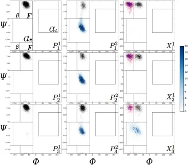
It is customary to use the dihedral angles (φ, ψ) in order to classify the different regions of the Ramachandran plots [74]. See Fig.1 for a schematic of the relevant dihedral angles.
On these plots, both PPII and PPI helices are to be found in the so-called F-region. In
order to include all the observed dihedral fluctuations, we have extended this region to
cover −110◦ < φ < −20◦ and (50◦ < ψ < 180◦ or −180◦ < ψ < −120◦). The β region consists of two parts: (−180◦ < φ < −110◦, 50◦ < ψ < 180◦ or −180◦ < ψ < −120◦) and (160◦ < φ < 180◦ and 120◦ < ψ < 180◦) regions. The α region is also divided into two parts: αR defined by −120◦ < ψ < 50◦ and −160◦ < φ < −20◦ and αL defined
by −50◦ < ψ < 120◦ and 20◦ < φ < 160◦. These regions are all clearly marked on the Ramachandran plots shown in Figs.4,5. We note that these definitions are tailored for the
proline-rich peptides considered here. The boundaries used here may not necessarily be
the optimal choice for other host/guest peptide systems, which typically may be expected
to be much more flexible.
Although this classification provides for a clear separation of the dihedrals for most
residues, it turns out, that for the amino acid guests there may be considerable overlap
between the F andβregions of the Ramachandran plots:de factothese may run together, without a clear separation between the two. In order to handle this situation, we used
a clustering technique to classify the secondary structure, rather than simply identifying them by region as defined above. For the most part, these two methods give the same
answer, although we can expect the clustering technique to be somewhat more accurate.
Specifically, for our purposes we used a central clustering method or vector quantization
[9] technique, with the stochastic implementation of the Expectation Maximization (EM)
[20] method in an algorithm that is reminiscent of the widely used K-means algorithm
algorithm in a stochastic manner. We initially make use of the regions as defined above
to identify the secondary structure of each sampled conformation. Five regions (clusters)
are considered, and associated with the F,β, αR,αLand “N” regions. The latter is used
to represent the population that falls outside the first 4 regions. For the most part, with
the exception of Gly, this population is negligible. Then, an association function zi c is
defined which gives the probability that the conformation belongs to a given cluster (or
region c). By iteration, these association functions are optimized for each conformation using a a Gibbs measure ∝ exp(−s(Dic)2), with s representing a modulating parameter and Dci the distance of (φ,ψ) of the ith conformation to the reference point of the cluster
c. The latter is defined as the average of all the (φ, ψ) dihedrals of the conformations weighted by zi
c. Then, the parameter s can be increased for improved accuracy once the
desired convergence has been reached using a smaller value ofs. We useds= 20rad−2. In the final step, a random number generator based on the optimized association functions
assigns each conformation to a single cluster.
When considering the guest amino acids, the so-called PPII content is simply given
by its F population. For proline, both C and T isomers fall into the F region. Hence,
since C isomers are associated with the PPI structure, such structures are not counted
when determining the PPII content. Thus, for a conformation to be classified as PPII,
Chapter 3
Results
Polyproline-rich peptides are characterized by a large number of stable structures based
on the cis/trans isomerization. For ease of discussion, we present the ABMD free energy
results and the aspects related to the guest/host and secondary structure (Ramachandran
plots) in separate sections.
3.1
Free energy and structure of polyproline-rich
peptides
To understand the structure of polyproline peptides with distributed guests, it is helpful
to review the results for pure polyproline peptides. Previously, we investigated the free
energy landscapes of such peptides as a function of different collective variables [47,48,51].
All of these simulation yield consistent results, showing that pure polyproline peptides are
characterized by a large number of stable structures based on the cis/trans isomerization of the ω torsion angle. The simulations show, in agreement with experiments, that the PPII structure is preferred over PPI. While there is some variation in this for small
peptides, the free energy differences between PPII and PPI increases as a function of the
peptide length.
As a first step to understanding the structure of polyproline-rich peptides with the
so-called triplet motif, we considered pure hydroxyl polyproline in implicit water. Figure
so that the free energy minima are distributed in a triangular or wedge-like shape, with
minima at (-n,n-1) ((n,n-1)) associated with pure PPII (PPI) structures. There are a
large number of such minima, which of course increase with the length of the peptide,
indicating that hydroxyl polyprolines too are characterized by a large number of stable
isomers. As with pure polyproline, hydroxyl polyproline favors the PPII-type structures
(i.e., structures associated with Ω ≤ 0. In fact, with pure hydroxyl proline, this trend is considerably more pronounced. Thus, in Fig.2 we see that qualitatively the depth
of the minima between PPII and PPI appear to be approximately equal for lengths
less than hexamers. For heptamers and above, it is the PPII-type structures that are
strongly favored. Presumably, this is because the hydroxyl groups on the sidechains can maximize their interaction with the waters in the more open PPII structure. This trend
is also reflected in the free energy differences between PPII and PPI given in Table
I, which shows that PPII is favored over PPI by a factor of about two for hydroxyl
proline over pure proline. Finally, we have also considered the free energies of nanomers
Ace−(P ro−P ro−HP ro)3−N me and Ace−(P ro−HP ro−HP ro)3−N me, which
constitute two mixed peptides. Qualitatively, these free energy maps resemble those of
the other proline/hydroxyl proline peptides with free energy differences of 12 and 10.3
kcal/mol, respectively. These numbers are intermediate to the other free energy results
indicating that the addition of the hydroxyl group stiffens the peptide.
We now turn to the (Ω,Λ) free energy landscapes of the (P P X)3 and (P XX)3
pep-tides. Plots for selected peptides are shown in Fig.3. Qualitatively, the behavior for all the
guests is very similar. As for the pure proline and hydroxyl proline case, many minima
are observed indicating a number of stable isomers. However, the distribution of these
minima is strongly skewed in the PPII direction. Moreover, given that a small number
of these minima are considerably deeper than the others, this implies that number of
stable conformers is considerably reduced when compared to that of the pure proline
case. This is particularly true for the two-guest case, where it is often numerically hard
to associate an unambiguous minima with a pure PPI structure. This result is not too surprising, given that the presence of other amino acids always favor the trans isomers.
Table II summarizes the estimated free energy differences between the PPII and PPI
structures for the different single- and double-guest peptides. Clearly, when to compared
to pure proline, the addition of other guests overwhelmingly favors the formation of a
Finally, we have also carried out a population analysis of the most probable structures
for the single- and double-guest peptides using HT-REMD. These results are summarized
in Table III, IV. In brief, for single-guest peptides the all-trans PPII structure is one of the
most probable structures often being either first or second. Another common structure
involves peptides characterized by a single cis prolyl bond. Structures with two cis bonds
are somewhat less common; usually the cis-bonds are separated by two or more bonds in
the trans configuration. Finally, Leu and Phe appear to be somewhat different insofar as
the most common structures involve more than two cis bonds. We note that as compared
to the case of other single-guest systems, the total percentage that is accounted for by
these structures is relatively low indicating that these systems are characterized by a very broad distribution of isomers. Turning to the case of double-guest systems, these
are primarily dominated by the all-trans configuration and configurations with only a
single cis bond. Generally speaking, the given top four configuration account for 54-100%
of the observed structures, showing that these systems strongly favor a small number of
PPII-like configurations with only a few interspersed cis bonds. This is to be contrasted
with the results for the single-guest peptides, where the top four configurations account
for 22- 94 % of the population, indicating a somewhat broader distribution of structures.
3.2
Secondary structural characteristics and
Ra-machandran plots
Having collected 104 equilibrium samples for each of the (P P X)
3 and (P XX)3 peptides
with the HT-REMD simulations, we have analyzed their structural properties with a
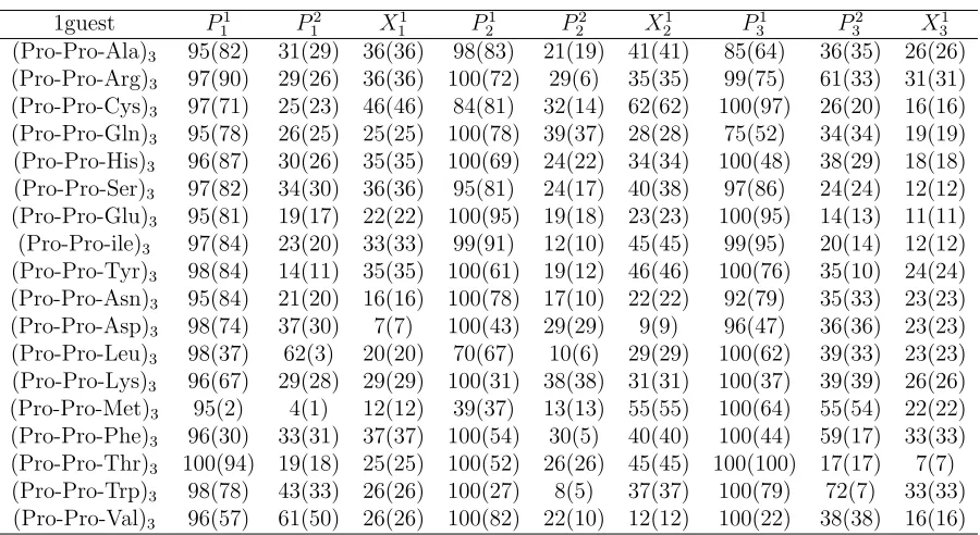
focus on the PPII content. The results are summarized in Tables 5-xx and Figs.4-5.
Ramachandran plots for selected residues are shown in Figs.4,5. On Fig.4a, we have
marked the relevant regions of these plots, i.e., theF, β, αR, and αL regions. On these
plots, each pixel represents a 1◦×1◦ bin, whose intensity represents its relative population, ranging from 1 to 20 samples out of 104 conformations. Pixels with more than 20 samples
are colored the same as the ones with 20 samples. Data within the different regions of
the Ramachandran plots are color-coded for clarity.
Figure 4 shows typical Ramachandran plots for polyproline peptides with one- and
two-distributed guests. In many respects the results obtained are similar to those
ex-pected, similar behavior is observed for residues with similar content within the peptides,
i.e., P1
1, P21, P31, and X11, X21, X31, etc all (roughly) display similar characteristics as far
as the Ramachandran plots are concerned. Concerning peptides with single-distributed
guests, these results most resemble previous results for peptides with an isolated guest.
Thus,P1
1,P21 andP31 all represent proline residues followed by another proline and almost
all the data falls into the F-region. This is also borne out by the statistics given in Table
5, which shows that over 90% of these residues fall into the F-region. The exceptions here
are the P31 residue of peptides with guests Ala, Asn, Asp and Gln, all of which have a small αR presence which brings down the corresponding F-region content. For data that
falls into the F-region, we have also probed its cis/trans content. Typically, the trans content drops by 15−30% when only the PPII content is considered (i.e., cis isomers excluded). Turning to P2
1,2,3 which represents the proline followed by a guest X 6=P, all
behave in the same way: there is a significant decrease in the F-region content in favor
of an strong increase in αR-region content. In terms of the guests, they all have a strong
presence in the β-F region, which effectively run together. The exception here is Gly, which (as noted in other studies [50, 52]) is characterized by a large population falling
outside of this region. In addition, all the X3 residues have an αR content. Presumably
the last group of residues (P31−P32 −X31) are slightly different insofar as there is a lot more fraying and motion associated with peptide C-terminal.
Turning to the peptides with two guests, we find much more consistent behavior
which probably reflects the greater stiffness of these peptides. Essentially, guests X11,,22,3
all have their residues fall into the β, F and αR regions, and no others (Gly excepted).
As seen in other systems with multiple guests [52], there is αR content of the X11,2,3
guest that is higher than for the X2
1,2,3 residue. This is also reflected in the drop in
the relative drop of the F-region content, which is given in Table VI. Finally, we note
that the prolines P11,2,3 all have both F- and αR-region content, so that qualitatively
they resemble P12,2,3 of the single-guest peptides. This is actually expected, since there is a strong correlation between the secondary structure of prolines followed by a guest, i.e., the P − X bond, which previously has been explored in terms of an odds-ratio analysis for proline-rich peptides with a single amino acid guest [50]. Given the qualitative
resemblance between the Ramachandran plots, we have carried out a similar odds-ratio
analysis for our distributed guest system.
P2
1,2,3−X11,2,3 (single guest) andP11,2,3−X11,2,3(double guest) residues, which in turn yields
a set of conditional probabilities. For instance, most of guest residues are in either F, β
or αR regions, while the proline has significant αR and F components. Hence, one can
calculate conditional probabilities p(X1
1,2,3 ∈F|P12,2,3 ∈F) and p(X11,2,3 ∈F|P12,2,3 ∈αR).
It is further possible to take into account the cis/trans isomerization of proline residue. All
in all, the relevant populations for theP −X correlations areFtF, Ftβ,FcF,Fcβ,FtαR,
FcαR, αtF, αtβ, αtαR, αcF, αcβ and αcαR. Given these correlations, we calculated the
odds-ratio based on the correlation between the F-regions of proline and guest residue,
in analogy with previous analysis [50, 52]. Thus, we set the property to be 1 (or 0) for
P12,2,3 ∈ F (or P12,2,3 ∈/ F) for the first index and X1,2,3 ∈ F ( or X1,2,3 ∈/ F) for the
second index, so thatp00=pαα+pαβ+pβα+pββ,p01=pαF +pβF,p10 =pFα+pF β and
p11=pF F.
Results for the OR analysis are given in Tables VII, VIII. Roughly speaking, Table VII
shows that there is significant variation in the OR for the different P-X residues within
a given peptide. In terms of single- and double-guests, the correlations (and therefore
the OR) tends be larger for the double-guests. Presumably, this is a reflection of possible
conformers for the peptides, which are now strongly skewed towards PPII-like structures.
For comparison purposes, we also give the OR results forP −X correlations involving a single-guest inside a much longer proline-rich peptide [50]. Quantitatively, these results are different from those of the distributed guest peptides. One important reason for this is
due to the presence of a significant αRcomponent in the Ramachandran plots associated
with the guests, which often drives up the p00 term considerably. For a single, isolated
guest in the center of an otherwise all-proline peptide the αR population tends to be
Chapter 4
Discussion
As previously noted, the structure of polyproline peptides consists not only of pure PPII
and PPI helices, but rather it is characterized by a broad distribution of stable structures
based on the cis/trans isomerization of the ω torsion angle. In an aqueous environment, the PPII-like structures are generally preferred. Here, we have investigated how these
structures change in the presence of distributed guests based on repeated (Pro-Pro-X) and (Pro-X-X) motifs, and with the use of hydroxyl proline. In terms of the latter,
the use of hydroxyl proline enhances the formation of PPII-like structures, especially
for the longer peptides. A similar effect is observed for the distributed guests systems.
Especially for the case of peptides with double guests. While the free energy plots are
still characterized by a large number of minima, the depth of these minima is strongly
skewed in favor of small number of PPII-like structures. Thus, both the use of hydroxyl
proline and distributed guests should lead to much stiffer helical structures. We believe
that one can take advantage of this in two ways. First, because of the presumed stiffness
pure polyproline it has often used used as a molecular ruler in single-molecule FRET
experiments. However, pure polyproline has been shown to be less than ideal because of the interspersed cis dihedral angles. Our results indicate that systems with distributed
guests such as Arg should be considerably better molecular rulers. Furthermore, the use
of hydroxyl proline should act to enhance this effect even more. Second, the enhanced
stiffness of proline peptides with distributed Arg may help explain why these peptides
are also good at penetrating cell membranes. Proline peptides when combined with Arg
as a distributed guests acts as a very stiff helix that needs to insert itself into the cell
Arg-rich TAT peptide [29] reveal that several peptides need to act cooperatively together
in order to “thin” the cell membrane locally via the electrostatic interaction between
the peptides and the charged headgroups of the distal lipid bilayer. This then opens up
a transient pore through which the peptides and water can translocate. We speculate
that the proline-based peptides with distributed Arg acts in a similar manner, with the
PPII-like structure helping with the initial insertion process.
Turning to the secondary structure of the proline-rich peptides, these display similar
characteristics as the comparable structures with isolated single- and multiple guests. We
have carried out a residue-by residue population analysis of the Ramachandran plots, with
a focus on the F and PPII content. A proline which is followed by another proline has almost all of its population fall into the F-region. By contrast, most of the variation
in the secondary structure is associated with the proline next to a guest X, as well in
the guest residues themselves. In terms of the local correlations between the proline and
guest residues, we note that these are quantified in terms of the OR or ∆∆G results. Based on the numerical values, one can classify the amino acid guests as having strong,
intermediate or weak correlations by means of an (arbitrary) delineation. Using the
PPII-based OR, we define the distributed guests to be strongly correlated if their OR >2; as intermediate if 1 < OR ≤ 2; and as being weakly correlated if 0 < OR ≤ 1. Table VII summarizes the resulant groupings according to their nature. Previous work on single, isolated guest systems shows that charged amino acid guests give strong/intermeadiate
correlations. In terms of polar amino acids, Gln and Ser have strong correlations, while
the rest tend to be characterized by intermeadiate values. Hydrophobic amino acids give
intermediate to weak correlations. Note that Val, Tyr and Ile all have an OR less than
unity, indicating a de facto anti-correlation.
Roughly speaking, systems involving distributed guests are characterized by larger
ORs, and are therefore expected to be more strongly correlated. The biggest changes
here are seen in terms of the polar and hydrophobic amino acids. While many of these
amino acids are characterized by intermediate or weak correlations when a single, isolated guest is involved, their characteristics change to intermediate or strong when these are
distributed throughout the peptide. In terms of single versus double distributed guests,
the latter tend to be larger. Thus, while Tyr and Trp are still weakly correlated for
chosen cutoffs, which may change (for instance) in the presence of explicit waters or other
solvents. Hence, for those guest systems that fall near the cutoffs, the results should only
Chapter 5
Summary
In summary, we have investigated the structural and free energy characteristics of
proline-rich peptides having distributed multiple guests in an implicit solvent environment. The
peptides investigated are all based on repeating triplets of the form (Pro-Pro-X) and
(Pro-X-X), where X is a natural amino acid. The choice of these kinds of proline-rich
amino acids is motivated both their natural occurence, and as a model for guest-host systems with repeated guests. The investigations are all based on classical molecular
dynamics simulations using ABMD combined with replica-exchange methods. The free
energy results show that while these kinds of systems are still characterized by a broad
distribution of conformers, the most probable states tend to shifted and strongly skewed
in favor of a small number of PPII-like structures. This tendency tends to be enhanced
for the systems with two guests. We have also carried out a secondary structural analysis
of these proline-rich systems with distributed guests. Qualitatively, the Ramachandran
plots resemble those of previously investigated single- and multiple isolated guest
sys-tems. We have quantified both the F and PPII content of these Ramachandran plots in
terms a residue-by residue population analysis. In addition, we have also examined the odd-ratio for the P-X bonds, which is a statistical measure of its correlations. Peptides
with distributed guest are characterized (primarily) by enhanced ORs, which reflect the
tendency of the guest amino acids to strongly influence its neighbouring proline,
REFERENCES
[1] A. A. Adzhubei and M. J. E. Sternberg. Left-handed polyprline ii helices commonly occur in globular proteins. J. Mol. Biol., 229:472–493, 1993.
[2] A. A. Adzhubei and M. J. E. Sternberg. Conservation of polyproline ii helices in homologous proteins: Implications for structure prediction by model building. Protein Science, 3:2395–2410, 1994.
[3] V. Babin, V. Karpusenka, M. Moradi, C. Roland, and C. Sagui. Adaptively Bi-ased Molecular Dynamics: An umbrella sampling method with a time-dependent potential. Int. J. of Q. Chem., 109:3666–3678, 2009.
[4] V. Babin, C. Roland, T. A. Darden, and C. Sagui. The free energy landscape of small peptides as obtained from Metadynamics with umbrella sampling corrections. J. Chem. Phys., 125:2049096, 2006.
[5] V. Babin, C. Roland, and C. Sagui. Adaptively Biased Molecular Dynamics for free energy calculations. J. Chem. Phys., 128:134101, 2008.
[6] V Babin and C Sagui. Conformational free energies of methyl-α-l-iduronic and methyl-β-d-glucronic acids in water. J. Chem. Phys., 132:104108, 2010.
[7] L. D. Barron, E. W. Blanch, and L. Hecht. Unfolded proteins studied by raman optical activity. Adv. Protein Chem., 62:51 – 90, 2002.
[8] R. B. Best, N. Buchete, and G. Hummer. Are current molecular dynamics force fields too helical? Biophysical Journal: Biophysical Letters, 95:L07 – L09, 2008.
[9] J. M. Buhmann. Stochastic algorithms for exploratory data analysis: Data clustering and data visualization. In Learning in Graphical Models, pages 405–420. Kluwer, 1998.
[10] Giovanni Bussi, Alessandro Laio, and Michele Parrinello. Equilibrium free energies from nonequilibrium Metadynamics. Phys. Rev. Lett., 96:090601, 2006.
[12] A. Chakrabartty, T. Kortemme, and R. L. Baldwin. Helix propensities of the amino acids measured in alanine-based peptides without helix-stabilizing side-chain inter-actions. Protein Sci., 3:843 – 852, 1994.
[13] B. W. Chellgren and T. P. Creamer. Effects of h2o and d2o on polyproline ii helical
structure. J. Am. Chem. Soc., 126:14734 – 14735, 2004.
[14] B. W. Chellgren and T. P. Creamer. Short sequences of non-proline residues can adopt the polyproline ii helical conformation. Biochemistry, 43:5664 – 5669, 2004.
[15] B. W. Chellgren, A. F. Miller, and T. P. Creamer. Evidence for polyproline ii helical structure in short polyglutamine tracts. J. Mol. Biol., 361:362 – 371, 2006.
[16] G. Cohen, R. Ren, and D. Baltimore. Modular binding domains in signal transduc-tion proteins. Cell, 80:237–248, 1995.
[17] T. P. Creamer. Left-handed polyproline ii helix formation is (very) locally driven. Proteins, 33:218 – 226, 1998.
[18] L. Crespo, G. Sanclimens, B. Montaner, R. Perez-Tomas, M. Royo, M. Pons, F. Al-bericio, and E. Giralt. Peptide dendrimers based on polyproline helices. J. Am. Chem. Soc., 124:8876–8883, 2002.
[19] D. S. Daniels and A. Schepartz. Intrinsically cell-permeable miniature proteins based on a minimal cationic PPII motif. J. Am. Chem. Soc., 129:14578, 2007.
[20] A. P. Dempster, N. M. Laird, and D.B Rubin. Maximum likelihood from incomplete data via the em algorithm. J. Roy. Statist. Soc. Ser. B, 39:1 – 38, 1977.
[21] S. Doose, H. Neuweiler, H. Barsch, and M. Sauer. Probing polyproline structure and dynamics by photoinduced electron transfer provides evidence for deviations from a regular polyproline type ii helix. Proc. Natl. Acad. Sci. USA., 104:17400–17405, 2007.
[22] Y. Duan, C. Wu, S. Chowdhury, M. C. Lee, G. Xiong, W. Zhang, R. Yang, P. Cieplak, R. Luo, T. Lee, J. Caldwell, J. Wang, and P. Kollman. A point-charge force field for molecular mechanics simulations of proteins based on condensed-phase quantum mechanical calculations. J. Comput. Chem., 24:1999 – 2012, 2003.
[23] A. W. F. Edwards. The measure of association in a 2 x 2 table. Journal of the Royal Statistical Society. Series A (General), 126(1):109–114, 1963.
[25] Daan Frenkel and Berend Smit. Understanding Molecular Simulation. Computa-tional Science Series. Academic Press, 2002.
[26] C. J. Geyer. Markov chain monte carlo maximum likelihood. InComputing Science and Statistics: The 23rd symposium on the interface, pages 156 – 163, Fairfax, 1991. Interface Foundation.
[27] J. Graf, P. H. Nguyen, G. Stock, and H. Schwalbe. Structure and dynamics of the homologous series of alanine peptides: A joint molecular dynamics/nmr study. JACS, 129:1179 – 1189, 2007.
[28] N. Helbecque and M. H. Loucheux-Lefebvre. Critical chain length for polyproline-ii structure formation in h-gly-(pro)n-oh. Int. J. Pept. Protein Res., 19:94–101, 1982.
[29] H. D. Herce and A. E. Garcia. Molecular dynmaics simulations suggest a mechanism for translocation of the HIV-1 TAT peptide across lipid membranes. Proc. Natl. Acad. Sci., 104:20805 – 20810, 2007.
[30] V. Hornak, R. Abel, A. Okur, B. Strockbine, A. Roitberg, and C. Simmerling. Com-parison of multiple amber force fields and development of improved protein backbone parameters. Proteins, 65:712 – 725, 2006.
[31] Thomas Huber, Andrew E. Torda, and Wilfred F. van Gunsteren. Local elevation: a method for improving the searching properties of molecular dynamics simulation. J. Comput. Aided. Mol. Des., 8:695–708, 1994.
[32] S. Kakinoki, Y. Hirano, and M. Oka. On the stability of polyproline-i and ii struc-tures of proline oligopeptides. Poly. Bul., 53:109–115, 2005.
[33] T. A. Keiderling and Q. Xu. Unfolded peptides and proteins studied with infrared absorption and vibrational cd spectra. Adv. Protein Chem., 62:91 – 162, 2002.
[34] M. A. Kelly, B. W. Chellgren, A. L. Rucker, J. M. Troutman, M. G. Fried, A. F. Miller, and T. P. Creamer. Host-guest study of left-handed polyproline ii helix formation. Biochemistry, 40:14376–14383, 2001.
[35] J. E. Kohn, I. S. Millett, J. Jacob, B. Zagrovic, T. M. Dillon, N. Cingel, R. S. Dothager, S. Seifert, P. Thiyagarajan, T. R. Sosnick, M. Zahid Hasan, V. S. Pande, S. Ruczinski, I. Doniach, and K. W. Plaxco. Random-coil behavior and the dimen-sions of chemically unfolded proteins. Proc. Nat. Acad. Sci., 101:12491 – 12496, 2004.
[37] T. Lelievre, M. Rousset, and G. Stoltz. Computation of free energy profiles with parallel adaptive dynamics. J. Chem. Phys., 126:134111, 2007.
[38] P. C. Lyu, M. I. Liff, L. A. Marky, and N. R. Kallenbach. Side chain contributions to the stability of alpha-helical structure in peptides. Science, 250:669 – 673, 1990.
[39] M. W. MacArthur and J. M. Thornton. Influence of proline residues on protein conformation. J. Mol. Biol., 218:397 – 412, 1991.
[40] J. MacQueen. Some methods for classification and analysis of multivariate obser-vations. In Proceedings of the Fifth Berkeley Symposium on Mathematical Statistics and Probability, Volume 1: Statistics., pages 281–297. Univ. of Calif. Press, 1967.
[41] J. Makowska, S. Rodziewicz-Motowidlo, K. Baginska, M. Makowski, J. A. Vila, A. Liwo, L. Chmurzynski, and H. A. Scheraga. Further evidence for the absence of polyproline ii stretch in the xao peptide. Biophysical Journal, 92:2904 – 2917, 2007.
[42] J. Makowska, S. Rodziewicz-Motowidlo, K. Baginska, J. A. Vila, A. Liwo, L. Chum-rzynski, and H. A. Scheraga. Polyproline ii conformation is one of many local con-formational states and is not overall conformation of unfolded peptides and proteins. Proc. Natl. Acad. Sci. USA., 103:1744–1749, 2006.
[43] I. H. McColl, E. W. Blanch, L. Hecht, N. R. Kallenbach, and L. D. Barron. Vibra-tional raman optical activity characterization of poly(l-proline) ii helix in alanine oligopeptides. J. Am. Chem. Soc, 126:5076 – 5077, 2004.
[44] M. Mezei, P. J. Fleming, R. Srinivasan, and G. D. Rose. Polyproline ii helix is the preferred conformation for unfolded polyalanine in water. PROTEINS, 55:502 – 507, 2004.
[45] D. L. Minor and P. S. Kim. Context is a major determinant of beta-sheet propensity. Nature, 371:264 – 267, 1994.
[46] R. Mohana-Borges, N. K. Goto, G. J. A. Kroon, H. J. Dyson, and P. E. Wright. Structural characterization of unfolded states of apomuglobin using residual dipolar couplings. J. Mol. Biol., 340:1131 – 1142, 2002.
[47] M. Moradi, V. Babin, C. Roland, T. Darden, and C. Sagui. Conformations and free energy landscapes of polyproline peptides. Proc. Natl. Aca. Sci. USA, 106:20746, 2009.
[49] M. Moradi, V. Babin, C. Roland, and C. Sagui. Are long-range structural correlations behind the aggregation phenomena of polyglutamine diseases? PLoS Comput. Biol., 8:e1002501, 2012.
[50] M. Moradi, V. Babin, C. Sagui, and C. Roland. A statistical analysis of the PPII propensity of amino acid guests in proline-rich peptides. Biophysical J., 100:1083 – 1093, 2011.
[51] M. Moradi, J.-G. Lee, V. Babin, C. Roland, and C. Sagui. Free energy and structure of polyproline peptides: an ab initioand classical molecular dynamics investigation. Int. J. Quantum Chem., 110:2865 – 2879, 2010.
[52] Mahmoud Moradi, Volodymyr Babin, Celeste Sagui, and Christopher Roland. PPII propensity of multiple-guest amino acids in a proline-rich environment. The Journal of Physical Chemistry B, 115(26):8645–8656, 2011.
[53] P. Mukhopadhyaya, G. Zubera, and D. N. Beratan. Characterizing aqueous solution conformations of a peptide backbone using raman optical activity computations. Biophys. J., 95:5574 – 5586, 2008.
[54] H. Okabayashi, T. Isemura, and S. Sakakibara. Steric structure of l-proline tides. ii. far-ultraviolet absorption spectra and optical rotations of l-proline oligopep-tides. Biopolymers, 6:323–330, 1968.
[55] A. Onufriev, D. Bashford, and D. A. Case. Modification of the generalized Born model suitable for macromolecules. J. Phys. Chem. B, 104:3712–3720, 2000.
[56] A. Onufriev, D. Bashford, and D. A. Case. Exploring protein native states and large-scale conformational changes with a modified generalized Born model. Proteins, 55:383–394, 2004.
[57] T. Pawson. Protein modules and signalling networks. Nature, 373:573–580, 1995.
[58] E. C. Petrella, L. M. Machesky, D. A. Kaiser, and T. D. Pollard. Structural re-quirements and thermodynamics of the interaction of proline peptides with profilin. Biochemistry, 35:16535 – 16543, 1996.
[59] A. L. Rucker and T. P. Creamer. Polyproline ii helical structure in protein unfolded states: Lysine peptides revisited. Protein Sci., 11:980, 2002.
[61] R. Schweitzer-Stenner. Distribution of conformations sampled by the central amino acid residue in tripeptides inferred from amide i band profiles and nmr scalar coupling constants. J. Phy. Chem. B, 113:2922 – 2932, 2009.
[62] Z. Shi, C. A. Olson, G. D. Rose, R. L. Baldwin, and N. R. Kallenbach. Polyproline ii structure in a sequence of seven alanine residues. Proc. Nat. Acad. Sci., 99:9190 – 9195, 2002.
[63] Z. Shi, R. W. Woody, and N. R. Kallenbach. Is polyproline ii a major backbone conformation in unfolded proteins? Adv. Protein Chem., 62:163 – 240, 2002.
[64] B. W. Silverman. Density Estimation for Statistics and Data Analysis. Monographs on statistics and applied probability. Chapman and Hall, 1986.
[65] C. K. Smith and L. Withka, J. M. Regan. A thermodynamic scale for the beta-sheet forming tendencies of the amino acids. Biochemistry, 33:5510 – 5517, 1994.
[66] L. Stryer and R. P. Haugland. Probing polyproline structure and dynamics by pho-toinduced electron transfer provides evidence for deviations from a regular polypro-line type ii helix. Proc. Natl. Acad. Sci. USA., 58:719–726, 1967.
[67] M. L. Tiffany and S. Krimm. New chain conformations of poly(glumatic acid) and poly(lycine). Biopolymers, 6:1379 – 1382, 1968.
[68] M. L. Tiffany and S. Krimm. Extended conformations of polypeptides and proteins in urea and guandine hydrochloride. Biopolymers, 12:575 – 587, 1973.
[69] J. A. Vila, H. A. Baldoni, D. R. Ripoll, and H. A. Scheraga. Fast and accurate computation of the 13c chemical shifts for an alanine-rich peptide. Proteins, 57:87 – 98, 2004.
[70] J. A. Vila, H. A. A Baldoni, D. R. R. Ripoll, A. Ghosh, and H. A. A. Scheraga. Polyproline ii helix conformation in a proline-rich environment: A theoritical study. Biophysical Journal, 86:731–742, 2004.
[71] F. Wang and D. P. Landau. Phys. Rev. Lett., 115:2050 – 2053, 2001.
[72] S. J. Whittington, B. W. Chellgren, V. M. Hermann, and T. P. Creamer. Urea promotes polyproline ii helix formation: Implications for protein denatured states. Biochemistry, 44:6269 – 6275, 2005.
Appendix A
Tables
Table A.1: Comparison of free energy difference f(P P I)−f(P P II) (kcal/mol) in im-plicit water for short polyproline and hydroxyl proline peptides from (Ω,Λ) plots. Data for the pure proline is taken from Ref. [48].
n 3 4 5 6 7 8 9