ESTIMATION AND MEASUREMENT OF BIOLOGICAL TISSUES USING OPTICAL SIMULATION METHOD
G. Jagajothi
Periyar Maniammai University
Electronics and Communication Engineering Vallam, Thanjavur-613 403, India
S. Raghavan
National Institute of Technology
Electronics and Communication Engineering Ministry of Human Resource and Development Trichirapalli-620 015, India
Abstract—This paper mainly deals with the optical properties of biological tissues that are measured using laser reflectometry method. The result is compared with the phantom and simulation values to get accurate result. The surface Backscattering was determined by laser reflectometry. The tissue equivalent phantom would be prepared with the help of white paraffin wax mixed with various colour pigments in multiple proportions. A familiar Monte Carlo Simulation is used for the analysis of the optical properties of the tissue. The normalized backscattered intensity (NBI) signals from the tissue surface, measured by the output probes after digitization are used to reconstruct the reflectance images of tissues in various layers below the skin surface. This method was useful to trace the abnormal in the tissue.
1. INTRODUCTION
The simulation is based on the random walks that photons make as they travel through tissue, which is chosen by statistically sampling the probability distributions for step size and angular deflection per scattering event. After propagating many photons, the net distribution of all the photon paths yields an accurate approximation to reality. In this method, we need to develop a constructive map from the measured
boundary data to conductivity images [1, 2]. Early detection of tumors in the human breast and other biological tissues by optical tomography [3, 4] have been carried out. The optical properties like absorption coefficient (µa), scattering coefficient (µs) and anisotropy parameter (g) are determined by various procedures [5, 6]. An important application of lasers in medicine warrants thorough understanding and knowledge about complex photon tissue interaction mechanisms. The spatial distribution of the backscattered component provides information on variation in internal composition of the tissue.
This diagnosing technique needs a thorough evaluation in terms of their potential limitations and patient safety prior to putting them into clinical practice. In this context, tissue equivalent optical phantoms can play an important role in evaluating a new optical diagnostic technique [5]. Since most of the imaging techniques are time consuming, biological tissues lose their optical characteristics [6– 9] with time once they were harvested away from the human body.
Monte Carlo Simulation is a statistical technique for simulating random process and has been applied to light-tissue interactions under a wide variety of situations [10–15]. Photon interaction with matter via scattering and absorption is stochastic in nature and can be described using Monte Carlo Method by appropriate weight absorption and scattering events [16]. Laser irradiation of skin using homogeneous and layered geometries [17–20] has been effectively simulated using the Monte Carlo Simulation.
2. MATERIALS AND METHODS
2.1. Preparation of Biological Human Tissues
The human tissues such as skin muscle, vessel, nerve, bone, tendon and fat were brought from a private hospital. These tissues were placed both in saline and formaldehyde solution to study the optical properties. Each sample contained both saline and formaldehyde solutions. We have collected 25 samples from various patients. Before measurement the thickness of the tissues was measured and completely mobbed with distilled water. Now, the tissue is ready for taking measurements. After 2 hours of harvest these measurements were taken.
2.2. Laser Reflectometry
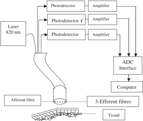
scattered light reemerges from the skin’s surface by diffuse reflectance [22–25]. The schematic of the multiprobe laser reflectometry is shown in Fig. 1.
Laser 820 nm
ADC
Interface
Computer
Photodetector Amplifier
Tissue
Afferent fibre 3-Efferent fibres Photodetector Amplifier
Photodetectorr Amplifier
. . .
Figure 1. Multiprobe laser reflectometry.
Laser light beam of a 0.1 cm diameter from a laser diode module, which is a compact semiconductor laser diode of 3mW power, operating at 820 nm, has been guided to the tissue surface by an optical fiber of 0.1 cm active diameter and 100 cm length. The diffusely backscattered light from the tissue surface is collected by three optical fibres of the same dimensions as that of the source fibre. These three fibres have been arranged parallel to each other in the measurement probe head with center-to-center separation of 0.2 cm. The diffusely backscattered light [26–29] signals from the tissue surface, collected by optical fibres have been converted into proportional current by three high speed, low noise silicon p-i-n diodes. The current outputs of these photo detectors were converted into their proportional voltage by three operational amplifiers in the current voltage converter mode. These have been in turn digitized by a 12-bit analog-to-digital converter and interfaced to a computer for storage and further analysis.
2.3. Data Acquisition
displayed on the monitor for analysis. While the cursor was moved across the forearm outline, the scanning probe was placed gently on the corresponding location on the human forearm and the data were collected for each and every finger. The data acquisition program to move the cursor across the hand outline was written in C.
3. RESULTS
The software system was implemented using MATLAB 7.1 Neural Network was used to compare the simulation result with experimental result of the tissue and it has a user-friendly interface facilitating interactions relevant to the imaging. Then the accuracy of the system was verified by comparing the theoretical and practical data from the developed software. The wounded region (abnormal) in the different layers of the organ was identified and detected by using the laser reflectometry technique.
The data acquisition has been carried out with the help of grid pattern, which completely covers the entire surface obtained. Then the simulation result is compared with that of the practical data obtained from laser reflectometry technique to verify the accuracy of the system. In this ANN of photon scattering, with and without abnormal tissue placed at various locations is compared. From the simulation result, correlation coefficient between nervous thickness and diffuse reflectance has been found out. Various size of skin lesions and with various diameter ring light source are correlated and results are obtained. We have obtained various optical properties such as reflectance, absorption and transmission for different inputs with refractive index (n), absorption coefficient (µa), scattering coefficient (µs), anisotropy (g), size of a layer (d) and number of incident photons for both normal and abnormal tissues.
Tumor Phantom
T R
Tumor Phantom Absorber
Figure 2. Comparison between measured and simulated result for a rotating phantom and tumor. (a) Geometry (b) Cross Section.
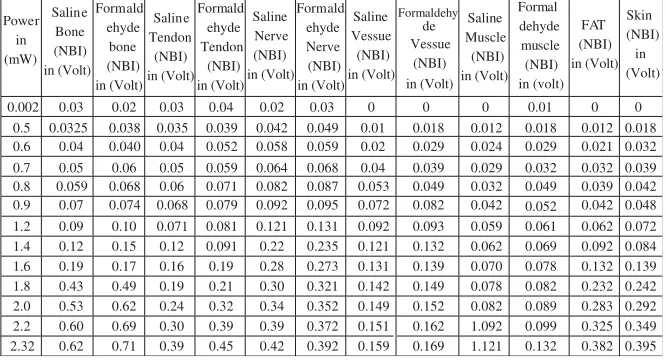
Table 1. Various human biological tissues optical properties using 820 nm Laser.
Power in (mW) Saline Bone (NBI) in (Volt) Formald ehyde bone (NBI) in (Volt) Saline Tendon (NBI) in (Volt) Formald ehyde Tendon (NBI) in (Volt) Saline Nerve (NBI) in (Volt) Formald ehyde Nerve (NBI) in (Volt) Saline Vessue (NBI) in (Volt) Formaldehy de Vessue (NBI) in (Volt) Saline Muscle (NBI) in (Volt) Formal dehyde muscle (NBI) in (volt) FAT (NBI) in (Volt) Skin (NBI) in (Volt)
0.002 0.03 0.02 0.03 0.04 0.02 0.03 0.01
0.5 0.0325 0.038 0.035 0.039 0.042 0.049 0.01 0.018 0.012 0.018 0.012 0.018 0.6 0.04 0.040 0.04 0.052 0.058 0.059 0.02 0.029 0.024 0.029 0.021 0.032 0.7 0.05 0.06 0.05 0.059 0.064 0.068 0.04 0.039 0.029 0.032 0.032 0.039 0.8 0.059 0.068 0.06 0.071 0.082 0.087 0.053 0.049 0.032 0.049 0.039 0.042 0.9 0.07 0.074 0.068 0.079 0.092 0.095 0.072 0.082 0.042 0.052 0.042 0.048 1.2 0.09 0.10 0.071 0.081 0.121 0.131 0.092 0.093 0.059 0.061 0.062 0.072 1.4 0.12 0.15 0.12 0.091 0.22 0.235 0.121 0.132 0.062 0.069 0.092 0.084 1.6 0.19 0.17 0.16 0.19 0.28 0.273 0.131 0.139 0.070 0.078 0.132 0.139 1.8 0.43 0.49 0.19 0.21 0.30 0.321 0.142 0.149 0.078 0.082 0.232 0.242 2.0 0.53 0.62 0.24 0.32 0.34 0.352 0.149 0.152 0.082 0.089 0.283 0.292 2.2 0.60 0.69 0.30 0.39 0.39 0.372 0.151 0.162 1.092 0.099 0.325 0.349 2.32 0.62 0.71 0.39 0.45 0.42 0.392 0.159 0.169 1.121 0.132 0.382 0.395
0
0 0 0 0
method. By this procedure the optical parameters of phantoms which are the same as that of tissues have been determined. Photons interacted with matter via scattering and absorption events. Laser irradiation of skin using homogeneous and layered geometries has been effectively simulated using MC method.
Table 1 shows the tissues obtained from various patients. we have developed a simulation software for enhancing the fast and accuracy of the system. Using this user could identify the status of the various optical parameters and also the level of tissue simulation during the light propagation. Hence this simulation would emulate the overall system perfect and gave more detailed information about these procedures. These values have been matched with each other in the same number of photons. Using this simulation programme the optical
0 1 2 2.5
0 SALINE NBI
* ANN NBI Formaldehyde NBI
FAT NBI *ANN NBI SKIN NBI
Δ Δ ANN NBI
ANN NBI
input power in mW input power in mW
0.5 1.5 0 0.5 1 1.5 2 2.5
NBI NBI 0 0.1 0.2 0.3 0.4 0.5 0.6 0.7 0.05 0.1 0.15 0.2 0.25 0.3 0.35 0.4
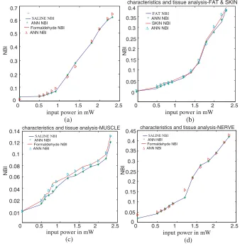
characteristics and tissue analysis-FAT & SKIN
(a) (b)
0
SALINE NBI
* ANN NBI Formaldehyde NBI
Δ
SALINE NBI
* ANN NBI Formaldehyde NBI
ANN NBI ΔANN NBI
0 1 2 2.5
input power in mW input power in mW
0.5 1.5 0 0.5 1 1.5 2 2.5
(c) (d) 0.01 0.02 0.04 0.06 0.08 0.1 0.12 0.14 0.05 0.1 0.15 0.2 0.25 0.3 0.35 0.4 0.45 NBI
characteristics and tissue analysis-MUSCLE characteristics and tissue analysis-NERVE
SALINE NBI * ANN NBI Formaldehyde NBI
ΔANN NBI
0
SALINE NBI * ANN NBI Formaldehyde NBI
ΔANN NBI
0 1 2
input power in mW input power in mW
0.5 1.5 0 0.5 1 1.5 2 2.5
(e) (f)
2.5 0.01
0.02 0.04 0.06 0.08 0.1 0.12 0.14
NBI
0.16 0.18
-0.1 0.1 0.2 0.3 0.4 0.5 0.6
NBI
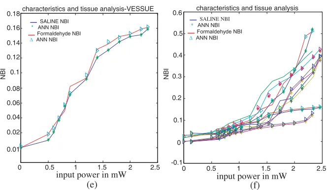
characteristics and tissue analysis-VESSUE characteristics and tissue analysis
Figure 3. Comparison of both saline and formaldehyde tissues with trained ANN results.
parameters were obtained. In this procedure, the simulation study has been carried out for 106 number of photons to propagate inside the three different layers. Based on the various input optical parameters such as photon count 10, absorption coefficient 1.0 cm,scattering coefficient 0.0, anisotropy coefficient 0.9 and number of layers 3. We have two approaches for analyzing the effect of an absorbing inclusion with different absorption coefficient on the detection of diffuse photons 1.Run a Monte Carlo Simulation for each absorption coefficient 2. Utilize the history file of detected photons achievements the path length each detected photon spent in each piece-wise constant region. To begin with, the initial position and direction of the photon is defined. The experimental values were trained by using ANN and plotted both the saline and formaldehyde solutions. Thus it is concluded that abnormal tissue graphs are slightly deviated from normal graph. The results are shown in Figs. 3(a)–(f).
4. DISCUSSION
this experimental set up. The back scattered intensity was very feeble Hence it was amplified by 106 ohm resistor through OP-AMP (TL 084-4 quadrant) circuit set up separately. To operate this circuit, external dual power supply of ±10 voltage was supplied to get the accurate value. The error values were gradually detected and made it them as zero. Then the set up was ready to measure the data from different human tissues samples that have been obtained from a private hospital to compare it. After that the optical properties of all samples were analysed and measured. These measured values were trained by using Feed Forward Backpropagation LM algorithm. Graphs of both saline and formaldehyde solutions tissues are shown in Fig. 4. Like wise we have analysed and plotted graphs for all tissues using this technique. Finally, These graphs were linearly match with the ANN value. In these figures, one sample put as reference and the remaining samples were compared with ANN simulation. Abnormal tissues were just deviated from the normal tissues which are measured after 2 hr of harvest. Then the accuracy of the system was verified by comparing the theoretical and practical data from the developed software. The wounded region (abnormal) in the different layers of the organ was identified and detected by using the laser reflectometry technique. The ANN graph used as reference and this was compared with both the normal and abnormal tissue. Hence, we compared all tissues to find the accuracy of the system. It should also be emphasized that the measurements were carried out on tissue stored at various low temperatures for various lengths of time. No significant variation in the values for µs, µa and g for these tissue samples was observed, as it was an evident from the values for the standard deviation for the measured values of these parameters. However these tissues might still not be truly representative of the native tissue. It was pertinent to emphasize that all the measurements were made on paired malignant and normal samples from individual patients. The results therefore strongly indicate of a significant difference in optical transparent parameters between malignant and normal tissue. The wounded region (abnormal) in the different layers of the organ was identified and detected by using the laser reflectometry technique.
The photons absorbed at various depths of the tissue have been computed. The points that were already at the grid points, without any interpolation of these have been carried out. To understand the accuracy of the estimates for µs and µa from the diffuse reflectance data, we first made measurement of tissue phantoms with known optical transport parameters.
biological tissues that depend on their composition and blood flow. The incidence light on the phantom is in the form of thin sheath of light but the transmitted component is received after a fixed separation. Because of this, the contribution due to scattering at the detector is increased. The reconstructed images of spatial variation of the backscattered intensity or depth variation of photons provided valuable data on the type, size and locations of abnormal tissue. In this paper, we consider locations and sizes of anomalies as the core information to search for and focus our attention on the fast and accurate estimation of them. MC Simulation is a statistical technique for simulating random processes and has been applied to light tissue interactions under wide variety of simulations.
ACKNOWLEDGMENT
The authors would like to thank All India Council For Technical Education (AICTE), Govt. of India, New Delhi for having supported the financial assistance for this work.
REFERENCES
1. Kwon, O. and Jeong et al., “Estimation of anomaly location and size using electrical impedance tomography,” IEEE Trans.
on Biomedical Engg., Vol. 50, 89–96, 2003.
2. Anderson, R. R. and J. A. Parrish, “The optics of human skin,”
J. Invest. Dermotol., Vol. 77, 13–19, 1981.
3. Van Gemert, T. M. J., S. L. Jacques, and H. J. C. Sterenborg, “Skin optics,”IEEE Trans. on Biomed. Engg., Vol. 36, 1146–1154, 1989.
4. Schmitt, J. M., G. X. Zhou, and E. C. Walker, “Multilayer model of photon diffusion in skin,”J. Opt. Soc. Am., Vol. A7, 2141–2153, 1990.
5. Hintz, S. R., D. A. Benaron, J. P. Vanhouten, J. L. Duckworth, H. S. Lic, D. K. Stevenson, and W. F. Cheong, “Stationary head band for clinical time of flight optical imaging at the bedside,”
Photochem. Photobiol., Vol. 68, 361–369, 1998.
6. Fantini, S., S. A. Walker, M. A. Franceschini, M. Kaschke, P. M. Schlag, and K. T. Moesta, “Assessment of the size, position and optical properties of breast tumors in viva by noninvasive optical methods,”Appl. Opt., Vol. 37, 1982–1989, 1998.
optical tomography of the human forearm,” Phys. Med. Boi., Vol. 46, 1117–1130, 2001.
8. Li, H., Y. Song, K. L. Worden, X. Jiang, A. Constantinescu, and R. P. Mason, “Non-invasive investigation for blood oxygenation dynamics of tumors by near-infrared spectroscopy,” Appl. Opt., Vol. 39, 5231–5243, 2000.
9. Hampel, U., E. Scheicher, H. Zepnick, and R. Freyer, “Clinical NIR spectroscopy and optical tomography of testis,” Proc.
SPIE2001, Vol. 4432, 210–220, 2001.
10. Jiao, S., G. Yao, and L. V. Wang, “Depth resolved two-dimensional stoke vectors of backscattered light and Mulller matrices of biological tissue measured with optical coherence tomography,”Appl. Opt., Vol. 39, 6318–6324, 2000.
11. Chacko, S. and M. Singh, “3-D reconstruction of transillumination tomographic images of human breast phantoms by red and infrared lasers,” IEEE Trans. Biomed. Eng., Vol. 47, 131–135, 2000.
12. Cubeddu, R., A. Pifferi, P. Taroni, A. Torricerlli, and G. Valentinil, “Imaging with diffusing light: An experimental study on the effect of the background optical properties,” Appl. Opt., Vol. 37, 3564–3573, 1998.
13. Schmitt, J. M., G. X. Zhou, and E. C. Walkker, “Multilayer model of photon diffusion in skin,”J. Opt. Soc. Amer. A, Vol. 7, 2141– 2153, 1990.
14. Colak, S. B., M. B. Van Mark, G. W. Hooft, J. H. Hoogenraad, E. S. Van der Linden, and F. A. Kuijpers, “Clinical optical tomography and NIR spectroscopy for breast cancer detection,”
IEEE J. Select Topics Quantum Electron., Vol. 5, 143–1158, 1999.
15. Chacko, S. and M. Singh, “Multi-layer imaging of human organs by measurement of laser back-scattering radiation,” Med. Biol.,
Eng. Comput., Vol. 37, 278–284, 1999.
16. Colak, S. B., M. B. Van Mark, G. W. Hoof, J. H. Hoogenraad, E. S. Van der Linden, and F. A. Kuijpers, “Clinical optical tomography and NIR spectroscopy for breast cancer detection,”
IEEE J. Select Topics Quantum Electron., Vol. 5, 1143–1158,
1999.
17. Cubeddu, R., A. Pifferi, P. Taroni, A. Torricelli, and G. A. Valentini, “Solid tissue phantom for photon migration studies,”Phys. Med. Biol., Vol. 42, 1971–1979, 1997.
the cerebro spinal fluid layer on light on light distribution in the tissue,”Appl. Op., Vol. 39, 4721–4729, 2000.
19. Flock, S. T., M. S. Patterson, B. C. Wilson, and D. R. Wyman, “Monte Carlo modeling of light propagation in highly scattering tissue—I: Model predictions and comparison with diffusion theory,” IEEE Trans. Biomed., Vol. 36, 1162–1168, 1989.
20. Grosenick, D., H. Wabnitz, H. Hrinneberg, and K. T. Oesta, “Development of a time-domain optical mannography and first invivo applications,” Appl. Opt., Vol. 38, 2927–2943, 1999. 21. Van Stavren, H. J., C. J. M. Moses, J. Van Maries, S. A. Prahl,
and M. J. C. Van, “Light scattering in intra lipid 10% in the wavelength range of 400–1100 nm,” Appl. Opt., Vol. 30, 4507– 4514, 1991.
22. Farrell, T. J., M. S. Patterson, and M. Essenpresis, “Influence of layered tissue architecture on estimates of tissue optical properties obtained from spatially resolved diffuse reflectometry,”Appl. Opt., Vol. 37, 1958–1972, 1998.
23. Chinn, S. R., E. A. Swanson, and J. G. Fujimoto, “Optical coherencetomography using a frequency tunable optical source,”
Opt. Lett., Vol. 22, 340–342, 1997.
24. Pougue, B. W., et al., “Three dimensional simulation of near infrared diffusion in tissue: Boundary condition and geometry analysis for finite-element image reconstruction,” Appl. Optics, Vol. 40, 588–599, 2001.
25. Mitic, G., J. Kober, J. Otto, E. Piles, E. Solkner, and W. Zinth, “Time gated transillumination of biological tissues and tissue like phantoms,” Appl. Opt., Vol. 33, 6699–6709, 1994.
26. Anderson-Engles, S., R. Berg, S. Svanberg, and O. Jarlman, “Time resolved transillumination for medical diagnostics,” Opt.
Lett., Vol. 15, 1179–1181, 1990.
27. Torricelli, A., A. Pifferi, P. Taroni, E. Giambattistelli, and R. Cubeddu, “Invivo optical characterization of human tissues from 610 to 1010 nm by time resolved reflectance spectroscopy,”
Phys. Med. Biol., Vol. 46, 2227–2237, 2001.
28. Arridge, S. R., Z. P. Vander, D. T. Delpy, and M. Cope, “Reconstruction methods of infra-red absorption imaging,”Proc. SPIE, Vol. 1431, 204–215, 1991.
29. Shanthi, S. and M. Singh, “Laser reflectance imaging of human organs and comparison with perfusion images,” Med. Biol.Eng.