Antigen (VCA) IgG, VCA IgM, and EBV Nuclear Antigen 1 IgG
Chemiluminescent Immunoassays for Detection of EBV Antibodies
and Categorization of EBV Infection Status Using
Immunofluorescence Assays as the Reference Method
Isabel Corrales,aEstela Giménez,aDavid Navarroa,b
Microbiology Service, University Clinic Hospital Institute for Clinical Research (INCLIVA),a
and Department of Medicine, School of Medicine, University of Valencia,b Valencia, Spain
Commercial immunoassays for detecting IgG and IgM antibodies against Epstein-Barr virus (EBV), viral capsid antigens (VCA), and IgGs toward EBV nuclear antigen-1 (EBNA-1) are routinely used in combination to categorize EBV infection status. In this study, we evaluated the performances of the Architect EBV VCA IgG, VCA IgM, and EBNA-1 IgG chemiluminescent micropar-ticle assays (CMIAs) in EBV serological analyses using indirect immunofluorescence assays and anticomplement immunofluo-rescence assays as the reference methods for VCA IgG, VCA IgM, and EBNA-1 IgG antibody detection, respectively. A total of 365 serum samples representing different EBV serological profiles were included in this study. Thevalues (concordances between the results) obtained in the Architect CMIA and those in the reference assays were 0.905 (P<0.0001) for VCA IgM, 0.889 (P< 0.0001) for VCA IgG, and 0.961 (P<0.0001) for EBNA-1 IgG. The sensitivities and specificities were, respectively, 91.08% and 99.48% for VCA IgM, 99.23% and 86.27% for VCA IgG, and 96.77% and 99.16% for EBNA-1 IgG. The sensitivities and specifici-ties of the Architect CMIA panel were, respectively, 99.15% and 98.6% for diagnosing a primary infection, 97.62% and 93.39% for diagnosing a past EBV infection, and 92.42% and 97.82% for diagnosing the absence of an EBV infection. In summary, we demonstrated that the Architect EBV antibody panel performs very well for EBV antibody detection and correctly categorizes clinically relevant EBV infection states.
C
ommercial immunoassays for detecting IgG and IgM anti-bodies against Epstein-Barr virus (EBV) and viral capsid an-tigen (VCA) and IgG antibody toward EBV nuclear anan-tigen-1 (EBNA-1) are routinely used in combination to diagnose primary EBV infection (i.e., acute infectious mononucleosis [IM]) and to categorize EBV infection status. The latter is particularly relevant in solid-organ transplant patients in order to assess the risk of posttransplantation lymphoproliferative disease (EBV-seronega-tive patients receiving an allograft from EBV-seroposi(EBV-seronega-tive donors) (1). Abbott Diagnostics (Wiesbaden, Germany) recently launched the Architect EBV antibody panel, which includes three two-step chemiluminescent microparticle immunoassays (CMIAs) for qualitatively detecting VCA IgG, VCA IgM, and EBNA-1 IgG an-tibodies on its automated random-access platform Architecti2000SR. In this study, we evaluated the performances of the Ar-chitect EBV VCA IgG, VCA IgM, and EBNA-1 IgG CMIAs in EBV serological analyses using indirect immunofluorescence (IIF) as-says and anticomplement immunofluorescence (ACIF) asas-says as the reference methods for VCA IgG, VCA IgM, and EBNA-1 an-tibody (Ab) detection, respectively (1).
MATERIALS AND METHODS
Serum specimens.A total of 365 serum samples representing different EBV serological profiles commonly encountered in clinical practice (as determined by IIF and ACIF) were included in this study. These speci-mens were selected from sera submitted to our laboratory between Janu-ary 2010 and March 2012 for routine EBV-specific antibody testing. Most of the serum samples (82%) belonged to children or young adolescents (median age, 8 years; range, 1 to 14 years; 61% male and 39% female) with fever, rash, or clinical suspicion of IM. In our laboratory, we routinely test
these samples with the Liaison VCA IgM, VCA IgG, and EBNA-1 IgG chemiluminescent assays (CLIAs) (DiaSorin, Saluggia, Italy) (2,3). In addition, sera from patients with a high suspicion of EBV-related IM are screened for the presence of heterophilic antibodies (HAs) as described below (2,3). The proportions of different EBV serological patterns in-cluded in this study do not represent the frequencies at which they are observed in routine laboratory EBV testing (1). The serum sample ali-quots used were stored at⫺20°C immediately after separation. The spec-imens were retrieved for EBV antibody testing by IIF and by the Architect EBV antibody panel. The EBV antibody profiles of the sera (according to IIF/ACIF methods) included (i) VCA IgG-negative (IgG⫺)/VCA IgM⫺/ EBNA-1 Ab⫺ (n ⫽ 68), (ii) VCA IgG⫺/VCA IgM-positive (IgM⫹)/ EBNA-1 Ab⫺(n⫽25), (iii) VCA IgG⫹/VCA IgM⫹/EBNA-1 Ab⫺(n⫽ 116), (iv) VCA IgG⫹/VCA IgM⫺/EBNA-1 Ab⫹(n⫽89), (v) VCA IgG⫹/ VCA IgM⫺/EBNA-1 Ab⫺(n⫽31), (vi) VCA IgG⫹/VCA IgM⫹/EBNA-1 Ab⫹(n⫽25), and (vii) VCA IgG⫺/VCA IgM⫺/EBNA-1 Ab⫹(n⫽11).
Architect Epstein-Barr virus chemiluminescent microparticle im-munoassays.These CMIAs are two-step chemiluminescent immunoas-says that use peptide-coated microparticles (VCA p18 or EBNA-1 p72) and acridine-labeled anti-IgG or anti-IgM conjugates for qualitatively de-tecting VCA IgM, VCA IgG, and EBNA-1 IgG antibodies. Samples were processed on a fully automated random-access analyzer (the Architect
Received25 February 2014 Accepted3 March 2014
Published ahead of print12 March 2014
Editor:R. L. Hodinka
Address correspondence to David Navarro, david.navarro@uv.es.
Copyright © 2014, American Society for Microbiology. All Rights Reserved.
doi:10.1128/CVI.00104-14
on August 17, 2020 by guest
http://cvi.asm.org/
i2000SR). The chemiluminescent signals were measured by a photomul-tiplier tube and expressed as relative light units (RLU); the Architecti system calculates each result by using the ratio of the sample RLU to the cutoff RLU (S/CO). The criteria for interpretation of individual parame-ters were as follows: for VCA IgM,⬍0.50 RLU was considered negative, 0.50 to 1 RLU equivocal, andⱖ1 RLU positive; for VCA IgG,⬍0.75 RLU was considered negative, 0.75 to 1 RLU equivocal, andⱖ1 RLU positive; and for EBNA-1 IgG,⬍0.50 RLU was considered negative, 0.50 to 1 RLU equivocal, andⱖ1 RLU positive. Sera were batched and tested simultane-ously over several consecutive days.
Immunofluorescence assays.IIF assays for VCA IgG and IgM were performed with the Merifluor EBV VCA IgG and VCA IgM IIF assays (Meridian Bioscience Inc.). The Merifluor EBV VCA IgM and EBV VCA IgG IIF assay methods are qualitative. EBV-infected lymphocytes from Burkitt lymphoma were incubated with patient serum. After being washed, cells complexed with bound anti-VCA antibodies were incubated with either anti-human IgM or anti-human IgG labeled with fluorescein. The sample is considered to be positive if approximately 10% to 20% of the cells in each field show apple-green fluorescence upon observation. ACIF was used to detect EBNA-1 antibodies (Merifluor EBV nuclear an-tigen test; Meridian Bioscience Inc.). Heat-inactivated patient serum was applied to the fixed antigens (EBV-infected lymphocytes from Burkitt lymphoma) on glass wells of a microscope slide. Following a washing step, guinea pig complement was added to react with any antigen-antibody complexes. After a 30-min incubation, the slides were washed. Fluoresce-in-conjugated goat antibody against the C3 component of guinea pig complement was added to react with the antigen-complement complexes. Positive reactions appeared as 20 to 30% of the cells exhibiting bright apple-green fluorescence against a background of counterstained EBNA-1 negative-control cells. These procedures were conducted and interpreted following the manufacturer’s instructions. IIF assays were read bv a single person (I.C.). The reader was blinded to the CLIA results.
Detection of heterophilic antibodies.HAs were detected by a differ-ential agglutination assay (I.M. kit; Microgen, Surrey, Great Britain).
Interpretation of Epstein-Barr virus serostatus.The criteria used to define the EBV serostatus were based on consensus EBV-specific antibody profiles (1). The VCA IgG⫺VCA IgM⫺EBNA-1 IgG⫺profile corre-sponded to an EBV-seronegative status. The VCA IgG⫹/VCA IgM⫹/ EBNA-1 IgG⫺and VCA IgG⫺/VCA IgM⫹/EBNA-1 IgG⫺/HA⫹patterns were interpreted as compatible with a primary EBV infection. The VCA IgG⫹/VCA IgM⫺/EBNA-1 IgG⫹profile represented a past EBV infection. The remaining EBV-specific antibody profiles were considered indeter-minate and were analyzed separately.
Statistical analysis.Kappa () statistics were used to evaluate the de-gree of consensus between the results of the Architect EBV assays and those of the immunofluorescence (IF) assays. These analyses were per-formed with the aid of the SPSS statistical package (version 20.0).
RESULTS
Single-parameter performance of the Architect immunoassays. Most of the sera (n⫽341) yielded S/CO values either above or below the established cutoff for the respective assay. Twenty-four serum samples gave S/CO values within the gray zone (GZ) in one or two EBV antibody assays: 17 (4.66%) in the VCA IgM assay, 4 (1.10%) in the EBV IgG assay, and 4 (1.10%) in the EBNA-1 IgG assay. Sera giving GZ S/CO values were excluded from the analyses described below. The concordances between the results obtained in the Architect CMIAs and those in the reference assay were good for all three parameters ( ⫽0.905 andP⬍0.0001 for VCA IgM;
⫽0.889 andP⬍0.0001 for VCA IgG; ⫽0.961 andP⬍0.0001 for EBNA-1 IgG). The single-parameter performances of the Ar-chitect CMIAs compared with those of the reference assay are shown inTables 1and2. The EBNA-1 IgG assay best matched the IF assay (ACIF assay), with only 6 discordant results, thus yielding
excellent sensitivity and specificity values (Table 2). The VCA IgG assay gave the lowest specificity, whereas the VCA IgM assay gave the lowest sensitivity.
Performance of the Architect assays for diagnosing clinically relevant Epstein-Barr virus infection statuses.The ability of the Architect assays to correctly identify the absence of a previous EBV infection or evidence of either a primary or a past EBV infection was assessed. Sera that displayed an isolated VCA IgM⫹profile were considered to correspond to a primary EBV infection only when they also tested positive for HA; the raw data are shown in Table 3. The concordances between the EBV antibody profiles obtained with the Architect panel and those with the reference assay were 97.4% for primary EBV infection, 97.5% for past EBV infection, and 92.4% for EBV seronegativity. The sensitivities and specificities of the Architect CMIA panel were, respectively, 99.15% and 98.6% for diagnosing a primary infection, 97.62% and 93.39% for diagnosing a past EBV infection, and 92.42% and 97.82% for diagnosing the absence of an EBV infection.
Performance of the Architect assays with indeterminate Ep-stein-Barr virus serological profiles.We also evaluated the per-formance of the Architect assays with sera displaying unresolved EBV serological profiles (n⫽73;Table 3). The overall agreement between the Architect panel and the reference method for these sera was modest (65.7%). The following findings were of particu-lar interest. (i) Two VCA IgM⫹/VCA IgG⫺/EBNA-1 IgG⫺/HA⫺ serum samples (n⫽8) were categorized as primary EBV infec-tions by the Architect panel. These sera belonged to two children (2 and 3 years old) with a high-level suspicion of EBV-related IM. One of the 8 serum samples tested negative for all three parameters in the Architect panel. This serum was obtained from an adult patient with persistent fever and an absence of clinical signs com-patible with IM. (ii) Concordant results between the IF assays and the Architect CMIAs were obtained for 23 of 25 serum samples with an isolated VCA IgG profile. The remaining two samples were interpreted by the Architect panel as compatible with a past infection. None of the 25 serum samples tested positive in the HA assay. (iii) Eleven of 21 serum samples displaying a VCA IgM⫹/ VCA IgG⫹/EBNA-1 IgG⫹/HA⫺profile were interpreted as corre-sponding to past EBV infections by the Architect panel. Most (n⫽
8) of these serum samples were obtained from children and young adults with a low-level suspicion of IM (persistent fever). (iv) The Architect panel interpreted one of three VCA IgM⫹/VCA IgG⫹/ EBNA-1 IgG⫹/HA⫹serum samples as corresponding to a primary
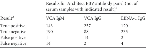
TABLE 1Single-parameter performances of the Architect EBV chemiluminescent immunoassay in comparison with those of the reference immunofluorescence methods
Resulta
Results for Architect EBV antibody panel (no. of serum samples with indicated result)b
VCA IgM VCA IgG EBNA-1 IgG
True positive 143 257 120
True negative 190 88 235
False positive 1 14 2
False negative 14 2 4
a
Sera tested by the Architect EBV panel were categorized as true or false
positives/negatives on the basis of their agreement or disagreement, respectively, with immunofluorescence assay results.
bSera displaying values within the gray zone of the respective assay were excluded from
the analyses.
on August 17, 2020 by guest
http://cvi.asm.org/
EBV infection. (v) Four of 11 isolated EBNA-1 IgG profile serum samples tested positive for both EBNA-1 IgG and VCA IgG in the Architect panel.
DISCUSSION
In this study, we evaluated the performance of the Architect EBV panel for EBV antibody detection using IF assays as a reference. IF assays have historically been considered the gold standard for EBV antibody testing, despite the fact that the reading of the assay re-sults is subjective. Thus, some researchers advocate for the use of immunoblot assays as the reference method (1). Regarding the choice of IF assays as gold standard methods, one must bear in mind that while these methods are considered to be more specific than enzyme immunoassays (EIAs), their sensitivities have occa-sionally been reported to be lower than those of EIAs, especially for VCA IgM and VCA IgG antibody detection (1).
We performed a single-parameter analysis and assessed the ability of the Architect antibody panel to correctly categorize clin-ically relevant EBV infection staging (primary infection to diag-nose EBV IM, past infection to exclude EBV IM and to ascertain previous viral exposure in transplant recipients or donors, and EBV seronegativity in both immunocompetent and transplant
pa-tients to show EBV susceptibility) (1). Finally, we evaluated the performance of the Architect panel with sera displaying unsolved EBV serological profiles. Interestingly, we obtained a re-markably low rate of indeterminate results (within the GZ of the assay) using the EBNA-1 IgG CMIA (1.10%) compared with those reported for other chemiluminescent assays (30% for the Dia-Sorin CLIA and 4% for the Immulite 2000 CLIA) (4). The fre-quencies with which sera gave S/COs within the GZ in the VCA IgG assay of 1.10% and in the VCA IgM assay of 4.66% were comparable to those of the above-mentioned CLIAs (4).
Single-parameter analyses revealed very good concordance be-tween the Architect EBV assays and the reference methods, which translated into excellent sensitivities and specificities for all three assays. The performance of several commercially available CLIAs compared to an IF assay as the reference method was assessed in two previous studies (4,5). The data reported in those studies are summarized inTable 2. In this context, the Architect VCA IgM and EBNA-1 IgG assays appeared to perform better than the CLIAs from DiaSorin and Siemens. The sensitivity of the Architect VCA IgG assay was superior to those reported for the other two CLIAs (described above), whereas its specificity was slightly lower (4,5). These differences could be attributable to several factors, including the use of different antigen preparations, solid phases, IF assays, and serum dilutions.
In this study, the overall agreements between the expected EBV status, as determined by the IF assays, and that obtained with the Architect antibody panel were excellent (97.4% for primary EBV infection, 97.5% for past EBV infection, and 92.4% for EBV sero-negativity). The most frequent EBV staging discrepancy between the Architect antibody panels and the IF assays was the occurrence of an isolated VCA IgG pattern in 5 patients with an expected EBV-seronegative profile. Although we assumed these to be Ar-chitect assay-specific false positives, we cannot rule out the possi-bility of them being true positives because follow-up specimens from these patients were not available. As stated above, it has been reported that EIAs might display better sensitivities than IF assay for VCA antibody detection (1). The above figures are in the upper range (74 to 95%) reported for several commercially available CLIAs or EIAs that were compared with immunofluorescence, Western blot, or line blot assays as reference assays (4–10).
Indeterminate, unresolved EBV antibody profiles occur
rela-TABLE 2Performances of three commercially available chemiluminescent immunoassays in detecting VCA IgG, VCA IgM, and EBNA-1 IgG antibodies compared with those of immunofluorescence assays as reference
Immunoassay
Sensitivity (%) Specificity (%)
VCA IgM VCA IgG EBNA-1-IgG VCA IgM VCA IgG EBNA-1-IgG
Architecta 91.0 99.2 96.7 99.4 86.2 99.1
DiaSorin
Meridianb 92.2 86.9 90.6 98.0 94.4 32.2
In-housec 94.4 90.2 95.9 76.9 95.8 83.3
Siemensd 77.9 94.4 93.8 95.8 100 76.6
aSera tested by the Architect EBV panel were categorized as true or false positives/negatives on the basis of their agreement or disagreement with immunofluorescence assay results.
Sera displaying values within the gray zone of the respective assay were excluded from the analyses.
bThe performances of the Liaison VCA IgM, VCA IgG, and EBNA-1 IgG chemiluminescent assays (CLIAs) (DiaSorin, Saluggia, Italy) were evaluated using theIF assays from
Meridian (like in the current study) as a reference (4).
cThe performances of the Liaison VCA IgM, VCA IgG, and EBNA-1 IgG chemiluminescent assays were evaluated using in-houseIF assays as a reference (5). d
The Immulite 2000 CLIA (Siemens, Germany) was evaluated using theIF assays from Meridian (like in the current study) as a reference (4).
TABLE 3Performance of the Architect EBV chemiluminescent immunoassay in categorizing sera with several EBV serological profiles, as determined by immunofluorescence reference methods
IF antibody profile (VCA IgM/VCA IgG/EBNA-1 IgG) (no. of serum samples)
Results (no. of serum samples) for the indicated Architect panel EBV antibody profile (VCA IgM/VCA IgG/EBNA-1 IgG)
⫺/⫺/⫺ ⫹/⫺/⫺ ⫹/⫹/⫺ ⫹/⫹/⫹ ⫺/⫹/⫹ ⫺/⫹/⫺ ⫺/⫺/⫹
⫺/⫺/⫺(66) 61 0 0 0 0 5 0
⫹/⫺/⫺(8)a
1 5 2 0 0 0 0
⫹/⫺/⫺(13)b 3 8 2 0 0 0 0
⫹/⫹/⫺(110) 0 2 108 0 0 0 0
⫹/⫹/⫹(21)b 0 0 0 10 11 0 0
⫹/⫹/⫹/(3)a
0 0 1 2 0 0 0
⫺/⫹/⫹(84) 0 0 0 1 82 1 0
⫺/⫹/⫺(25) 0 0 0 0 2 23 0
⫺/⫺/⫹(11) 2 0 0 0 4 0 5
a
Sera tested positive for heterophilic antibodies (HAs).
bSera tested negative for HAs.
on August 17, 2020 by guest
http://cvi.asm.org/
tively frequently in routine laboratory EBV serological testing (1, 8). The presence of an isolated VCA IgM profile in the absence of HAs most commonly reflects either the presence of a primary infection (particularly in children⬍5 years of age) or a false-pos-itive result. In our series, 2 of 13 serum samples with this pattern were categorized as primary infections by the Architect panel. These sera belonged to two children with a high-level suspicion of EBV-related IM. Thus, although speculative, these may have been false-negative VCA IgG results that were obtained using the refer-ence method (1).
The presence of an isolated VCA IgG profile occurs in 2 to 8% of immunocompetent patients tested for the EBV antibody (8, 11), and most of these cases are thought to correspond to past infections. We included 25 serum samples with this EBV antibody profile, none of which tested positive in the HA assay. Two of these 25 samples also tested positive for EBNA-1 IgGs in the Architect panel. These serum samples were drawn from two adult patients with low-level suspicion of IM, meaning that the results might have represented true positives for EBNA-1 IgGs (1).
VCA IgG, VCA IgM, and EBNA-1 IgG antibodies are detected concomitantly in 3 to 6.4% of samples subjected to routine EBV testing (8, 9, 12). In most cases, this profile reflects a past EBV infection, although it may also correspond to a relatively recent EBV-related primary infection, an EBV reactivation episode, a cross-reaction with CMV IgM in the setting of a primary CMV infection, or a state of polyclonal stimulation by a heterologous infectious agent (1,13,14). In this study, 11 of 21 samples with a VCA IgM⫹/VCA IgG⫹/EBNA-1 IgG⫹/HA⫺profile were inter-preted as corresponding to past EBV infections by the Architect panel. Most of these sera were obtained from children and young adults with a low-level suspicion of IM, so they might have been true-negative VCA IgM results. In addition, one out of three VCA IgM⫹/VCA IgG⫹/EBNA-1 IgG⫹/HA⫹serum samples was inter-preted as corresponding to a primary EBV infection by the Archi-tect panel, which we assume was a true negative for EBNA-1 IgG⫹, given that this serum sample belonged to a patient with clear signs and symptoms of IM.
The presence of an isolated EBNA-1 IgG profile has been re-ported in 1.4 to 5.3% of sera tested with commercial EIAs (8,15). Although classic texts consider this to be implausible, it has been shown in most cases to correspond to past infections (8,15). In this study, 4 of 11 serum samples that gave an isolated EBNA-1 IgG profile with the IF assay were EBNA-1 IgG and VCA IgG positive in the Architect panel, again suggesting a plausible possi-bility of these being true positives for VCA IgG.
This study had several limitations. First, no follow-up speci-mens were available from the patients. Second, further testing (VCA IgG avidity or real-time PCR assays) was not performed on sera displaying unresolved EBV antibody profiles. Third, a defin-itive clinical diagnosis was not available for most of the patients. Fourth, sera giving discordant results in the index and reference assays were not reassayed using a second reference method (West-ern or line blot assays) to resolve the discrepancies. Fifth, poten-tially cross-reactive sera, such as those obtained during primary cytomegalovirus or human herpesvirus 6 infection or parvovirus B19 infection (16,17), were not included in our study panel. Any of these factors may have caused some misclassifications (partic-ularly with sera displaying unresolved EBV antibody profiles) and thus might have skewed our calculations of the sensitivities and specificities. In addition, evaluation of the diagnostic accuracy of
the Architect EBV panel under routine laboratory conditions, which was not performed in the current study, seems crucial for ascertaining the actual clinical value of the antibody panel. Studies addressing this issue are under way. Despite these limitations, the data presented here indicate that the Architect EBV antibody panel performs very well for EBV antibody detection and correctly categorizes clinically relevant EBV infection states.
ACKNOWLEDGMENTS
Abbott Diagnostics provided the reagent kits for the serological analyses free of charge.
David Navarro received honoraria from Abbott Diagnostics for his attendance at two conferences. The other authors have no conflicts of interest.
REFERENCES
1.Hess RD.2004. Routine Epstein-Barr virus diagnostics from the labora-tory perspective: still challenging after 35 years. J. Clin. Microbiol.42:
3381–3387.http://dx.doi.org/10.1128/JCM.42.8.3381-3387.2004. 2.Bravo D, Muñoz-Cobo B, Costa E, Clari MA, Tormo N, Navarro D.
2009. Evaluation of an immunofiltration assay that detects immunoglob-ulin M antibodies against the ZEBRA protein for the diagnosis of Epstein-Barr virus infectious mononucleosis in immunocompetent patients. Clin. Vaccine Immunol.16:885– 888.http://dx.doi.org/10.1128/CVI.00123-09. 3.Meza LD, Sancho-Tello S, Muñoz-Cobo B, Costa E, Bravo D, Pazos JM, Corrales I, Marcano X, Tohalino M, Navarro D.2012. Performance of an immunofiltration assay detecting IgM antibodies against ZEBRA and viral capsid p18 proteins (Immunoquickfiltration EBV M) for the diagno-sis of heterophile antibody-negative primary Epstein-Barr virus infection in children. J. Clin. Virol. 53:270 –271.http://dx.doi.org/10.1016/j.jcv .2011.12.008.
4.de Ory F, Guisasola ME, Sanz JC, Garcia-Bermejo I.2011. Evaluation of four commercial systems for the diagnosis of Epstein-Barr virus primary infections. Clin. Vaccine Immunol. 18:444 – 448. http://dx.doi.org/10 .1128/CVI.00486-10.
5.Gärtner BC, Gärtner BC, Hess RD, Bandt D, Kruse A, Rethwilm A, Roemer K, Mueller-Lantzsch N.2003. Evaluation of four commercially available Epstein-Barr virus enzyme immunoassays with an immunoflu-orescence assay as the reference method. Clin. Diagn. Lab. Immunol.10:
78 – 82.http://dx.doi.org/10.1128/CDLI.10.1.78-82.2003.
6.Binnicker MJ, Jespersen DJ, Harring JA, Rollins LO, Beito EM.2008. Evaluation of a multiplex flow immunoassay for detection of Epstein-Barr virus-specific antibodies. Clin. Vaccine Immunol.15:1410 –1413.http: //dx.doi.org/10.1128/CVI.00082-08.
7.Debyser Z, Reynders M, Goubau P, Desmyter J.1997. Comparative evaluation of three ELISA techniques and an indirect immunofluores-cence assay for the serological diagnosis of Epstein-Barr virus infection. Clin. Diagn. Virol. 8:71– 81. http://dx.doi.org/10.1016/S0928-0197(97) 00014-7.
8.Klutts JS, Ford BA, Perez NR, Gronowski AM. 2009. Evidence-based approach for interpretation of Epstein-Barr virus serological patterns. J. Clin. Microbiol.47:3204 –3210.http://dx.doi.org/10.1128/JCM.00164-09. 9.Lupo J, Germi R, Semenova T, Buisson M, Seigneurin JM, Morand P.
2012. Performance of two commercially available automated immunoas-says for the determination of Epstein-Barr virus serological status. Clin. Vaccine Immunol.19:929 –934.http://dx.doi.org/10.1128/CVI.00100-12. 10. Kreuzer C, Nabeck KU, Levy HR, Daghofer E.2013. Reliability of the Siemens Enzygnost and Novagnost Epstein-Barr virus assays for routine laboratory diagnosis: agreement with clinical diagnosis and comparison with the Merifluor Epstein-Barr virus immunofluorescence assay. BMC Infect. Dis.13:260.http://dx.doi.org/10.1186/1471-2334-13-260. 11. De Paschale M, Agrappi C, Manco MT, Mirri P, Viganò EF, Clerici P.
2009. Seroepidemiology of EBV and interpretation of the “isolated VCA IgG” pattern. J. Med. Virol.81:325–331.http://dx.doi.org/10.1002/jmv .21373.
12. Nystad TW, Myrmel H.2007. Prevalence of primary versus reactivated Epstein-Barr virus infection in patients with VCA IgG-, VCA IgM- and EBNA-1-antibodies and suspected infectious mononucleosis. J. Clin. Vi-rol.38:292–297.http://dx.doi.org/10.1016/j.jcv.2007.01.006.
13. Gulley ML, Tang W.2008. Laboratory assays for Epstein-Barr
on August 17, 2020 by guest
http://cvi.asm.org/
related disease. J. Mol. Diagn. 10:279 –292. http://dx.doi.org/10.2353 /jmoldx.2008.080023.
14. Odumade OA, Hogquist KA, Balfour HH, Jr.2011. Progress and prob-lems in understanding and managing primary Epstein-Barr virus infec-tions. Clin. Microbiol. Rev.24:193–209.http://dx.doi.org/10.1128/CMR .00044-10.
15. García T, Tormo N, Gimeno C, Navarro D.2008. Assessment of Epstein-Barr virus (EBV) serostatus by enzyme immunoassays: plausibility of the isolated EBNA-1 IgG positive serological profile. J. Infect.57:351–353. http://dx.doi.org/10.1016/j.jinf.2008.07.017.
16. Berth M, Bosmans E.2009. Acute parvovirus B19 infection frequently causes false-positive results in Epstein-Barr virus- and herpes simplex vi-rus-specific immunoglobulin M determinations done on the Liaison plat-form. Clin. Vaccine Immunol. 16:372–375. http://dx.doi.org/10.1128 /CVI.00380-08.
17. Costa E, Tormo N, Clari MA, Bravo D, Muñoz-Cobo B, Navarro D.
2009. Performance of the Epstein-Barr virus and herpes simplex virus immunoglobulin M assays on the Liaison platform with sera from patients displaying acute parvovirus B19 infection. Clin. Vaccine Immunol.16:
1247–1248.http://dx.doi.org/10.1128/CVI.00142-09.