Western University Western University
Scholarship@Western
Scholarship@Western
Electronic Thesis and Dissertation Repository
8-2-2016 12:00 AM
Characterization and personalization of botulinum toxin type A
Characterization and personalization of botulinum toxin type A
therapy for upper limb tremor in Parkinson disease and Essential
therapy for upper limb tremor in Parkinson disease and Essential
tremor patients using multi-sensor kinematic technology
tremor patients using multi-sensor kinematic technology
Olivia Samotus
The University of Western Ontario
Supervisor Dr. Mandar Jog
The University of Western Ontario Graduate Program in Neuroscience
A thesis submitted in partial fulfillment of the requirements for the degree in Master of Science © Olivia Samotus 2016
Follow this and additional works at: https://ir.lib.uwo.ca/etd
Part of the Nervous System Diseases Commons, Neurosciences Commons, and the Other Analytical, Diagnostic and Therapeutic Techniques and Equipment Commons
Recommended Citation Recommended Citation
Samotus, Olivia, "Characterization and personalization of botulinum toxin type A therapy for upper limb tremor in Parkinson disease and Essential tremor patients using multi-sensor kinematic technology" (2016). Electronic Thesis and Dissertation Repository. 4035.
https://ir.lib.uwo.ca/etd/4035
This Dissertation/Thesis is brought to you for free and open access by Scholarship@Western. It has been accepted for inclusion in Electronic Thesis and Dissertation Repository by an authorized administrator of
i
Abstract
Tremor commonly affects the upper extremities in essential tremor (ET) and Parkinson disease
(PD) patients where many experience functional disability and ultimately seek therapy. As ET and
PD tremor features overlap and clinical assessment is challenging due to its highly complex
nature, misdiagnosis is common resulting in unsuitable therapies and prognosis. Current treatment
options for ET and PD tremor include pharmacotherapy, focal therapy with botulinum toxin type
A (BoNT-A) injections, and surgical interventions which provide modest relief of tremor.
However, such therapies are commonly associated with significant adverse events and lack
long-term efficacy and tolerability. Hence lack of standardized, objective measures of tremor and
suboptimal treatment options are two significant unmet needs faced by neurologists today. The
hypothesis of this thesis was to determine whether joint tremor amplitude can differentiate
between ET and PD tremor types and can be applied towards improving BoNT-A tremor therapy.
The first objective was to apply motion sensor kinematic technology to investigate the role of
paired tasks in modulating tremor biomechanics in 24 ET and 28 PD participants. Paired tasks
involved variating limb positioning while at rest, posture, and under weight-bearing conditions.
Motion sensor devices were placed over the wrist, elbow and shoulder joints capturing joint
angular tremor amplitude in multiple degrees of freedom (DOF). Kinematic measures of tremor
allowed detailed segmentation of tremor into directional components, which cannot be performed
visually. The relationship of joint tremor severity between paired tasks and across all tasks
generated unique tremor profiles and provided a simple method to differentiate ET and PD tremor
types. The second objective was to apply tremor kinematics to better tailor BoNT-A injection
parameters. Participants were injected in the upper limb, which exhibited their most bothersome
tremor, every 16 weeks, a total of 3 injection cycles, and attended follow-up visits six weeks
following treatment, for a total of 6 study visits. Clinical rating scales and kinematic recordings
were conducted at each visit.Dosing was based on clinician’s experience and kinematic data, and
muscle site of injection was determined kinematically. A significant decrease in mean clinical
tremor rating scores during rest and action tasks and significant improvement in arm function was
observed at week 6 and continued throughout the study in both ET and PD individuals. Ten PD
participants and eight ET participants reported mild weakness in injected muscles that had no
interference with arm function. Kinematic technology is a promising method for standardizing
ii
Keywords
Parkinson’s disease, Essential tremor, kinematics, upper limb, biomechanics, focal therapy,
botulinum toxin type A
List of Abbreviations
Abd/Add Abduction/adduction shoulder degree of freedom
BG Basal ganglia
BoNT-A Botulinum toxin type A; incobotulinumtoxinA
DOF Degree of freedom
ET Essential tremor
F/E Flexion/extension degree of freedom at wrist/elbow/shoulder
FTM Fahn-Tolosa-Marin Tremor Rating Scale
L1 “Load-1” kinematic scripted task
L2 “Load-2” kinematic scripted task
MMT Manual muscle testing
P1 “Posture-1” kinematic scripted task
P2 “Posture-2” kinematic scripted task
PD Parkinson’s disease
PIGD Postural instability and gait disturbance
P/S Pronation/supination degree of freedom at wrist
R1 “Rest-1” kinematic scripted task
R2 “Rest-2” kinematic scripted task
RMS Root mean square
R/U Radial/ulnar degree of freedom at wrist
QUEST Quality of Life in Essential Tremor Questionnaire
iii
The Co-Authorship Statement
This integrated thesis contains two peer-reviewed publications, in chapters 3 and 4. Chapter
3, entitled “Functional ability improved in Essential Tremor by incobotulinumtoxinA
injections using kinematically determined biomechanical patterns – A made to measure
therapy” was written by Olivia Samotus et al. Olivia Samotus was involved in study
supervision, coordination, writing and editing of the entire manuscript, analysis and
interpretation of data, and statistical analysis. Fariborz Rahimi was involved in the study
concept and design. Jack Lee was involved in editing of the manuscript and obtaining of
funding. Mandar Jog was the senior responsible author involved in study concept and design,
study supervision, editing of manuscript, and obtaining of funding.
Chapter 4, entitled “Effective management of upper limb parkinsonian tremor by botulinum
toxin type A injections using sensor-based biomechanical patterns” was co-authored by
Fariborz Rahimi and Olivia Samotus et al. Fariborz Rahimi was denoted as a co-author due
to the involvement in study concept and design. Olivia Samotus was involved in study
supervision and coordination, and writing and editing of the entire manuscript. Olivia was
also involved in statistical analysis and interpretation of the data. Jack Lee was involved in
editing of the manuscript and obtaining of funding. Mandar Jog was the senior responsible
author involved in study concept and design, study supervision, editing of manuscript, and
iv
Acknowledgements
I would like to firstly acknowledge and thank the patience and time provided by the study
participants as the advancement of therapy and knowledge would not be possible without
their contribution. Being able to make a positive difference and really change someone’s life
for the better has shown me different aspects and perspectives of life which I am ever so
grateful for.
With the passion, the dedication, and the brilliance to think simply outside the box, I would
like to thank my supervisor, Dr. Jog, who has given me every opportunity to learn, to grow
and for letting me realize science never sleeps.
I would like to extend my appreciation to the lab members, especially Jack Lee and Hadi
Moradi, who have been a pillar of support in helping me maintain the integrity and quality of
the research we continue to conduct. The success and development of our technology would
not be where it is today without them.
I would like to acknowledge the time, effort and support provided by my advisory committee
members, Dr. Tom Miller, Dr. Mark Speechley, and Dr. Susanne Schmid, and by my defence
committee members, Dr. Seyed Mirsattari, Dr. Scott Adams, and Dr. Raj Rajakumar, for
v
Table of Contents
Abstract ... i
List of Abbreviations ... ii
The Co-Authorship Statement ... iii
Acknowledgements ... iv
List of Tables ... viii
List of Figures ... ix
List of Appendices ... x
Preface... xi
1. Introduction ... 1
1.1. Parkinson’s disease: the importance of tremor and its pathogenesis ... 1
1.1.1. Classification of PD tremor ... 4
1.1.2. Current treatments of PD tremor... 5
1.1.2.1. Pharmacotherapy agents ... 6
1.1.2.2. Botulinum toxin type A focal tremor therapy ... 8
1.1.2.3. Surgical intervention ... 9
1.1.2.3.1. Deep brain stimulation ... 9
1.2. Essential tremor: symptoms and etiology ... 12
1.2.1. Classification of ET ... 13
1.2.2. Current treatments of ET ... 14
1.3. Rationale... 18
1.3.1. Using technology to differentiate ET and PD tremor types ... 18
1.3.2. Using technology to personalize therapy ... 20
1.4. Summary ... 21
1.5. References ... 22
2. Differentiating parkinsonian tremor and essential tremor by the variation in tremor amplitude denoted in paired rest, postural and weight-bearing tasks ... 27
2.1. Introduction ... 27
2.2. Methods, ... 28
2.2.1. Study timeline ... 28
vi
2.2.2.1. Study criteria for ET participants ... 29
2.2.2.2. Study criteria for PD participants ... 29
2.2.3. Kinematic assessment ... 30
2.2.3.1. Kinematic experimental tasks ... 30
2.2.3.2. Kinematic sensor set-up ... 32
2.2.3.3. Kinematic tremor analysis output ... 33
2.2.4. Statistical analyses ... 34
2.3. Results ... 34
2.3.1. Study demographics ... 34
2.3.2. Effect of task variation on joint tremor severity ... 34
2.3.2.1. Essential Tremor ... 34
2.3.2.2. Parkinson Disease ... 37
2.3.2.3. Comparing Essential Tremor and Parkinson Disease profiles ... 39
2.3.3. Effect of task variation on wrist tremor composition ... 42
2.4. Discussion ... 43
2.5. Conclusion ... 45
2.6. References ... 46
3. Functional ability improved in Essential Tremor by incobotulinumtoxinA injections using kinematically determined biomechanical patterns – A made to measure therapy ... 48
3.1. Introduction ... 48
3.2. Methods ... 50
3.2.1. Study Timeline ... 50
3.2.2. Study Criteria ... 50
3.2.3. Kinematic Assessment ... 52
3.2.3.1. Kinematic Experimental Tasks ... 52
3.2.4. Injection Determination ... 53
3.2.5. Clinical Scale Assessment ... 53
3.2.6. Statistical Analyses ... 54
3.3. Results ... 55
3.3.1. Participant Demographics ... 55
vii
3.3.3. Clinical and Kinematic Efficacy Results ... 62
3.4. Discussion ... 66
3.4.1. Study Limitations ... 70
3.5. Conclusion ... 71
3.6. References ... 71
4. Effective management of upper limb parkinsonian tremor by botulinum toxin type A injections using sensor-based biomechanical patterns ... 73
4.1. Introduction ... 73
4.2. Methods ... 75
4.2.1. Study Criteria and Timeline ... 75
4.2.2. Study Inclusion/Exclusion Criteria ... 75
4.2.3. Clinical Scale Assessment ... 77
4.2.4. Kinematic Assessment ... 77
4.2.5. Injection Determination ... 77
4.2.6. Statistical Analyses ... 77
4.3. Results ... 78
4.3.1. Participant Demographics ... 78
4.3.2. Selecting Kinematically-Based IncobotulinumtoxinA Injection Parameters ... 80
4.3.3. Clinical and Kinematic Efficacy Results ... 85
4.3.4. Side Effects ... 89
4.4. Discussion ... 90
4.4.1. Study Limitations ... 92
4.5. Conclusion ... 92
4.6. References ... 93
5. General Discussion and Conclusion ... 95
5.1. Technology for differentiating pathological tremor forms ... 95
5.2. Personalization of BoNT-A therapy using kinematics ... 96
5.3. Future directions ... 97
viii
List of Tables
Table 1-1. Treatment options for ET. ... 15
Table 3-1. ET participant demographics and baseline UPDRS, QUEST and FTM parts A to C scores... 55
Table 3-2. Total injected dosage and number of muscles injected as determined by injector across all participants ... 59
Table 3-3. Mean injected dosage per arm muscle treated at each treatment time-point. ... 61
Table 4-1. PD participant demographics and baseline UPDRS scores. ... 79
Table 4-2. Total injected dosage and number of muscles injected as determined by injector across all participants ... 82
Table 4- 3: Mean injection dosage by arm muscle treated at each treatment time-point. ... 84
ix
List of Figures
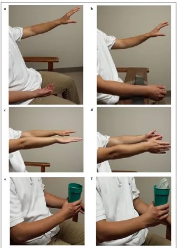
Figure 2-1. Images depicting three static, scripted task variations during rest, posture, and weight-bearing paired tasks. ... 31
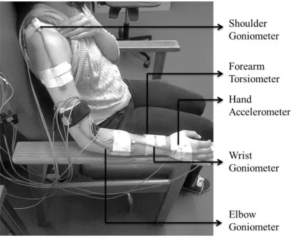
Figure 2-2. Images depicting sensor placement. ... 33
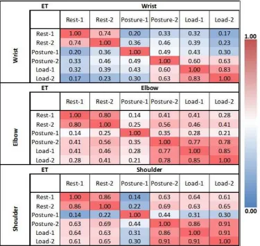
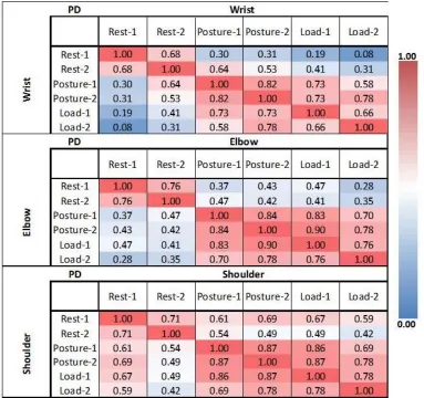
Figure 2-3. Pearson’s coefficient correlation heat map of mean tremor amplitude in the wrist (top), elbow (middle) and shoulder (bottom) across all scripted tasks in ET participants. .... 36
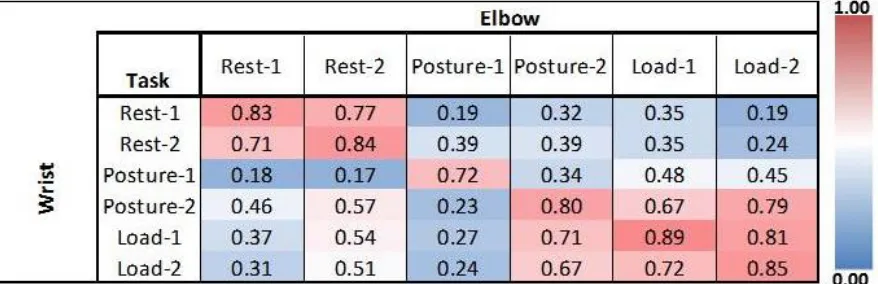
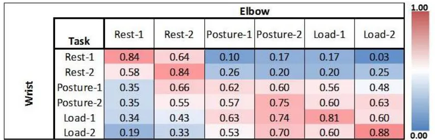
Figure 2-4. Pearson’s coefficient correlation map of mean wrist and elbow tremor amplitudes across all scripted tasks in ET participants. ... 37
Figure 2-5. Pearson’s coefficient correlation heat map of mean tremor amplitude in the wrist (top), elbow (middle) and shoulder (bottom) across all scripted tasks in PD participants. .... 38
Figure 2-6. Pearson’s coefficient correlation map of mean wrist and elbow tremor
amplitudes across all scripted tasks in PD participants. ... 39
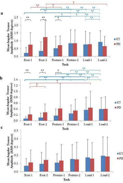
Figure 2-7. Mean joint tremor amplitude in ET and PD participants quantified in each
scripted task. Error bars represent standard deviation of population. ... 40
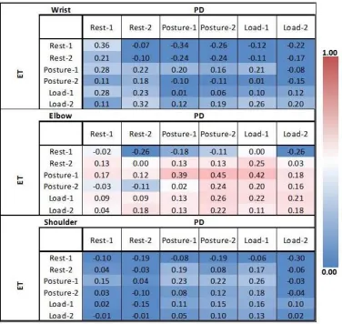
Figure 2-8. Pearson’s coefficient correlation map of mean tremor amplitude at the wrist (top), elbow (middle) and shoulder (bottom) across all scripted tasks between ET and PD
participants. ... 41
Figure 2-9. Task variation significantly modulated the mean percent distribution of wrist tremor in the F/E DOF between all paired tasks in ET participants. ... 42
Figure 2-10. Task variation significantly modulated the mean percent distribution of wrist tremor in the F/E DOF between all paired tasks in PD participants. ... 43
Figure 3-1. CONSORT flow diagram displaying the progress of the study’s design. ... 52
Figure 3-2. Sample kinematic data showing (A) presence of tremor in the wrist, elbow and shoulder joints and (B) the ideal injection parameters determined using the kinematics with the injector’s best clinical judgement. ... 58
Figure 3-3. IncobotulinumtoxinA treatments significantly reduced severity of tremor and provided functional benefit for fine and gross motor tasks with mild muscle weakness in treated muscles. ... 66
Figure 4-1. CONSORT Flow Diagram Displaying the Progress of the Study Design. ... 77
Figure 4-2. Sample participant kinematic data readout of tremor generated from the wrist, elbow and shoulder joints and individualized muscle selection based on both kinematic tremor profile and the injector’s best clinical judgement. ... 81
Figure 4-3. IncobotulinumtoxinA treatments significantly reduced tremor severity and improved arm function in the treated arm of PD participants reported qualitatively and
x
List of Appendices
Appendix A: Ethics Initial Approval ... 99
Appendix B: Letter of Information ... 100
Appendix C: Fahn-Tolosa-Marin Rating Scale ... 110
Appendix D: Unified Parkinson’s Disease Rating Scale ... 114
Appendix E: Quality of Life in Essential Tremor Questionnaire ... 116
Appendix F: Manual Muscle Testing ... 118
xi
Preface
Tremor is the most common movement disorder encountered by clinicians today and is one of
the most challenging symptoms to treat. Despite the high prevalence of tremor, limited
progress has been made in treating tremor due to the relatively unclear understanding of many
underlying conditions, such as PD and ET. The prevalence of ET markedly increases with age
approximately affecting 6.3% globally and 14.3% in Canada of those aged > 65 years old1 and
more than 70% of PD patients have tremor as the presenting feature. As there is no
standardized technique to distinguish among common tremor types, misdiagnosis rates in PD
and ET are as high as one-third and hence lies a great interest in developing simple, objective
techniques to distinguish such tremor types. All traditional pharmacological agents
recommended for tremor therapy in PD and ET patients are approved to treat other conditions
(e.g. anticonvulsants, beta-blockers) and thus produce suboptimal tremor relief coupled with
significant adverse effects. A focal therapy with BoNT-A injected into tremulous muscles has
shown past success in tremor relief although prominent muscle weakness has resulted in early
discontinuation of therapy. This thesis demonstrates that accurate and intelligent kinematic
measurement addresses the significant unmet need for more objective measures to differentiate
tremor types and facilitate advances in tremor therapy.
Chapter 1 is an account of what has been published regarding upper limb tremor in PD and ET and current practices involved with tremor therapy. The aim of this thesis was to develop
wearable technology that was feasible to differentiate tremor profiles and to improve focal
therapy by individualization of BoNT-A injection parameters.
Chapter 2 is a report of the differentiation of ET and PD tremor types in the upper limb by weight-loading and paired task variations using angle-based kinematic technology.
Chapter 3 and 4 summarize the use of kinematic technology to measure tremor biomechanics and to enhance efficacy and tolerability of BoNT-A for ET and PD tremor. Tremor kinematic
measures from each individual were utilized for muscle selection and calculating BoNT-A
dosages thereby ultimately personalizing therapy.
1
1. Introduction
1.1.Parkinson’s disease: the importance of tremor and its pathogenesis
More than 10 million people worldwide suffer from a major neurodegenerative disease
including Alzheimer’s disease, multiple sclerosis, and PD, which is expected to grow
significantly as the aging population lives longer and grows over the next few decades.
Affecting 1% of the population older than 60 years, PD is a chronic, neurodegenerative
disease with a broad spectrum of motor and non-motor features.2 The prevalence of PD
increases with age and is projected to continue to rise in parallel with our aging population.
While the neuropathology is generally well understood, the etiology remains a mystery.
A 60-80% loss of dopaminergic neurons within the substantia nigra pars compacta (SNc) of
the lateral ventral tier located in the basal ganglia (BG) and presence of Lewy bodies are
significant pathological findings in PD.2,3 A lack of dopamine producing neurons leads to
dopamine depletion in the striatum, particularly in the dorsolateral putamen. Genetic
mutations, mishandling of misfolded proteins ubiquitin–proteasome, the autophagy–
lysosomal systems, mitochondrial dysfunction and increased oxidative stress are identified as
contributing mechanisms underlying the death of dopaminergic and non-dopaminergic cells
in PD.4 Early in the disease, dopamine deficiency is the predominant neurochemical
abnormality and is thought to be strongly linked to bradykinesia and rigidity, unlike the
generation of tremor which remains unclear. As the disease progresses, involvement of
non-dopaminergic brain regions results in levodopa (dopamine)-resistant motor and non-motor
symptoms.2 Even though the first descriptive account of PD was written in 1817 by James
Parkinson, there is still an uncertain understanding of the mechanisms behind the occurrence
of dopaminergic deficiencies in PD.5 A better understanding of underlying mechanisms could
help offer new possibilities for symptomatic treatments aimed to improve function and
quality of life in PD patients.
PD presents in a sporadic, idiopathic fashion.6 It is marked by primary motor symptoms of
bradykinesia (slow voluntary movement), muscle rigidity and tremor often with postural
2
symptoms experienced by PD patients may include mood disorders, cognitive disorders and
sleep disorders. The progression of PD and the manifestation of motor and non-motor
symptoms vary considerably from patient to patient although 69% of patients reveal tremor
at disease onset and 75% of patients will develop tremor during the course of their disease.7,8
Tremor is clinically and pathologically distinctive from the other parkinsonian features.
Tremor expression and severity may be associated with a genetic background and can also be
associated with disease course and prognosis.8 PD patients with a tremor-dominance show
evidence of a milder disease course and a better prognosis in terms of life expectancy
compared to patients with predominant PIGD symptom profiles.8,9 The PIGD subtype has a
larger annual increase in symptom severity, faster progression to Hoehn and Yahr grade 4,
worse cognitive performance, and a higher degree of disability at 5 and 8 years of disease,
compared to the tremor-dominant subtype.7 Despite these differences, both PD subtypes have
a similar disease duration at the time of death and thus tremor is not predictive of longer
survival rates.9 It is unclear whether resting, postural/action tremor or a combination of the
two tremors best describes this tremor-dominant subtype.
Tremor pathogenesis in both PD and ET are linked with elevated activity within the
cerebello-thalamo-cortical circuit. However, in PD, it is thought that dopaminergic
dysfunction of the pallidum, particularly originating from the loss of dopaminergic
projections from the retrorubral area, generates this increased activity.7,9 Tremor severity is
not related to the amount of dopamine deficiency in the SNc determined pathologically.10
Anatomically, the BG and cerebellar circuits are connected. The cerebellar nuclei project
glutamatergic, excitatory projections to the posterior part of the ventrolateral thalamus
whereas the globus pallidus interna (GPi) within the BG projects GABAergic, inhibitory
projections to the anterior part of the ventrolateral thalamus. Hence, motor cortical activity is
facilitated by activity of the cerebellar outputs and inhibited by GPi outputs. In addition, the
cerebellar nuclei have GABAergic, inhibitory projections to the inferior olive which sends
excitatory projections to Purkinje cells in the cerebellar cortex and cerebellar nuclei.7 BG and
thalamus activity are synchronous with tremor, though the region in which this occurs varies.
3
neurons.11 These findings propose that the driving force of rest tremor is not within the BG
but is from the posterior ventrolateral thalamus.12 A metabolic imaging study using positron
emission tomography (PET) outlined the sensorimotor cortex, rostral cerebellum, dentate
nucleus, and putamen regions synchronous with tremor, which correlated with clinical tremor
scores, in those with the tremor-dominant subtype.13 In 2011, Mure and colleagues reported
that posterior ventrolateral stimulation reduced tremor amplitude and reduced the metabolic
activity in both the cortico-cerebellar circuit and within the BG suggesting a relationship to
tremor severity and a convergence in the motor cortex.13 Currently, there are three
hypotheses regarding the cause of PD resting tremor: the thalamic pacemaker hypothesis, the
basal ganglia pacemaker hypothesis, and the dimmer-switch hypothesis.7,9
The basis of the thalamic pacemaker hypothesis originates from studies observing the
intrinsic biophysical properties of guinea pig thalamic neurons which are single cell
oscillators at distinct frequencies, 9-10 Hz and 4-6 Hz, when neurons are slightly depolarized
producing low-threshold calcium spikes (LTS) or hyperpolarized, respectively.7 These
frequency ranges within the ventrolateral thalamus are similar to the frequency of PD tremor
and the presence of LTS in the thalamus was also identified in PD patients. However, the
presence of LTS may not be tremor-related as LTS was observed in both PD subtypes and
tremor-dominant patients exhibited “tremor-locked bursts” without LTS. In contrast,
“tremor-locked bursts” may be formed by LTS in the thalamocortical circuit.14
It is thought
that LTS may be generated by the increased inhibitory activity from the GPi to the thalamus
which would suggest tremor production occurs in the anterior ventrolateral thalamus.14 This
hypothesis contrasts the effect of tremor suppression by targeting the posterior ventrolateral
thalamus with deep brain stimulation (DBS). As there may be other brain regions that
hyperpolarize thalamic neurons, this hypothesis does not explain why stimulating the BG
reduces tremor. The advantage of this model is that it illustrates the fundamental role of the
4
The BG pacemaker hypothesis identifies the BG as the area of excessive synchronization
which results in tremor. This hypothesis proposes that striatal inhibition of globus pallidus
externa (GPe) neurons and the excitatory subthalamic nucleus (STN) generate the pacemaker
by forming a feedback system that engages in synchronized bursting.7,15 Previously described
with in vitro data, the frequency of BG oscillations is between 0.4 and 1.8 Hz, which is lower
than the associated frequency ranges of PD tremor.7 A computational model demonstrated
that STN and GPe are prone to LTS bursts due to the increased feedback of the dopamine
depleted BG circuitry. This model definably explains the role of dopamine depletion within
the BG but fails to elucidate the causal role of tremorgenesis by the cerebellothalamocortical
circuit.
The dimmer-switch hypothesis is based on imaging studies in tremor-dominant and
non-tremor PD patients and suggests that the BG triggers non-tremor episodes and the
cerebellothalamocortical circuit modulates tremor amplitude. Data indicates that dopamine
depletion in the pallidum correlates with tremor severity and that the tremor-dominant
subtype has increased functional connectivity between the BG and the
cerebellothalamocortical circuit. This model explains why DBS in the BG (GPi or STN) or in
the ventrolateral thalamus can suppress tremor. 7
1.1.1. Classification of PD tremor
Tremor is an involuntary, rhythmic movement affecting one or more body parts but most
commonly affects the upper limbs and hands. Tremor varies depending on the circumstances
under which they occur. PD patients with tremor can demonstrate different types of tremor:
at rest, with posture, action/kinetic or orthostatic. Typical PD tremor is observed unilaterally
or asymmetrically in the upper limb at rest, while the body part is completely supported by
gravity, with a frequency between 4 and 6 Hz. However, in more advanced disease, tremor
may appear bilaterally in the upper limbs. Approximately 34-60% of PD patients may also
present with a tremor produced by a voluntary contraction called a postural tremor, when
5
movement including non-target directed and intention tremor where the amplitude increases
towards the target. Postural and action tremors are typically delayed compared to ET, and is
described as a re-emergent tremor (with a mean delay of ±10 seconds);16 tremor frequency is
higher than 1.5 Hz and is within a similar frequency range as a resting tremor.16 As the
frequency of resting and re-emergent tremors is similar, this suggests that both tremors
originate from similar pathophysiological processes. Aside from tremor in the arms, patients
may exhibit rest tremor in the lips, jaw or in the lower extremities.
Isolated postural and action tremors can occur in PD with a frequency varying between 4 and
9 Hz.9 Postural tremor without parkinsonian features is often diagnosed as ET and is
observed more frequently in those patients with a family history of PD.16 Some PD patients
have postural tremor similar to ET for many years before the onset of PD features suggesting
that ET is a potential risk factor for PD.4 Orthostatic tremor, a position-dependent postural
tremor, can be isolated or co-exist with resting tremor with different frequency ranges (4-6,
8-9 or 13-18 Hz) and responds to dopaminergic treatment suggesting that it is a manifestation
of PD rather than an association of ET and PD tremor types.9 In some cases, postural and
kinetic tremors can be more disabling than rest tremor and may present as an initial symptom
prior to diagnosis.4
Tremor is a unique symptom of PD in that the rate of tremor progression and severity is
unlike the other PD symptoms such as bradykinesia, rigidity, balance and gait. Tremor is
complex to visually assess and the manifestations of tremor can challenge proper diagnosis
and ultimate prognosis triggering high misdiagnosis rates when using the current
classification system by the Movement Disorders Society (MDS).17
1.1.2. Current treatments of PD tremor
Investigations into understanding the pathogenesis of PD and providing new insights into the
6
neurosurgery has driven the focus of scientific advancement. While some of these advances
are translating novel therapeutics into clinical practice, true pathogenesis targeted treatments
are still lacking. This may be in part due to the need for better animal models that can better
mimic the progression of the disease. There is a lack of success for neuroprotective strategies
due to the scarce understanding of the various genetic, environmental, and other mechanisms
contributing to neurodegeneration in PD. Efficacy of therapeutics is tested using
evidence-based medicine designed to control for placebo effects. However, there are limitations to
placebo-controlled randomized trials due to the inhomogeneous population of included
patients, the short-term nature of these studies and specified patient demographics, which is
not representative of clinical practice. This leads many physicians to rely on their own
experience and best clinical judgement in selecting the optimal treatment option and for the
therapeutic plans with patients.
Various treatment options are available to alleviate symptoms associated with PD. First line
therapy is the use of oral pharmacological agents while surgical intervention is generally
reserved for drug refractory PD. Generally, initiation of therapy is often delayed until
symptoms are functionally disabling and interfere with activities of daily living (ADLs) or
the patient’s vocation.18
Choice in therapeutics heavily depends on the patient’s age,
dominant symptoms, disease stage and cognitive state. Treatments are titrated and adjusted to
changes in patient symptomatologies. Approximately 10% of patients are selected for
surgical therapies.6
1.1.2.1. Pharmacotherapy agents
Pharmacological treatments seek to correct the imbalance within the BG due to the loss of
dopamine producing neurons. There are many classes of dopamine replacement medications
7
If motor symptoms are mild and require therapy, dopamine agonists (pramipexole and
ropinirole), levodopa, or a monoamine oxidase type B inhibitor (MAOBI; selegiline or
rasagiline) can be administered to produce symptomatic benefit.2 Past studies show the use of
beta-blockers, often propranolol, may improve PD tremor and motor function;
anticholinergic medications are more effective than placebo for improving motor function
but are inconclusive as a tremor therapy and frequent adverse events, such as cognitive
impairments, dizziness, fatigue, tachycardia, lead to discontinuation.2 Clozapine has been
shown to improve bothersome or disabling PD tremor when used for tremor resistant to other
therapies though significant adverse events such as agranulocytosis, myocarditis, seizures
and sedation can occur.19 Six, small class 3 randomized-control trials have shown mixed
evidence for the possible efficacy of amantadine as a monotherapy or as an adjunct therapy
due to the poor quality of studies.2
Levodopa or dopamine agonists are prescribed for patients with more severe symptoms and
impairment in ADLs. Levodopa remains the gold standard in providing the greatest
symptomatic benefit for PD and is linked to lower incidences of freezing, hallucinations,
somnolence, impulse control disorders, and edema than initial treatment with dopamine
agonists. However, dopamine agonists greatly benefit patients younger than 60 years of age
or with younger-onset of disease and are associated with fewer dopaminergic motor
complications associated with levodopa, such as dyskinesia which can be more disabling
than the PD symptoms.2,20 A meta-analysis showed the long-term benefits of
levodopa-sparing versus levodopa as an initial treatment and concluded that levodopa alone was more
effective in reducing Unified Parkinson’s Disease Rating Scale (UPDRS) parts I-III scores
though levodopa alone group were at a higher risk of developing dyskinesia and wearing-off
phenomenon.21 Interestingly, a study demonstrated that more than triple of patients receiving
levodopa-sparing therapy discontinued prematurely due to adverse events compared to
patients receiving levodopa alone therapy.21 New evidence shows that the early advantage of
dopamine agonists over levodopa diminishes over time (estimated 10 years).19 Those with
tremor as the initial manifestation of PD or having a tremor-dominant subtype have a lower
8
patients would benefit from starting with levodopa compared with dopamine agonists. Thus,
levodopa and dopamine agonists appear to be reasonable options for initiating dopaminergic
replacement therapy but are associated with different efficacy and adverse effects.20
Unfortunately, levodopa and dopamine-replacement therapies do not target tremor and tend
to produce suboptimal tremor relief, and in some cases may worsen tremor. Additionally, it is
often tremor that is sub-optimally alleviated or is a persistent tremor despite improvement in
other cardinal PD symptoms, such as bradykinesia and rigidity, which are optimally managed
by levodopa therapy. Recent studies have reported that tremor is high up among the key
symptoms found to be one of the most bothersome symptoms experienced by patients.23,24
Many clinicians are inclined to simply increase medication dosages or introduce adjunctive
therapies. Dopamine agonists and anticholinergic medications can be used concomitantly
with levodopa to treat tremor but may be accompanied by neuropsychiatric and cognitive
side effects.18 In double-blind studies, levodopa combined with carbidopa known to increase
availability of levodopa in the brain, improves resting tremor by more than 50%, as well as
bradykinesia and rigidity compared to using levodopa alone.18 Efficacy of
levodopa/carbidopa formulations and dopamine agonists have been reported to reduce rest
and postural tremors to a similar level; however poor long-term efficacy and adverse events
arise.18 To reduce the incidence of adverse event profiles of oral, anti-tremor medications, a
focal therapy with BoNT-A has been suggested to be efficacious in providing tremor
reduction at the source, given the focal and asymmetric presentation of tremor.25
1.1.2.2. Botulinum toxin type A focal tremor therapy
Treating upper limb tremor with BoNT-A has not been widely adopted in clinical practice.
However, BoNT-A is used to treat a wide variety of symptoms related to PD such as foot and
hand dystonias, blepharospasm, lid apraxia, sialorrhea, hyperhidrosis, and jaw tremor. When
injected intramuscularly, BoNT-A inhibits the release of acetylcholine (ACh) at the
neuromuscular junction resulting in the blockade of neuromuscular conduction and reduced
9
synaptic fusion complex of ACh vesicles bound to the pre-synaptic membrane by soluble N
-ethylmaleimide-sensitive factor attachment protein receptor (SNARE) proteins. SNARE
proteins mediate the docking and exocytosis of ACh vesicles at the presynaptic nerve
terminal. The proteolytic action of BoNT-A cleaves SNAP-25, one of the three proteins
which form SNARE, preventing the release of ACh into the synaptic cleft.26 BoNT-A at the
neuromuscular junction interrupts neurotransmitter release thereby causing muscle paralysis.
Thus, when BoNT-A is injected into muscles contributing to tremor, effective alleviation of
tremor can be achieved.27
There are limited number of studies reporting the efficacy of BoNT-A for PD tremor25
though the success of BoNT-A lies in individualizing therapy by accurately distinguishing
tremulous muscles.27 However most past studies treating ET, dystonic tremor, or PD tremor
used randomized or fixed dosing regimens based on visual/clinical assessments, which has
led to early discontinuation of therapy due to bothersome, dose-dependent muscle
weakness.28–30 The use of accelerometry and surface electromyography to identify arm
muscles with tremorogenic activity reduced incidence of weakness and improved functional
outcomes in ET.31 However, accelerometry provides an overall tremor severity score and is
not capable of providing a more detailed breakdown regarding which degree of freedom
(DOF) tremor is acting in.
1.1.2.3. Surgical intervention
Stereotactic surgery is the next treatment option for patients with disabling tremor resistant to
pharmacotherapy. Tremor can be effectively reduced following DBS implantation or lesional
therapy.
1.1.2.3.1. Deep brain stimulation
DBS is a surgical technique involving the implantation of electrodes in specific brain regions.
10
known to be more effective than oral medication in reducing rest tremor severity and
frequency.33 A recent study has shown that patients with early motor complications who
underwent DBS and medical therapy have significant improvements in ADLs and relief of
motor disability for two years compared to patients with medical therapy alone.34 DBS
significantly improved survival of severe, late-stage patients and reduced admittance to
long-term care facilities compared to patients with no DBS.35
The electrodes are connected to a device called an implanted pulse generator (IPG) that
delivers electrical stimuli to brain regions in order to modulate or disrupt patterns of neuronal
activity associated to PD. Three specific sites in the brain are most commonly targeted for
DBS in PD: STN and GPi nuclei in the BG where much of the physiological modulation
occurs in PD, and in the ventro-intermediate thalamic nucleus (Vim).32 Bilateral DBS of the
STN (STN-DBS) is performed in patients who are levodopa responsive with persistent motor
symptoms despite optimal medical therapy. However, only a fraction of PD patients are
eligible for this neurosurgical intervention. Surgical selection criteria include patients
experiencing motor complications refractory to best medical treatment, who are
levodopa-responsive, less than 65 years of age, with no mental health issues (depression, dementia) and
have had PD for more than 7 years. In addition, intracranial hemorrhage, infection, and
post-operative seizures are the most common adverse events33 and impairments in gait, speech,
and balance have been shown to have less chance of improvement and in some cases may
worsen.6 When the DBS is turned on about 2-4 weeks following surgery, most patients may
require a reduction in medication dosages as the presence or severity of side effects such as
dyskinesias increase.
Electrical stimuli promote neurogenesis, increased blood flow to stimulated areas, and
astrocytes to release calcium and neurotransmitters including adenosine and glutamate.6
However, the exact mechanism of how DBS alleviates PD symptoms is not yet clear despite
these physical and physiological changes.32 High frequency STN-DBS suppresses the
11
reduces synchronous beta-frequency oscillations.36,37 However, the effect of how DBS
intervention improves appendicular symptoms such as tremor, rigidity, and bradykinesia
remains elusive.38
1.1.2.3.2. Lesional therapy
A surgical procedure involving lesioning the GPi reduces motor fluctuations, tremor and
dyskinesias as the GPi is thought to be overactive in PD. This procedure, pallidotomy, is
performed unilaterally as bilateral procedures rarely occur due to high incidence of severe
adverse events including cognitive impairment, dysarthria, and dysphagia.6 The reduction in
PD symptoms by pallidotomy is not as great as DBS intervention.32 Suppression of tremor,
rigidity, dyskinesia and bradykinesia has been reported for five years following surgery
however bradykinesia gradually reoccurs with freezing of gait at a 10-year follow-up.6 In
addition, cognitive and executive functions are reduced after five and 10 year follow-ups and
thus, pallidotomy is an alternative to DBS or when DBS is no longer effective.6
An invasive procedure called thermal radiofrequency thalamotomy involves the passage of a
probe into the VIM nucleus of the thalamus, an area understood to be involved in the tremor
cerebello-rubro-thalamo-cortical circuit, and performing a lesion to the side contralateral to
the tremor. Bilateral thalamotomy may affect executive function by disrupting
neuroanatomical pathways between subcortical and prefrontal cortical areas. Unilateral
thalamotomy significantly improves PD symptoms of tremor, rigidity, mobility, well-being,
and ADLs.6 The use of transcranial magnetic resonance imaging (MRI)-guided high intensity
focused ultrasound to perform thalamotomy has reduced complications involving
hemorrhages, motor deficits and post-operative cognitive impairment.39 However, side
effects including paraesthesia of lips, tongue and fingers, nausea and pain at one year
follow-up do arise.
Gamma knife is a non-invasive surgical approach involving the emission of highly focused
12
Improvements in tremor and ADLs have been demonstrated though benefits take several
months to appear. There are contrasting results on the effectiveness of gamma knife in
reducing rest, postural, action and head tremor. Gamma knife lacks the accuracy observed in
invasive procedures since there is no intraoperative electrophysiological confirmation of
target site.6 A large spectrum of possible complications involving the delayed effects of
radiation necrosis and thus such procedures are reserved for PD patients with severe
disabling symptoms who fail to adequately respond to pharmacotherapy.
1.2.Essential tremor: symptoms and etiology
ET is the most common adult-onset movement disorder after restless legs syndrome.1 The
prevalence of ET increases with age, affecting approximately 1% of the general population
and 2.3-14.3% of those aged 60 years and older.1 Approximately 70-80% of people with mild
ET do not seek therapy and go undiagnosed and thus, prevalence is underestimated. The
course of ET varies widely as in some patients the tremor remains mild while in others it
becomes progressively and functionally disabling that many seek effective treatment. The
slow, progressive nature of ET may evolve to more body parts/areas, increasing in amplitude
and frequency.40 The age of onset is a bimodal distribution with peaks at early childhood and
around the age of 60 years of age.41 A family history of ET and aging are risk factors for ET.
The etiology of ET is genetic by inheritance in a Mendelian autosomal dominant fashion in
approximately 50% of patients increasing the prevalence of ET within certain families 42
although, specific genes have not been identified.42 A positive family history is very common
and these ET patients have a younger age at onset.43 Non-genetic and environmental factors
are possible causes of ET yet are much less extensively studied.
ET is characterized by bilateral tremor during voluntary movement occurring most frequently
in the hands and arms in absence of other medications or neurological signs that might cause
tremor. Isolated head tremor with no signs of dystonia fall under ET. For definite certainty of
ET, ET must be present for over five years with thorough exclusion of other causes. ET is
13
disability and over the past 20 years, investigators have sought to better classify and define
ET as growing literature considers ET to be a family of diseases.44 ET as a monosymptomatic
disorder has been challenged, as disturbances in gait, mood, cognition and hearing have been
observed and linked to thalamic and cerebellar dysfunction.44 Problems with postural
stability and gait are evident in patients with longer disease time. Nevertheless, ET is a
clinical syndrome of action tremor in the upper limbs (at least 95% of patients) and less
commonly in the head (at least 34%), face/jaw (7%), voice (at least 12%), tongue (30%),
trunk (5%), and lower limbs (30%).44
The pathophysiology of ET remains largely unknown as there is no known pathological or
biochemical findings, no suitable animal model and hence most knowledge about ET is
gathered from clinical studies. The frequency of ET ranges between 4 and 12 Hz and is
thought to emerge from neuronal oscillation in the cortico-bulbo-cerebello-thalamo-cortical
loop but the cause is unknown.44 Low frequency repetitive transcranial magnetic stimulation
(rTMS) over the posterior cerebellar cortex modulates the cerebello-thalamo-cortical circuit
resulting in significant reduction in tremor severity for three weeks.45 Oscillation in the
cortico-bulbo-cerebello-thalamo-cortical loop is also observed in PD, Wilson disease and
rhythmic cortical myoclonus. 40 The frequency of ET is associated with the dysfunction in
the central component as several central lesions in the pons, internal capsule, cerebellum and
frontal/sub-cortex modify ET. The peripheral component of ET modifies amplitude and is
linked to adrenergic mechanisms in the muscle spindle.46 A recent morphometric study
reported all ET cases present with histological change, 24% of ET showed Lewy body
inclusions and 76% had cerebellar pathology (Purkinje cell loss).47 Some studies report
altered gamma amino-butyric acid (GABA) function in ET.40
1.2.1. Classification of ET
Classical ET is bilateral, largely symmetrical, postural and/or kinetic tremor – although one
limb may be more pronounced than the other. Postural tremor is provoked when arms are
fully extended in front of the body though more pronounced tremor is observed when the
shoulder is abducted, elbow is flexed and hands are held under the chin.40 Kinetic tremor is
14
is tested by asking the patient to go back and forth between touching their nose and the
target. Completing Archimedes spiral and line drawings, pouring, and writing tasks are other
methods for testing action tremor.
Many ET patients are incorrectly diagnosed with PD.40 PD tremor during the early course of
PD may present as an action tremor without rest tremor, bradykinesia or rigidity thus making
it extremely difficult to distinguish early PD from ET. However, postural tremor in ET
manifests immediately on positioning arms horizontally where PD postural tremor has
delayed onset. Additionally, classical PD tremor is at rest however one-third of ET patients
have rest tremor which typically manifests later in the disease course.40 Rest tremor can be
elicited by physical or emotional stress and thus definite rest tremor should be evident when
the patient is lying down with the arms fully supported. Rest tremor is not a feature of
cerebellar disorders. Cerebellar tremor is slower than ET with variable amplitude, irregular
rhythm and is primarily a proximal, upper limb tremor, in contrast to the distal tremor of ET.
Dystonia with associated tremor is typically jerky and irregular occurring commonly in the
head and is most prominent when the body part is positioned in a direction opposite to the
direction of the dystonic side and thus may be considered a postural/kinetic tremor. A
sensory trick alleviates dystonia but does not improve ET. Physiological tremor occurs under
stressful situations and is non-progressive though frequency ranges from 8 to 12 Hz. Writing
tremor may be a feature of ET but is better associated with the dystonic posturing of the hand
classifying it as a dystonic tremor. Thus, proper positioning and tests are required in these
movement disorders as ataxic or dystonic features can be misinterpreted for tremor.
1.2.2. Current treatments of ET
As the basic neuropathology and neurotransmitter deficits in ET are largely unknown, no
disease-specific drug is available and all traditional oral agents used in treating ET were
primarily developed for other diseases.48 Treatment options currently available for ET
include oral pharmacotherapy, botulinum toxin A (BoNT-A) injection, and stereotactic
15
is the only pharmacotherapy approved by the US Food and Drug Administration to date,
primidone, benzodiazepines, gabapentin, pregabalin, topiramate, and nimodipine are used in
the treatment of ET, with an average tremor reduction of 50% or less.48,50,51 Poor efficacy is
frequently coupled with dose limiting side-effects, such as drowsiness with primidone,
bradycardia, syncope, fatigue, and erectile dysfunction with propranolol. Additionally,
tolerance to the initial benefit often leads to the discontinuation of drugs in 56.3% of
patients.50DBS of the VIM nucleus of the thalamus may benefit patients with disabling ET,
though only a fraction of patients are eligible for this highly invasive procedure that may
produce lasting neurological side-effects, including paresthesias (6-36%), dysarthria
(3-18%), ataxia (6%), limb weakness (4-8%), balance disturbance (3-8%) and dystonia
(2-9%).49,50 Efficacy and risks of gamma knife thalamotomy is comparable to VIM-DBS.49
Ultrasound guided thalamotomy can be performed only unilaterally and again carries the
same potential risks as gamma-knife surgery and thus large, randomized, controlled trials are
required to assess the procedure’s efficacy and safety.39,52,53
A review of the current,
recommended treatment options for ET is summarized in Table 1-1.
Table 1-1. Treatment options for ET.
Drug Name Drug Class Level
Total Daily Dosage (mg/d) Sample size Efficacy (compared to baseline)
Adverse Effects Reference
Propranolol Beta blocker A 40 -
320 533
16
Primidone Anticonvulsa
nt A
50 -
1000 218
50% mean
improvement rated by clinical scales and accelerometry Ataxia, vertigo, nausea, vomiting, fatigue, malaise, dizziness, unsteadiness, confusion, impotence, rash 51,54,55
Atenolol Beta blocker B 50 -
150 79
25% mean improvement by clinical scales, 37% mean improvement by accelerometry
Light-headedness, nausea, cough, dry mouth, sleepiness, decreased pulse and blood pressure
51,54
Alprazolam Benzodiazepi
ne B
0.125 -
3 46
25-37% mean improvement by clinical scales
Mild sedative and fatigue effects
51,54
Gabapentin (monotherapy)
Anticonvulsa
nt B
1200 -
1800 61
Drowsiness, fatigue, dizziness, nervousness, shortness of breath, reduced libido
33% improvement by clinical scales, 77% improvement by accelerometry 51,54,55 Sotalol Beta-adrenergic receptor antagonist
B 75 -
240 50
28% mean improvement in clinical scales
Serious ventricular arrhythmias, dose-related QT interval prolongation, reduced alertness
51,52,54
Topiramate Anticonvulsa
nt B
25 -
300 335
29% improvement in clinical scales (mean dose=292 mg/d); 30% improvement in tremor (up to 400 mg/d) with a 32% attrition rate due to adverse events Dizziness, disorientation, paraesthesia, weight loss, memory difficult, appetite suppression, cognitive difficulties, upper respiratory tract infection, taste perversion, fatigue, nausea, headache, somnolence
17
Nimodipine
Calcium channel blockers
C 120 16
45% improvement by clinical scales and 53% improvement by accelerometry (n=14) Headache, heartburn, hypotension 51,54
Clonazepam Benzodiazepi
ne C 0.5 - 6 44
45% improvement by accelerometry Drowsiness, depression, cognitive and behavioural impairments 51,54
BoNT-A Neurotoxin C
50 - 300 U/arma 283 20-27% improvement by clinical scales (n=133) Dose-dependent muscle weakness occurred in 30% of patients, reduced grip strength, stiffness, cramping, pain at injection site 51,54,56 DBS Unilateral or bilateral VIM-DBS; STN-DBS
C 398
40-90%
improvement by clinical scales up to 3 years. Chronic stimulation gradually worsens efficacy leading to loss of tremor suppression in about 70% of patients Dysarthria, disequilibrium, paresthesias, weakness, headache, intracranial hemorrhage, subdural hemorrhage, lead dislodgement, generalized motor seizures. About 18% experience
equipment
malfunction or lead displacement 54,56 Thalamotomy Gamma-knife; MRI-guided Focused Ultrasound
C 181
55-90%
improvement by clinical scales
Hemiparesis, transient problems with speech, motor function, dysarthria, verbal/cognitive deficit, weakness,
18 a
BoNT-A units are in mouse units from mouse lethality assay
In addition to pharmacotherapy and surgical intervention for ET, BoNT-A injected into
tremulous muscles has demonstrated modest effect in reducing arm tremor in ET and
similarly for PD rest tremor. A randomized, double-blind study by Brin and colleagues
demonstrated in 133 ET patients a significant improvement in postural tremor up to 16 weeks
and kinetic tremor improvement for 6 weeks without any improvements in motor tasks and
functional disability.28 Common side effects such as hand muscle weakness restrict its
application as patients found the weakness more disabling than their tremor. The randomized
injection regimen is the probable cause of the resulting dose-dependent muscle weakness. In
fact, muscle weakness is often cited as the main and most severe adverse event of this
therapy reducing efficacy and ease of use in 45-60% of patients.29,30,57 Pacchetti and
colleagues identified tremulous wrist flexor/extensor and bicep muscles by more objective
measures such as accelerometry and surface EMG to better target BoNT-A; this resulted in
significant reduction in tremor amplitude and improved ADLs scores in 20 patients.31 Yet,
practice guidelines score a level C recommendation and believe BoNT-A has a modest effect
at best.48
1.3.Rationale
1.3.1. Using technology to differentiate ET and PD tremor types
There are no biochemical or radiological biomarkers in PD and ET and thus proper diagnosis
heavily relies on clinical observation. As ET and PD tremor have overlapping tremor features
and may occur in the same circumstances, reliable and accurate methods for differentiating
these tremor types are important. Possible biomarkers involving MRI, PET and SPECT
(MRgFUS) confusion, facial
paresis. About 7% experience
19
scans, movements about the wrist, olfactory deficits, and REM sleep disorders have been
proposed for PD but none are satifactory.58 The criterion for ET is based on clinical
assessment of tremor however patients usually present with more than one type of tremor and
it is not clear whether ET is separable from different types of tremor.59 A study failed to
differentiate ET and PD tremor by combining EMG and kinematic analysis.58 As both
diseases are incurable and therapy is focused on alleviating symptoms and improving quality
of life, there is a great interest in differentiating between these diseases for proper
symptomatic management and reducing the economic burden to healthcare utilization,
caregiver costs and lost productivity.60
To quantify tremor in the clinical setting, standardized clinical rating scales such as the
Fahn-Tolosa-Marin (FTM) tremor rating scale, the Washington Heights-Inwood Genetic Study of
ET rating scale, Whiget tremor rating scale and UPDRS part III for the tremor motor
symptoms can be used in ET and PD.61 The FTM uses a 5-point scale rating tremor severity
based on tremor amplitude from 0 (no tremor) to 4 (severe tremor) in each part of the body.62
UPDRS ratings use an integer scale (0-4) to assess severity of motor functions rather than a
quantitative, ratio-based approach.63 UPDRS maintains high subjectivity as intra-individual
ratings vary, limiting its accuracy and value as a measure for clinical diagnosis and in clinical
trials/studies.64 In addition, questions and phrases used in scales are ambiguous, the
sensitivity of scores to change in clinical trials, and the differences in ratings between
patients and assessors needs to be revised.61,65 Tremor rating scales also lack good inter-rater
reliability and continuous/repetitive monitoring capabilities remain a concern. Thus there is a
need for more objective outcomes that can be applicable to clinical practice.66
Several targeted and sensor-based methods for monitoring and for analysis of tremor have
been developed over the past decades. Advantages in these emerging techniques include high
accuracy, repetitiveness and reliability of measurements and that the devices are small,
non-invasive and are easy to use.67 The use of surface or needle-EMG involves electrical contacts
fixed on the skin or using a needle placed in a muscle to analyze duration of EMG activity
20
recordings can classify ET apart from parkinsonian tremor.67 However, EMG analysis takes a
skillset and may not be practical in clinical practice. Gyroscopes and accelerometers are low
in cost, small, and are convenient to measure useful parameters such as frequency and
amplitude of tremulous body segments. Unfortunately, such tools have really only been
applied for clinical management and have not been successful in distinguishing pathological
tremor types. Flexible angular sensors called goniometers measure joint angles and are
frequently used in sports and rehabilitation 67 Currently, goniometers have not yet been
applied in large scale studies of tremor and thus it is unknown whether these sensors can
detect differences between tremor characteristics in PD and ET patients.67 However, recent
studies have demonstrated that kinematic technology for tremor characterization is a reliable
and feasible methodology and have generated the ability to distinguish tremulous muscle
groups.68–71
Owing to the lack of standardized methodologies to measure upper limb tremor, this current
thesis focused on the use of wearable, motion sensor technology to quantify the tremor
biomechanics at each arm joint in PD and ET individuals while individuals were in their best
medically managed state. By deconstructing joint tremor amplitudes in each individual over
a series of tasks, differences in tremor profiles were established thereby enabling an
objective, simple method to distinguish between ET and PD tremor types.
1.3.2. Using technology to personalize therapy
Many tremor analysis tools are very expensive and are generally non-portable, such as an
optical motion capture system.67 Multi-sensor motion recordings, wearable
accelerometers/gyrostats, and tremor apps for smartphones (based on accelerometry) to
measure tremor have been established and are available for the general public. Such
technological advances allow in-home monitoring, individualized therapy titrations, and is a
diagnostic insight for clinicians.72 However, wearable devices are not reliable measures in
distinguishing tremulous muscles and do not provide accurate or diagnostically relevant
21
patient care or as outcome measures in clinical studies should exercise caution as there are a
limited number of studies investigating the validity of these devices for tremor analysis.
Current research focuses on the use of portable and simple to use devices that provide
applicable information for clinicians to better individualize therapy. One study extracted
tremor characteristics, such as frequency, direction and amplitude, from spiral drawing on a
digitizing tablet. Writing and spiral drawing tasks have recently been used as an objective
measure of proximal versus distal arm tremor severity.73 However tremor characteristics
during rest or other functional tasks could not be measured.72
While BoNT-A injection in the upper extremities is a viable treatment option as a
monotherapy or adjunctive therapy for ET and PD tremor, limited functional efficacy due to
dose-dependent wrist and finger muscle weakness remain a concern. The use of injection
protocols involving fixed BoNT-A dosages and predetermined muscle sites, regardless of
patient’s unique tremor characteristics are probable reasons for this problem.74,28
During a
tremor assessment, the characterization of the movement dynamics along the whole arm,
muscle selection, dosing and proper muscle localization during injection are important
considerations when utilizing BoNT-A to enhance arm function and to minimize likelihood
of weakness.27,68,69 While clinical knowledge and use of technologies, such as EMG or
ultrasound aid in muscle localization and injection, visually-guided assessments to
characterize tremulous movements at various joints are likely to fail due to the variability and
complexity of tremor and the difficulty in accurately separating multi-joint, whole arm
movements. Thus, the lack of adequate tremor assessment tools and poor injection guidelines
have limited the use of BoNT-A for ET and PD tremor.27,69,75 Ultimately, it is of great
interest to improve the efficacy and tolerability of BoNT-A therapy by applying kinematic
tremor biomechanics captured from each individual in order to better target BoNT-A to
muscles contributing to the overall joint tremor.
22
The focus of this thesis is to apply commercially available motion sensor technology to
measure the variation in joint tremor amplitude brought out by various scripted tasks to
generate unique, correlational tremor profiles of PD and ET tremor types. Kinematic tremor
assessments employing rest and postural paired tasks is simple, quick and could be translated
to the clinic setting as a diagnostic aid, The use of multi-sensor kinematic technology
addresses the significant unmet need in tremor therapy by pairing BoNT-A injections with
individualized, multi-joint movement patterns. This combination has shown to alleviate
tremor severity, improve the functional efficacy of BoNT-A while limiting the likelihood of
dose-dependent weakness perceivable by patients.
1.5.References
1. Louis ED, Ferreira JJ. How common is the most common adult movement disorder? Update on the worldwide prevalence of essential tremor. Mov Disord. 2010;25(5):534-541. doi:10.1002/mds.22838.
2. Connolly BS, Lang AE. Pharmacological treatment of Parkinson disease: a review. JAMA. 2014;311(16):1670-1683. doi:10.1001/jama.2014.3654.
3. Burke RE, O’Malley K. Axon degeneration in Parkinson’s disease. Exp Neurol. 2013;246:72-83. doi:10.1016/j.expneurol.2012.01.011.
4. Jankovic J. Parkinson’s disease: clinical features and diagnosis. J Neurol Neurosurg Psychiatry. 2008;79(4):368-376. doi:10.1136/jnnp.2007.131045.
5. Goetz CG. The history of Parkinson’s disease: Early clinical descriptions and neurological therapies. Cold Spring Harb Perspect Med. 2011;1(1).
doi:10.1101/cshperspect.a008862.
6. Tarazi FI, Sahli ZT, Wolny M, Mousa SA. Emerging therapies for Parkinson’s disease: From bench to bedside. Pharmacol Ther. 2014;144(2):123-133. doi:10.1016/j.pharmthera.2014.05.010.
7. Helmich RC, Toni I, Deuschl G, Bloem BR. The pathophysiology of essential tremor and Parkinson’s tremor. Curr Neurol Neurosci Rep. 2013;13(9):378.
doi:10.1007/s11910-013-0378-8.
8. Baumann CR. Epidemiology, diagnosis and differential diagnosis in Parkinson’s disease tremor. Parkinsonism Relat Disord. 2012;18:S90-S92. doi:10.1016/S1353-8020(11)70029-3.
9. Helmich RC, Hallett M, Deuschl G, Toni I, Bloem BR. Cerebral causes and consequences of parkinsonian resting tremor: a tale of two circuits? Brain. 2012;135(11):3206-3226. doi:10.1093/brain/aws023.
10. Hallett M. Parkinson’s disease tremor: pathophysiology. Park Realt Disord. 2012;18S1:S85-S86. doi:10.1016/S1353-8020(11)70027-X.
23
functionally identified cells. Brain. 1994;117:531-543. doi:10.1093/brain/117.3.531. 12. Zaidel A, Arkadir D, Israel Z, Bergman H. Akineto-rigid vs. tremor syndromes in
Parkinsonism. Curr Opin Neurol. 2009;22(4):387-393. doi:10.1097/WCO.0b013e32832d9d67.
13. Mure H, Hirano S, Tang CC, et al. Parkinson’s disease tremor-related metabolic network: Characterization, progression, and treatment effects. Neuroimage. 2011;54(2):1244-1253. doi:10.1016/j.neuroimage.2010.09.028.
14. Magnin M, Morel A, Jeanmonod D. Single-unit analysis of the pallidum, thalamus and subthalamic nucleus in parkinsonian patients. Neuroscience. 2000;96(3):549-564. doi:10.1016/S0306-4522(99)00583-7.
15. Plenz D, Kital ST. A basal ganglia pacemaker formed by the subthalamic nucleus and external globus pallidus. Nature. 1999;400(6745):677-682. doi:10.1038/23281. 16. Jankovic J, Schwartz KS, Ondo W. Re-emergent tremor of Parkinson’s disease. J
Neurol Neurosurg Psychiatry. 1999;67(5):646-650. doi:10.1136/jnnp.67.5.646. 17. Gövert F, Deuschl G. Tremor entities and their classification. Curr Opin Neurol.
2015;28:393. doi:10.1097/WCO.0000000000000211.
18. Schadt CR, Duffis EI, Charles PD. Pharmacological treatment of disabling tremor. Expert Opin Pharmacother. 2005;6(3):419-428. doi:10.1517/14656566.6.3.419. 19. Connolly BS, Lang AE. Pharmacological treatment of Parkinson disease: a review.
JAMA. 2014;311(16):1670-1683. doi:10.1001/jama.2014.3654.
20. Group PS. Pramipexole vs levodopa as initial treatment for Parkinson disease: A randomized controlled trial. Parkinson Study Group. JAMA. 2004;61:1044-1053. doi:10.1001/archneur.61.7.1044 61/7/1044 [pii].
21. Xie C, Zhang Y-Y, Wang X-D, et al. Levodopa alone compared with levodopa-sparing therapy as initial treatment for Parkinson’s disease: a meta-analysis. Neurol Sci. 2015;36(8):1319-1329. doi:10.1007/s10072-015-2253-7.
22. Kipfer S, Stephan MA, Schu WMM. Resting Tremor in Parkinson Disease. 2014;68(8):1037-1039.
23. Politis M, Wu K, Molloy S, G Bain P, Chaudhuri KR, Piccini P. Parkinson’s disease symptoms: the patient's perspective. Mov Disord. 2010;25(11):1646-1651.
doi:10.1002/mds.23135.
24. Louis ED, Machado DG. Tremor-related quality of life: A comparison of essential tremor vs. Parkinson’s disease patients. Parkinsonism Relat Disord. 2015;21(7):729-735. doi:10.1016/j.parkreldis.2015.04.019.
25. Mills R, Bahroo L, Pagan F. An update on the use of botulinum toxin therapy in Parkinson’s disease. Curr Neurol Neurosci Rep. 2015;15(1):511. doi:10.1007/s11910-014-0511-3.
26. Comella CL, Pullman SL. Botulinum toxins in neurological disease. Muscle Nerve. 2004;29(5):628-644. doi:10.1002/mus.20033.
27. Rahimi F, Bee C, Debicki D, Roberts AC, Bapat P. Effectiveness of BoNT A in Parkinson ’ s Disease Upper Limb Tremor Management. Can J Neurol Sci. 2013:663-669.
28. Brin MF, Lyons KE, Doucette J, et al. A randomized, double masked, controlled trial of botulinum toxin type A in essential hand tremor. Neurology. 2001;56(11):1523-1528. doi:10.1212/WNL.56.11.1523.