Scholarship@Western
Scholarship@Western
Electronic Thesis and Dissertation Repository
10-12-2017 10:00 AM
Characterization of plant-spider mite interactions and
Characterization of plant-spider mite interactions and
establishment of tools for spider mite functional genetic studies
establishment of tools for spider mite functional genetic studies
Nicolas Bensoussan
The University of Western Ontario
Supervisor Vojislava Grbić
The University of Western Ontario Graduate Program in Biology
A thesis submitted in partial fulfillment of the requirements for the degree in Doctor of Philosophy
© Nicolas Bensoussan 2017
Follow this and additional works at: https://ir.lib.uwo.ca/etd
Part of the Agronomy and Crop Sciences Commons, Biotechnology Commons, Cell Biology Commons, Entomology Commons, Genetics Commons, and the Molecular Genetics Commons
Recommended Citation Recommended Citation
Bensoussan, Nicolas, "Characterization of plant-spider mite interactions and establishment of tools for spider mite functional genetic studies" (2017). Electronic Thesis and Dissertation Repository. 5015.
https://ir.lib.uwo.ca/etd/5015
This Dissertation/Thesis is brought to you for free and open access by Scholarship@Western. It has been accepted for inclusion in Electronic Thesis and Dissertation Repository by an authorized administrator of
i
The two-spotted spider mite, Tetranychus urticae Koch (Acari: Tetranychidae), is one of the most polyphagous herbivores feeding on cell contents of over 1100 plant species including more than 150 crops. However, despite its important pest status and a growing understanding of the molecular basis of its interactions with plant hosts, knowledge of the way mites interface with the plant while feeding and the plant damage directly inflicted by mites is lacking. Likewise, while the use of the reverse genetic tools in plants facilitated our understanding of the establishment of defense mechanisms against spider mite herbivory, such tools are lacking for spider mite, preventing the expansion of functional analysis to both sides of the interacting organisms.
First, using various microscopy methods, I uncovered several key features of T. urticae feeding. By following the stylet path within the plant tissue, I determined that the stylet penetrates the leaf either in between epidermal pavement cells or through a stomatal opening, without damaging the epidermal cellular layer. Recordings of mite feeding events established that the duration of mite feeding ranges from several minutes to more than half an hour, during which time, mites consume a single mesophyll cell in a pattern that is common to both bean and Arabidopsis plant hosts. In addition, this study determined that leaf chlorotic spots, a common symptom of mite herbivory, do not form as an immediate consequence of mite feeding.
Second, using a soaking delivery method of dsRNA, I successfully triggered the RNAi response in TuVATPase and TuCOPB2 target genes, resulting in visible phenotypes that correlated with reduced mite fitness and silencing of the VATPase gene. In addition, using RNAi-associated phenotypes, I enhanced RNAi efficiency by mixing dies with dsRNA, to preselect mites that successfully ingested dsRNA, and have established a minimum size of 400 nucleotides of dsRNA to achieve a potent RNAi in spider mite.
ii
Keywords
iii
Co-Authorship Statement
A section of Chapter 1, including RNAi mechanism and RNAi in Tetranychid mites was published in a review article: International Organization for Biological and integrated Control. I am co-author with Dr. Vojislava Grbić (VG) who helped me for editing the manuscript.
A version of Chapter 2 is published in the journal: Frontiers in plant Science. I am co-author with Maria Estrella Santamaria (MES), Vladimir Zhurov (VZ), Isabelle Diaz (ID), Miodrag Grbić (MG), and Vojislava Grbić (VG). VG and MG conceived and together with ID planned the study. NB and MS performed experimental procedures, and collected data. NB, VZ, MG, and VG performed analysis and wrote the manuscript.
A version of the Chapter 3 was published in Plos One. For this section of the published manuscript, I am co-author with Takeshi Suzuki (TS), Maria Andréa Nunés (MAN) and Pengyu Jin (PJ) who contributed significantly. I performed gene expression analysis for each mite phenotype by qPCR, and synthesized dsRNA. TS developed the soaking method, and analyzed mortality and fecundity. MAN, PJ and I performed soaking of the 13 inbred lines, monitored mite fitness (mortality; fecundity) and analyzed data.
iv
Acknowledgments
I would like to express my gratitude to my supervisor Dr. Vojislava Grbić who guided me through my PhD. I appreciated her patience and encouragement for all these years. I would like to thank her for the trust and the autonomy she has given me. To Dr. Miodrag Grbić, for his help and creative mind that encouraged me to think bigger. I would like to thank my advisors, Dr. Mark Bernards and Dr. Denis Maxwell. Their help and opinions were valuable. To the Biotron and especially to Dr. Richard Gardiner, who helped me to develop my microcopy skills and histology techniques during my PhD.
I would like to thank all my lab members, past and present. To Biljana and Vlad for their help during my project and coffee breaks. To Kristie, Golnaz, Zaichao and Peng for their enthusiasm and time spent together. To Hooman, for his time spent in the lab and on the tennis court. To the former post docs, Maria, Andrea and Takeshi, for their support during my project and for the valuable skills I learned from them.
Thanks to the staff members, Carol, Diane and Arzie for their availability and support over the years.
I would like to thank my parents and family members for their understanding, patience and support. Thanks for helping during all these years, and belief in my capacity to achieve my goals.
v
Table of Contents
Abstract ... i
Co-Authorship Statement ... iii
Acknowledgments ... iv
List of Tables ... ix
List of Figures ... x
List of Appendices ... xii
List of Abbreviations ... xiii
Chapter 1 ... 1
1 General introduction ... 1
1.1 The two-spotted spider mite – Tetranychus urticae (Koch) ... 1
1.2 Feeding mechanism and digestive tract ... 3
1.3 Plant damage and cytological changes associated with feeding ... 6
1.4 Spider mite control strategy ... 8
1.5 RNA interference ... 9
1.6 RNAi pathways and biological function ... 10
1.7 Mechanism of RNAi through the exogenous application of dsRNA ... 11
1.8 RNAi as a tool for pest control ... 11
1.9 Factors influencing RNAi efficiency ... 13
1.9.1 dsRNA design ... 13
1.9.2 dsRNA delivery method ... 14
1.9.3 Gut Environment ... 16
1.9.4 RNAi in Tetranychid mites ... 16
1.10Rationale and specific goal of my research ... 18
1.11Reference ... 20
vi
2 Plant-herbivore interaction: dissection of the cellular pattern of Tetranychus urticae
feeding on the host plant ... 30
2.1 Introduction ... 30
2.2 Material and Methods ... 33
2.2.1 Plant growth and material rearing ... 33
2.2.2 Monitoring of T. urticae feeding ... 33
2.2.3 T. urticae cuticular preparations ... 33
2.2.4 Phalloidin staining ... 33
2.2.5 Scanning electron microscopy ... 34
2.2.6 Trypan blue staining of plant tissue and quantification of damage caused by T. urticae feeding ... 34
2.2.7 Histological analysis of T. urticae stylet path through the plant tissue .... 35
2.3 Results ... 36
2.3.1 Determination of the T. urticae feeding event ... 36
2.3.2 Determination of T. urticae Feeding Pattern on Plant Tissue ... 38
2.3.3 Tetranychus urticae Feeding ... 44
2.4 Discussion ... 48
2.4.1 Mechanism of T. urticae feeding ... 48
2.4.2 Mechanism of T. urticae Feeding: Comparison with other Cell-Content Feeding Herbivores and Implications for the Host Plant Defenses ... 51
2.5 Reference ... 56
Chapter 3 ... 61
3 RNA interference in the two-spotted spider mite Tetranychus urticae through soaking in solution of dsRNA. ... 61
3.1 Introduction ... 61
3.2 Materials and Methods ... 63
3.2.1 T. urticae rearing conditions ... 63
vii
3.2.3 dsRNA fragments ... 64
3.2.4 dsRNA preparation for TuVATPase ... 64
3.2.5 Soaking mites in solution of dsRNA ... 67
3.2.6 Analysis of RNAi efficiency in inbred lines ... 67
3.2.7 RT-qPCR ... 68
3.2.8 Data analysis of survival and fecundity ... 68
3.2.9 Data analysis of TuVATPase RNAi effect on inbred lines ... 69
3.2.10 Imaging ... 69
3.3 Results ... 69
3.3.1 Induction of RNAi in soaked mites ... 69
3.3.2 Differential RNAi response in T. urticae inbred lines ... 75
3.4 Discussion ... 83
3.5 Reference ... 86
Chapter 4 ... 89
4 Towards the establishement of an optimized method for gene silencing in the two-spotted spider mite Tetranychus urticae ... 89
4.1 Introduction ... 89
4.2 Materials and Methods ... 91
4.2.1 T. urticae rearing conditions ... 91
4.2.2 Preparation of developmentally synchronized mites ... 91
4.2.3 dsRNA fragments ... 92
4.2.4 dsRNA synthesis ... 95
4.2.5 Localization of COPB2 mRNA expression pattern using whole mount in situ hybridization ... 97
4.2.6 Soaking mite in solution of dsRNA ... 98
4.2.7 Data analysis of survival and fecundity ... 98
viii
4.2.9 Analysis of RNAi efficiency using various dsRNA fragment length ... 99
4.2.10 dsRNA-TuVATPase mix ... 100
4.2.11 Imaging ... 100
4.3 Results ... 100
4.3.1 Induction of RNAi in dsRNA TuCOPB2 soaked mites ... 100
4.3.2 Whole mount in situ hybridization ... 103
4.3.3 Testing the uniformity of the dsRNA uptake ... 103
4.3.4 The effect of dsRNA size on RNAi efficiency ... 106
4.4 Discussion ... 112
4.4.1 Induction of RNAi in dsRNA TuCOPB2 soaked mites ... 112
4.4.2 Optimization of dsRNA soaking method and RNAi design ... 113
4.5 Reference ... 116
Chapter 5 ... 119
5 Summary and Discussion ... 119
5.1 T. urticae feeding and plant damage ... 119
5.2 RNAi establishment and further optimization in T. urticae through soaking in dsRNA ... 122
5.3 Reference ... 125
6 List of Appendices ... 128
ix
List of Tables
Table 2.1: Distribution of trypan blue stained cells within bean and Arabidopsis leaf tissues resulting from T. urticae feeding for 10 min. ... 43
Table 3.1: Primers used in this study. ... 66
x
List of Figures
Figure 1.1: T. urticae life cycle. ... 2
Figure 1.2: Schematic representation of a sagittal section through T. urticae female. ... 4
Figure 1.3: Mechanism of RNA interference in eukaryotic cell. ... 12
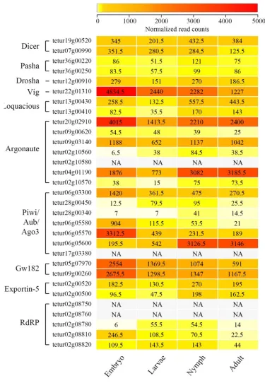
Figure 1.4: Heatmap representing expression patterns of RNAi-associated genes at different developmental stages of Tetranychus urticae, based on the number of mapped RNAseq reads (Illumina). ... 17
Figure 2.1 T. urticae feeding duration and mouth-part organs. ... 37
Figure 2.2: Mite feeding event and plant damage. ... 39
Figure 2.3: Bean and Arabidopsis leaves cell layer. ... 41
Figure 2.4: Plant damage associated with spider mite feeding. ... 42
Figure 2.5: Interface between T. urticae and plant tissue during feeding. ... 45
Figure 2.6: Model of the interactions between the plant and the cell-content feeding herbivores—the two-spotte spider mite and the aphid. ... 55
Figure 3.1: Fragment used for the synthesis of dsRNAs. ... 65
Figure 3.2: White phenotype associated with soaking in solution of dsRNA. ... 70
Figure 3.3: Dark phenotype associated with RNAi response to dsRNA. ... 72
Figure 3.4: Adult survivorship after soaking treatment in solution of dsRNA-TuVATPase separately for normal and dark-body mites. ... 73
Figure 3.5: Adult mite fecundity and TuVATPase gene expression after soaking treatment in solution of dsRNA-TuVATPase. ... 74
Figure 3.6: RNAi response in soaked T. urticae inbred lines. ... 77
Figure 3.7: Inbred line mortality after treatment with dsRNA-TuVATPase. ... 79
xi
Figure 3.9: Dark mite frequency in the inbred lines and London reference population after treatment with dsRNA. ... 81
Figure 3.10: Scatter 3D function displaying RNAi response parameter as a collection of points, plotted using three-dimensional cartesian coordinates. ... 82
Figure 4.1: Fragments used for synthesis of dsRNAs. ... 93
Figure 4.2: Schematic of the template preparation for dsRNA synthesis and mRNA DIG-labeled probesusing the PCR method. ... 96
Figure 4.3: Mite phenotype associated with RNAi response to dsRNA-TuCOPB2. ... 101
Figure 4.4: Adult survivorship after soaking treatment in solution of dsRNA-TuVATPase separately for normal and dark-body mites. ... 102
Figure 4.5: Adult mite fecundity after soaking treatment in solution of dsRNA-TuCOPB2. ... 104
Figure 4.6: Whole-mount in situ hybridization of COPB2 gene expression pattern in T. urticae female. ... 105
Figure 4.7: Correlation between blue mite and RNAi response after soaking for 24 hours in solution containing dsRNA with blue dye. ... 107
Figure 4.8: The effect of dsRNA-TuVATPase sequence sizes and variability of gene regions targeted on RNAi efficiency. ... 109
Figure 4.9: The effect of dsRNA-TuCOPB2 sequence sizes and variability of gene regions targeted on RNAi efficiency. ... 110
xii
List of Appendices
Appendix 1: Brightfield and confocal microscopy observation of fecal pellets and crystals of guanine in T. urticae. ... 128
Appendix 2: Representative picture of a cryosection of a mite feeding on a bean leaf previously frozen and embedded in optimal cutting temperature compound (OCT
compound). ... 129
Appendix 3: Representative picture of a microtome section of a mite feeding on a bean leaf previously frozen, fixed, dehydrated and embedded in paraffin. ... 130
xiii
List of Abbreviations
°C degree Celsius
µg microgram
ng nanogram
µl microliter
µm micrometer
bp base pair
cDNA complementary deoxyribonucleic acid
DIG digoxigenin
DNA deoxyribonucleic acid
dsRNA double stranded ribonucleic acid mRNA messenger ribonucleic acid PBS phosphate-buffered saline PCR polymerase chain reaction RNA ribonucleic acid
SEM scanning electron microscopy
RT-qPCR quantitative reverse transcription PCR
v/v volume/volume
DNase deoxyribonuclease BSA bovine serum albumin HRc high resolution camera SSC saline-sodium citrate buffer NBT nitro blue tetrazolium
Chapter 1
1
General introduction
1.1 The two-spotted spider mite –
Tetranychus urticae
(Koch)
The two-spotted spider mite, Tetranychus urticae (Acari: Tetranychidae) belongs to a group of plant pests whose members produce abundant silk (hence the name “spider mites”). The first specimen described was collected by the German arachnologist and entomologist Carl Ludwig Koch in 1836 on a stinging nettle, Urtica dioica (Koch, 1836). T. urticae belongs to the phylum of the Arthropoda, which are characterized by a segmented body, jointed appendages and presence of a chitinous exoskeleton. Within the arthropods, T. urticae belongs to the subphylum Chelicerata that represents the second largest group of arthropods. In the group of Acari, which is separated from spiders, phytophagous mites belong to the order of Trombidiformes, with about 22 000 species described (Walter, 2004). Despite its small size (about 0.5 mm long), T. urticae is an economically important agricultural pest with a global distribution (Bolland et al., 1998). The T. urticae genome was sequenced and annotated, and is characterized by a small size of 90 Mb (Grbić et al., 2011).
1.2 Feeding mechanism and digestive tract
Since the 1940s, extensive studies have been carried out to depict mite anatomy and body structures associated with feeding. Bearing in mind the minute size of the mite, most of the reports on anatomic features used histological cross sections coupled with electron microscopy. Although, some nomenclature may vary between authors, a good agreement among reports regarding features of the mouthparts is established. A simplified description of a mite’s body is described below and in Figure 1.2.
The mite body is divided in two functional domains: the gnathosoma, housing organs specialized in feeding (Becker, 1935), and the idiosoma, carrying the legs and organs dedicated in digestion, reproduction and excretion (Hammen, 1970). The gnathosoma is separated from the idiosoma by a flexible cuticule called circumcapitular furrow (Alberti, 2006; Grandjean, 1969). The gnathosoma is composed of two parts, the stylophore (STY; dorsal part of the gnathosoma), housing mobile elongated cheliceral digits (CHD; feeding tube), and the infracapitlum (ventral part of the gnathosoma), including the buccal cavity (BC), the pharynx (PH) and the pedipalps (PE) (André and Remacle, 1984; Mothes and Seitz, 1981b).
Figure 1.2: Schematic representation of a sagittal section through T. urticae female.
process, a mite presses the infracapitulum in the contact with the leaf surface, using its pedipalps. The chemo- and mechano-sensing receptors that are presents within sensorial organ such as the setae (Mills, 1973), presumably help mites find a suitable feeding spot. In preparation for feeding, the protractor muscles contract and each cheliceral digit runs into the infracapitulum groove (rostral gutter), which helps interlock them in the narrowest part of the buccal cavity to form a hollow tube-stylet (Andre and Remacle, 1984; Summers et al., 1973). Once protracted, the stylet length ranges from 100 µm in larval stages to about 150 µm in adult female mites (Avery and Briggs, 1968; Ekka, 1969; Sances et al., 1979). The function of this tube remains unknown and it is still the subject of controversy among authors whether it can be used as a piercing structure to deliver salivary secretion into plant tissue and/or is used for the food uptake as well (André and Remacle, 1984; Hislop and Jeppson, 1976; Summers et al., 1973). Alternatively, the buccal cavity has been postulated to be the orifice by which the nutrient extruded from the epidermis is absorbed (Alberti and Crooker, 1985; Nuzzacci and De Lillo, 1991b).
Regardless of the way mites ingest plant nutrients, the food is absorbed into the buccal cavity, through the pharynx (PH) that produces a sucking force and acts as a pump. The nutrients flow into the esophagus (ES), and are absorb in the ventriculus (VE) that consists of large digestive “floating” cells (FC) in the midgut of the mite. The midgut is composed of the ventriculus above the nervous system (CNM) connected to the esophagus and the excretory system (EX). Two large cavities, the caeca specialized in digestion, fill most of the lateral part of mite body (Alberti and Crooker, 1985; Alberti and Storch, 1973). Large digestive “floating” cells are pinched from the ventriculus epithelium and are presumably involved in the absorption of the gut content by phagocytosis and pinocytosis (Mothes and Seitz, 1981a). These floating cells are transported into the excretory organ responsible for the elimination of food residue and nitrogenous wastes (Blauvelt, 1945). Two type of digestion product are excreted (Gasser, 1951; Hazan et al., 1974). Black balls located in the ventriculus and in the caeca, and white pellets located only in the excretory organ. Recently, these two type of residues were visualized under confocal microscope (Occhipinti and Maffei, 2013). These authors demonstrated that the black pellets contain chlorophyll and its degradation products while the white pellets are composed of the guanine residue (see also personal observation, Appendix 1).
1.3 Plant damage and cytological changes associated with
feeding
T. urticae feeding causes extensive damage and important physiological changes in the host plant. They have been reported to feed on all aerial parts of the plant including cotyledon, leaves, stems, fruits and flowers. Although mites are mostly found at the abaxial side of the leaf (the underside that is shaded by the leaf itself), they are able to feed on both sides. The degree and the depth of injury are depending of several factors: 1) the length of the stylet, from 100 to 150µm 2) the feeding duration 3) mite population density and 4) host plant features (Avery and Briggs, 1968; Campbell et al., 1990; Ekka, 1969; Sances et al., 1979).
layer (Campbell et al., 1990; Sances et al., 1979). Similar conclusion were made with mites feeding on cucumber (Park and Lee, 2002). Also, a low mite density seems to affect mostly the spongy mesophyll, while with a high density of mites, both spongy and palisade mesophyll layers were affected (Sances et al., 1979).
Interestingly, the brown mites, Bryobia rubrioculus, feeding predominantly on the upper epidermis of almond leaves, caused damaged on the palisade layer, leaving the spongy layer unaffected (Summers and Stocking, 1972). The same authors evaluated the stylet length of B. rubrioculus to be 75 µm long while the palisade layer is 90 µm thick, leaving the spongy mesophyll layer unreachable for mites feeding from the upper epidermis. Also, it seems that, damage associated with feeding is localized within the mesophyll layer that is underneath the epidermis were the mite tends to settle. However, it is still unclear where the stylet is inserted in the epidermal cells. For instance, Avery and Briggs
(1968) didn’t observe epidermal damage and postulated that the stylet was inserted
between the epidermal cells along the anticlinal walls. Similarly, no evidence of tissue
damaged were observed to the lower epidermis of strawberry leaves (Sances et al., 1979).
However, Campbell (1990) observed the presence of stylet holes through the periclinal
walls of epidermal cells.
Although these studies represent the benchmark of T. urticae feeding pattern, these
observations were made days or weeks post feeding, at the time when plant responses to
mite feeding are triggered. As a consequence, these observations may be the combination
of direct damage caused by mite feeding and plant responses, leaving the direct feeding
injury unknown. For instance, changes to the stomatal apparatus reported by Sances et
al., (1979) was not caused by direct T. urticae feeding, but rather resulted from injury to
the spongy mesophyll layer that lead to reduced turgor in guard cells associated with
plant’s physiological response to mite feeding. Similarly, Mothes and Seitz (1982)
reported a strong reduction of leaf thickness in bean leaves by 50%, 6 days post feeding.
Likewise, several other reports (Avery and Briggs, 1968; Mothes and Seitz, 1981a;
Tanigoshi and Davis, 1978) showed an extensive cell death in leaf tissue upon prolonged
mite infestation, leaving the extent of leaf damage directly caused by mite feeding
chloroplast abnormalities in cells that appear adjacent to the punctured cell (that may
have been consumed by mite feeding) has been reported by Avery and Briggs (1968) and
Tanigoshi and Davis (1978). In contrast, no morphological distortion in mesophyll cells adjacent to mite-damaged cells were observed in strawberry, suggesting that plant responses may differ in different species and may be dependent on their physiological state (Campbell et al., 1990; Sances et al., 1979).
1.4 Spider mite control strategy
T. urticae, is one of the most polyphagous arthropods, feeding on more than 1100 plant species including more than 150 agricultural crops that belong to more than 140 different families (Jeppson et al., 1975; Migeon and Dorkeld, 2006-2017). Despite increasing effort from the scientific community and growers, spider mite control remains challenging. Three main factors contribute to spider mite spread: pesticide resistance, crop practice and global warming. The current Integrated Pest Management (IPM;
defined as a combination of existing pest management measures to decrease pests and reduce or minimize the use of pesticides) of spider mite relies on biological control using predatory mites and pesticides. However, while biological control is efficient under specific condition and low population density, T. urticae control requires utilization of pesticides. Unfortunately, T. urticae rapidly develops resistance to pesticides making mites the arthropod with the highest occurrence of pesticide resistance. This is due to its short life cycle, high fecundity and its specific mating system through which recessive resistance alleles are easily fixed via haploid males (Carrière, 2003; Denholm et al., 1998). Moreover, It has been evaluated that T. urticae can develop resistance to a new pesticide between 2 to 4 years (Van Leeuwen et al., 2015).
on spider mites but affect mite predators, resulting in spider mite expansion. Also, damage caused by spider mite infestation coupled with an increase of mite population in warm weather is predicted to exacerbate plant damage as spider mites reproduce faster in high temperature. Likewise, drought-stress of host plants increases spider mite oviposition by two times (Ximénez-Embún et al., 2016).
1.5 RNA interference
Prior to the discovery and the characterization of RNAi mechanisms by Fire and Mello in 1998, the phenomena was initially reported in the petunia by Jorgensen and his team in an attempt to overexpressed the chalcone synthase gene (chsA), involved in anthocyanin production (Napoli et al., 1990). Unexpectedly, transgenic petunia carrying the chsA gene under the control of the strong promoter (35S) yielded petunia plants with white flowers or partially white with a pigmented background instead of purple flowers. The authors demonstrated that both endogenous and exogenous chsA transcripts were suppressed and that the reduction of mRNA transcript was not related to the reduction of the transcription efficiency, suggesting a “co-suppression” of homologous mRNA. A similar phenomenon was observed in the fungus Neurospora crassa, in an effort to increase the production of an orange pigment. The transformation of N. crassa wild-type (orange phenotype) by the carotenogenic albino-3 (al-3) or albiono-1 (al-1)resulted in an albino phenotype in a few transformants in which transcripts levels were drastically reduced (Romano and Macino, 1992). The authors qualified this phenomenon in fungus as “quelling”.
Since the RNAi discovery, key elements underlying the RNAi mechanism were discovered. For instance, Hamilton (1999), identified small double stranded RNA molecules of about 25 nucleotides that were complementary to the targeted mRNA. They hypothesized that these small RNAs are the mediators of mRNA degradation. This hypothesis was verified in a series of experiments in which exogenous short-interfering dsRNA (siRNA) fragments of about 21 nucleotides long were able to trigger gene silencing in Drosophila embryo (Hammond et al., 2000) and mammalian cell lines (Elbashir et al., 2001). In the same time, the key enzyme responsible for the cleavage of dsRNA into a population of siRNAs was identified in Drosophila melanogaster. It is a type III RNAse enzyme that is referred to as Dicer (for its dicing activity) (Bernstein et al., 2001; Hammond et al., 2000). Further, two proteins, Argonaute 1 (AGO1) and Argonaute 2 (AGO2) were identified in Drosophila to form a complex with the Dicer enzyme, yielding the RISC complex (RNAi-induced silencing complex), hypothesized to directly mediate the “slicing” activity (Hutvágner and Zamore, 2002; Martinez et al., 2002; Nykänen et al., 2001). However, only Argonaute 2 has been demonstrated to have the endonuclease activity (Liu et al., 2004).
1.6 RNAi pathways and biological function
Three main RNAi pathways associated with small RNAs have been characterized, each displaying specific function in eukaryotic organisms.
Micro RNAs (miRNA) are derived from long hairpin RNA encoded by the host genome. Its processing yields a single small non-coding RNA of about 22 nucleotides (Bartel, 2004). This endogenous RNAi is involved in gene regulation and functions as a repressor of either gene translation (Translation Gene Silencing; TGS; Djuranovic et al., 2012) or at the post transcriptional level (Post-Transcriptional Gene Silencing; PTGS; Bushati and Cohen, 2007; Leucci et al., 2013; Pillai et al., 2017). The Piwi-interacting short RNAs (piRNAs) mediated pathway has been demonstrated to be involved in silencing of transposable elements, preserving genome integrity (Aravin et al., 2007; Lee, 2015; Sienski et al., 2012).
siRNA pathway can be manipulated through biotechnology to suppress a specific gene target by delivering dsRNA into the cell.
1.7 Mechanism of RNAi through the exogenous application
of dsRNA
When exogenous dsRNA is introduced, dsRNA molecules are processed by the enzyme Dicer-2 into a population of uniformly sized siRNA molecules of 20 to 25 bp long and loaded into the RISC complex. siRNAs are subsequently recruited by the Argonaute proteins into a RISC complex that either cleaves the homologous mRNA or prevents its translation (Figure 1.3). This mechanism is particularly interesting, since the application of the exogenous dsRNA can be used to knock-down, in theory, any gene. Since RNAi depends on the complementarity between the target and dsRNA, the RNAi response is expected to be specific. For more than a decade, RNAi has been used as a reverse genetic tool (Lawson and Wolfe, 2011), therapeutic treatment (Aagaard and Rossi, 2007) and recently in plant-pest control (Baum et al., 2007). The use of RNAi in the control of plant-pests is based on the premise that if the targeted mRNA encodes an essential protein, its silencing leads to lethality. The sequence specificity of a dsRNA fragment used for the RNAi should secure effectiveness against a particular species without affecting other organisms.
1.8 RNAi as a tool for pest control
Figure 1.3: Mechanism of RNA interference in eukaryotic cell. Exogenous dsRNA triggers RNAi by activating Dicer enzyme that is going to cleave long dsRNA fragment into a population of short interfering RNAs (siRNAs) of 20 to 25 base pairs. siRNAs are loaded into a RISC complex that induces either degradation of complementary mRNA or its inhibition. This phenomenon is called Post Translation Gene Silencing, (PTGS). (adapted from Bensoussan and Grbić, 2017).
Gut lumen
dsRNA
PTGS
RNA degradation
Inhibition of translation Targeted
mRNA
Dicer
siRNA
Risc
Reduce protein production Exogenous dsRNA
Cytoplasm
Baum and his team, designed an RNAi screen in which they tested 290 dsRNAs, targeting genes coding for proteins of vital function and supplied in artificial diet. They identified 14 genes that strongly responded to RNAi, causing increased larval mortality in the western corn rootworm, Diabrotica virgifera virgifera (WCR). Subsequently, corn plants were transformed to express dsRNA against the proton pump VATPase of WCR, one of the targets identified in the artificial diet screen. These plants showed significant reduction of root damage with low nodal injury upon WCR infestation, indicating the efficiency of the RNAi in crop protection (Baum et al., 2007). Although, these studies highlight the potential of RNAi as a promising tool to control pests, RNAi efficiency has been shown to be variable among insects and arthropods. Even though the RNAi machinery is conserved across insect and arthropods classes, RNAi responsiveness differs among orders (Bellés, 2010). For example, it has been reported that coleopterans are very sensitive to RNAi.
In contrast, RNAi responsiveness in lepidopterans has been reported to be low and variable between species, tissues, gene targets, delivery methods and required high concentration of dsRNA to trigger equivalent RNAi efficiency as in coleopterans (Ivashuta et al., 2015; Terenius et al., 2011). Moreover, factors including, but not limited to, RNAi design, dsRNA concentration, delivery method, targeted gene, life stage and expression level of RNAi machinery have been demonstrated to be critical (Bolognesi et al., 2012; Chu et al., 2014; Coleman et al., 2015; Huvenne and Smagghe, 2010; Ivashuta et al., 2015).
1.9 Factors influencing RNAi efficiency
1.9.1
dsRNA design
Factors including dsRNA fragment size, concentration and targeted mRNA region can
with optimal concentration of 0.01 µg/µl, after which further increase of dsRNA concentration didn’t increase RNAi potency.
In insects such as Drosophila, fragment sizes ranging from 21 to 592 base pairs have been tested for RNAi efficiency on S2 cells (Saleh et al., 2006). The authors clearly show a positive correlation between fragment size and increased RNAi efficiency. In addition, long dsRNAs were more efficient compare to siRNA when expressed in transgenic plants (Mao et al., 2011). Overall, most of the studies have shown that dsRNA of 100 to 600 bp are required for the successful RNAi. The region of mRNA sequence targeted by dsRNA can also influence RNAi efficiency. For instance, ingestion of dsRNA designed against the 3’ end of the apoptosis gene AeIAP1 of the mosquito Aedes aegypti, showed greater effect than dsRNA against the 5’ end region (Pridgeon et al., 2008). However, no differences where observed between dsRNA designed either against the 5’ or 3’ ends of the targeted gap gene hunchback, a key regulator in the anteroposterior patterning in the pea aphid, Acyrthosiphon pisum (Mao and Zeng, 2012).
These studies point out the variability of RNAi sensitivity when designing dsRNA intrinsic to the target and the organism. Also, they hint at a need to design several fragments, each targeting different parts of the mRNA, which can also be used to test the experimental reproducibility. Also, in silico analysis of the targeted sequence must be performed prior to synthesizing dsRNA to avoid off-target effects form sequence match between population of siRNA (product from dsRNA cleavage) and mRNA from undesired targets (Du et al., 2005; Jackson et al., 2003). Although, to date, no consensus about the degree of sequence mismatch between siRNA derived from dsRNA and off targeted mRNA has been established.
1.9.2
dsRNA delivery method
Carthew, 1998). The advantages associated with this method is the high silencing efficiency of the target gene and the control of the amount of dsRNA delivered into the body. However, variation of RNAi response has been observed when compared between delivery methods on the same organism (Terenius et al., 2011). Injection of dsRNA was more efficient to silence apn gene in Spodoptera litura compare to the interference from feeding on hairpin-loop transformed plants as a result of a lower concentration of delivered dsRNA with feeding (Rajagopal et al., 2002).
In contrast, ingestion of dsRNA through oral delivery was more efficient than injection when a continuous feeding was applied in the Cotton Bollworm, Helicoverpa armigera (Yang and Han, 2014). The development of the oral delivery method was introduced as an alternative of injection, that is time consuming and subject to mechanical damage that is often lethal (Liu et al., 2010), or that induces an immune response (Han et al., 1999). Also, injection of dsRNA or small RNAs were widely used as a reverse genetics tool, while non-invasive methods of dsRNA delivery, such as soaking or feeding, are more suitable for the development of RNAi as biopesticide. Moreover, oral delivery is convenient, less invasive, and can be used as high-throughput screen. The first demonstration of RNAi through oral delivery was done in C. elegans feeding on Escherichia coli transformed to express dsRNAs (Timmons and Fire, 1998).
Also, several studies have shown the potential of using artificial diet mixed with dsRNA for oral delivery. It was successful to trigger RNAi in whiteflies, aphids and honey bees (Aronstein et al., 2006; Ghanim et al., 2007; Whyard et al., 2009; Wuriyanghan et al., 2011). Oral delivery through feeding on transgenic plants expressing dsRNA was efficient to trigger RNAi in Coleopteran (Baum et al., 2007), Lepidoptera (Dai et al., 2008), Helicoverpa (Bally et al., 2016), and in Hemiptera (Zha et al., 2011).
solution of dsRNA resulted in 87% transcript level downregulation compare to 97% when injected, albeit rather associated with high mortality due to the mechanical stress.
1.9.3
Gut Environment
Although the oral delivery of dsRNA may provide an advantage to follow a natural ingestion route through the gut, the ability of dsRNA to be ingested and absorbed by epithelium gut cells can influence RNAi potency. Moreover, the spatio-temporal expression of key components of the RNAi machinery can also affect RNAi efficiency and its ability to spread through the body (Chintapalli et al., 2007; Rinkevich and Scott, 2013). Recently, there is increasing evidence of the presence of nuclease in the gut and hemolymph in arthropods that, this could result in dsRNA degradation prior to cell uptake and a lack of potent RNAi in some species (Allen and Walker, 2012; Garbutt et al., 2013; Wynant et al., 2014). Interestingly, the elimination of the nuclease activity in the potato beetle, Leptinotarsa decemlineata, increased RNAi efficiency (Spit et al., 2017). In addition to the presence of nucleases, extreme gut pH can provide a deleterious environment to dsRNA stability (Lomate and Bonning, 2016). However, an acidic gut environment has been shown to be required for SID-2 activity in dsRNA cell uptake in Drosophila S2 cells (McEwan et al., 2012).
1.9.4
RNAi in Tetranychid mites
Khila and Grbić (2007) demonstrated that parental RNAi can silence the expression of the homeobox gene, Distal-less (Dll). Injection of either Tu-Dll-specific dsRNA or siRNA into adult female mites resulted in offspring with truncated and fused leg segments. This study was the first to demonstrate that RNAi could be a valid reverse genetic approach in T. urticae. However, compared to other arthropods, for example Tribolium and Oncopeltus, dsRNA injection is not a viable delivery method for T. urticae, as females are less than 0.5 mm in length. Recently, Kwon et al., (2013) reported an alternative method for the administration of dsRNA to T. urticae. They developed a protocol for oral delivery of dsRNA using bean leaf discs that floated on a solution containing dsRNA. Using this method, they were successful in decreasing the expression levels of several genes involved in mite metabolism and physiology, resulting in mite mortality five days post feeding. Even though this method may not be practical for high throughput screens of potential mite targets, as it requires large quantities of dsRNA that must be replaced daily, it has been useful as a reverse genetics tool. For example, application of this dsRNA delivery method in the red mite, Pannonychus citri, has been successful in down-regulating the chitinase (PcCht1) gene by almost 60%. Associated with the reduced expression of this target gene was lethality that resulted from larval molting failure (Xia et al., 2016). Similarly, oral delivery of dsRNA to the carmine spider mite, Tetranychus cinnabarinus, was successful in down-regulating an esterase (TCE2) whose expression was implicated in acaricide resistance. The exogenous application of acaricides to the dsRNA-treated mites increased their effectiveness, indicating the correlation between the expression of TCE2 gene and acaricides resistance (Shi et al., 2016). These examples reinforce the feasibility of using RNAi as a reverse genetics tool for functional studies of mite biology, and as a tool for the development of new mite management strategies.
1.10 Rationale and specific goals of my research
chelicerate herbivore with tools that enable tracking of reciprocal responses in plant-spider mite interactions. However, despite their important pest status and a growing understanding of the molecular basis of interactions with plant hosts, knowledge of the way mites interface with the plant while feeding and the plant damage directly inflicted by mites is lacking.In addition, a critical step in the analysis of spider mite gene function is the availability of efficient reverse genetic tool. To further promote the establishment and use of T. urticae as a model herbivore, the objectives of my work were:
1) To characterize plant damage that directly results from mite feeding at a cellular level and duration of a feeding event.
2) To investigate the T. urticae stylet pathway while feeding on host plants.
3) To establish RNA interference in T. urticae through soaking of adult female mites in the solution of dsRNA against the target gene VATPase.
1.11 Reference
Aagaard, L., and Rossi, J. J. (2007). RNAi therapeutics: Principles, prospects and challenges. Adv. Drug Deliv. Rev. 59, 75–86.
Alba, J. M., Schimmel, B. C. J., Glas, J. J., Ataide, L. M. S., Pappas, M. L., Villarroel, C. A., et al. (2015). Spider mites suppress tomato defenses downstream of jasmonate and salicylate independently of hormonal crosstalk. New Phytol. 205, 828–840.
Alberti, G. (2006). On some fundamental characteristics in acarine morphology. Atti della Accad. Naz. Ital. di Entomol. R.A. LIII, 315–360.
Alberti, G., and Crooker, A. (1985). “Internal anatomy,” in Spider mites: their biology, natural enemies and control., eds. W. Helle and M. W. Sabelis (Elsevier Ltd), 29– 62.
Alberti, G., and Kitajima, E. W. (2014). Anatomy and Fine Structure of Brevipalpus Mites (Tenuipalpidae) - Economically Important Plant-Virus Vectors. Stuttgart: Schweizerbart.
Alberti, G., and Storch, V. (1973). Zur Feinstruktur der “Munddrüsen” von Schnabelmilben (Bdellidae, Trombidiformes). Z.Wiss.Zool. 186, 149–160.
Allen, M. L., and Walker, W. B. (2012). Saliva of Lygus lineolaris digests double stranded ribonucleic acids. J. Insect Physiol. 58, 391–396.
André, H. M., and Remacle, C. (1984). Comparative and functional morphology of the gnathosoma of Tetranychus urticae (Acari: Tetranychidae). Acarologia. 25, 179– 190.
Aravin, A. A., Hannon, G. J., and Brennecke, J. (2007). The piwi-piRNA pathway provides an adaptive defense in the transposon arms race. Science. 318, 761–764.
Aronstein, K., Pankiw, T., and Saldivar, E. (2006). SID-I is implicated in systemic gene silencing in the honey bee. J. Apic. Res. 45, 20–24.
Avery, D. J., and Briggs, J. B. (1968). The aetiology and development of damage in young fruit trees infested with fruit tree red spider mite, Panonychus ulmi (Koch). Ann. Appl. Biol. 61, 277–288.
Bally, J., McIntyre, G. J., Doran, R. L., Lee, K., Perez, A., Jung, H., et al. (2016). In-plant protection against Helicoverpa armigera by production of long hpRNA in chloroplasts. Front. Plant Sci. 7, 1453.
Baum, J. A., Bogaert, T., Clinton, W., Heck, G. R., Feldmann, P., Ilagan, O., et al. (2007). Control of coleopteran insect pests through RNA interference. Nat Biotech. 25, 1322–1326.
Becker, E. (1935). Die Mundwerkzeuge de Tetranychus telarius (L.) und deren Funcktionin Beziehung zur chemischen Bekampfung des letzteren. Rev. Zool.14, 637–554.
Bellés, X. (2010). Beyond Drosophila: RNAi in vivo and functional genomics in insects. Annu. Rev. Entomol. 55, 111–128.
Bensoussan, N., and Grbić, V. (2017). RNA interference in the two-spotted spider mite Tetranychus urticae. Int. Organ. Biol. Integr. Control. 124, 200–206.
Bernstein, E., Caudy, A. A., Hammond, S. M., and Hannon, G. J. (2001). Role for a bidentate ribonuclease in the initiation step of RNA interference. Nature. 409, 363– 366.
Blauvelt, W. (1945). The internal anatomy of the common spider mite (Tetranychus telarius L.). Agri. Exp. Stn. 270, 1–35.
Bolland, H. R., Gutierrez, J., and Flechtmann, C. H. (1998). World catalogue of the spider mite family:(Acari: Tetranychidae). Brill. Leiden; Boston.
Bolognesi, R., Ramaseshadri, P., Anderson, J., Bachman, P., Clinton, W., Flannagan, R., et al. (2012). Characterizing the mechanism of action of double-stranded RNA activity against western corn rootworm (Diabrotica virgifera virgifera LeConte). PLoS One 7, e47534.
Boudreaux, H. B. (1963). Biological Aspects of Some Phytophagous Mites. Annu. Rev. Entomol. 8, 137–154.
Brusca, R. C., and Brusca, G. J. (2003). Invertebrates. Sunderland, MA: Sinauer Associates.
Bushati, N., and Cohen, S. M. (2007). microRNA Functions. Annu. Rev. Cell Dev. Biol. 23, 175–205.
Campbell, E. M., Budge, G. E., and Bowman, A. S. (2010). Gene-knockdown in the honey bee mite Varroa destructor by a non-invasive approach: studies on a glutathione S-transferase. Parasit. Vectors. 3, 73.
Campbell, R. J., Grayson, R. L., and Marini, R. P. (1990). Surface and ultrastructural feeding injury to strawberry leaves by the twospotted spider mite. Hortscience. 25, 948–951.
Chintapalli, V. R., Wang, J., and Dow, J. A. T. (2007). Using FlyAtlas to identify better Drosophila melanogaster models of human disease. Nat Genet. 39, 715–720.
Chu, C.C., Sun, W., Spencer, J. L., Pittendrigh, B. R., and Seufferheld, M. J. (2014). Differential effects of RNAi treatments on field populations of the western corn rootworm. Pestic. Biochem. Physiol. 110, 1–6.
Coleman, A. D., Wouters, R. H. M., Mugford, S. T., and Hogenhout, S. A. (2015). Persistence and transgenerational effect of plant-mediated RNAi in aphids. J. Exp. Bot. 66, 541–548.
Conte, D., MacNeil, L. T., Walhout, A. J. M., and Mello, C. C. (2015). “RNA Interference in Caenorhabditis elegans,” in Current Protocols in Molecular Biology (Hoboken, NJ, USA: John Wiley & Sons, Inc.), 26.3.1-26.3.30.
Dai, H., Ma, L., Wang, J., Jiang, R., Wang, Z., and Fei, J. (2008). Knockdown of ecdysis-triggering hormone gene with a binary UAS/GAL4 RNA interference system leads to lethal ecdysis deficiency in silkworm. Acta Biochim. Biophys. Sin. (Shanghai). 40, 790–5.
Denholm, I., Horowitz, A. R., Cahill, M., and Ishaaya, I. (1998). “Management of Resistance to Novel Insecticides" in Insecticides with Novel Modes of Action: Mechanisms and Application,” eds. I. Ishaaya and D. Degheele (Berlin, Heidelberg: Springer Berlin Heidelberg), 260–282.
Ding, S. W. (2010). RNA-based antiviral immunity. Nat Rev Immunol. 10, 632–644.
Ding, S. W., and Voinnet, O. (2007). Antiviral Immunity Directed by Small RNAs. Cell. 130, 413–426.
Djuranovic, S., Nahvi, A., and Green, R. (2012). miRNA-Mediated gene silencing by translational repression followed by mRNA deadenylation and decay. Science. 336, 237–240.
Du, Q., Thonberg, H., Wang, J., Wahlestedt, C., and Liang, Z. (2005). A systematic analysis of the silencing effects of an active siRNA at all single-nucleotide mismatched target sites. Nucleic Acids Res. 33, 1671–1677.
Ekka, I. (1969). The Rearing of the Two-Spotted Spider Mite (Tetranychus urticae Koch) on Artificial Diets. Ph.D thesis. McGill University.
Elbashir, S. M., Harborth, J., Lendeckel, W., Yalcin, A., Weber, K., and Tuschl, T. (2001). Duplexes of 21-nucleotide RNAs mediate RNA interference in cultured mammalian cells. Nature. 411, 494–498.
Garbutt, J. S., Bellés, X., Richards, E. H., and Reynolds, S. E. (2013). Persistence of double-stranded RNA in insect hemolymph as a potential determiner of RNA interference success: Evidence from Manduca sexta and Blattella germanica. J. Insect Physiol. 59, 171–178.
Gasser, R. (1951). Zur Kenntnis der gemeinen Spinnmilbe Tetranychus Urticae Koch: Mitteilung 1: Morphologie, Anatomie, Biologie und Oekologie. Rull Soc Ent Suisse. 24, 217–262.
Ghanim, M., Kontsedalov, S., and Czosnek, H. (2007). Tissue-specific gene silencing by RNA interference in the whitefly Bemisia tabaci (Gennadius). Insect Biochem. Mol. Biol. 37, 732–738.
Grandjean, F. (1969). Stasis. Actinopiline. Explanation of my mites classification into three major groups. Terminology of -soma. Acarologia. 11, 796–827.
Grbić, M., Van Leeuwen, T., Clark, R. M., Rombauts, S., Rouzé, P., Grbić, V., et al. (2011). The genome of Tetranychus urticae reveals herbivorous pest adaptations. Nature. 479, 487–492.
Hamilton, A. J. (1999). A Species of small antisense RNA in posttranscriptional gene silencing in plants. Science. 286, 950–952.
Hammen, L. van der. (1970). General notes on the fondamental structure of the gnathosoma. Acarologia 12, 16–22.
Hammen, L. van der (1980). Glossary of acarological terminology. Vol. 1: General terminology. The Hague: Dr. W. Junk, B. V.
Hammond, S. M., Bernstein, E., Beach, D., and Hannon, G. J. (2000). An RNA-directed nuclease mediates post-transcriptional gene silencing in Drosophila cells. Nature. 404, 293–296.
Han, Y. S., Chun, J., Schwartz, A., Nelson, S., and Paskewitz, S. M. (1999). Induction of mosquito hemolymph proteins in response to immune challenge and wounding. Dev. Comp. Immunol. 23, 553–562.
Hazan, A., Gerson, U., and Tahori, A. S. (1974). Spider mite webbing. I. The production of webbing under various environmental conditions. Acarologia. 16, 68–84.
Helle, W., and Sabelis, M. W. (1985). Spider mites: their biology, natural enemies and control. eds. W. Helle and M. W. Sabelis Amsterdam: Elsevier Ltd.
Hislop, R. G., and Jeppson, L. R. (1976). Morphology of the mouthparts of several species of phytophagous mites. Ann. Entomol. Soc. Am. 69, 1125–1135.
Huvenne, H., and Smagghe, G. (2010). Mechanisms of dsRNA uptake in insects and potential of RNAi for pest control: A review. J. Insect Physiol. 56, 227–235.
Ivashuta, S., Zhang, Y., Wiggins, B. E., Ramaseshadri, P., Segers, G. C., Johnson, S., et al. (2015). Environmental RNAi in herbivorous insects. RNA. 21, 840–850.
Jackson, A. L., Bartz, S. R., Schelter, J., Kobayashi, S. V, Burchard, J., Mao, M., et al. (2003). Expression profiling reveals off-target gene regulation by RNAi. Nat Biotech. 21, 635–637.
Jeppson, L. R., Keifer, H. H., and Baker, E. W. (1975). Mites injurious to economic plants. Berkeley: University of California Press.
Jonckheere, W., Dermauw, W., Zhurov, V., Wybouw, N., Van den Bulcke, J., Villarroel, C. A., et al. (2016). The Salivary Protein Repertoire of the Polyphagous Spider Mite Tetranychus urticae : A Quest for Effectors. Mol. Cell. Proteomics. 15, 3594–3613.
Kant, M. R., Sabelis, M. W., Haring, M. A., and Schuurink, R. C. (2008). Intraspecific variation in a generalist herbivore accounts for differential induction and impact of host plant defences. Proc. R. Soc. B Biol. Sci. 275, 443–452.
Kennerdell, J. R., and Carthew, R. W. (1998). Use of dsRNA-mediated genetic interference to demonstrate that frizzled and frizzled 2 act in the wingless pathway. Cell. 95, 1017–1026.
Khila, A., and Grbić, M. (2007). Gene silencing in the spider mite Tetranychus urticae: dsRNA and siRNA parental silencing of the Distal-less gene. Dev. Genes Evol. 217, 241–251.
Koch, C. L. (1836). Tetranychus urticae. Dtsch. Crustac. Myriapoda, Arachn.1, 10.
Kwon, D. H., Park, J. H., and Lee, S. H. (2013). Screening of lethal genes for feeding RNAi by leaf disc-mediated systematic delivery of dsRNA in Tetranychus urticae. Pestic. Biochem. Physiol. 105, 69–75.
Lawson, N. D., and Wolfe, S. A. (2011). Forward and reverse genetic approaches for the analysis of vertebrate development in the zebrafish. Dev. Cell. 21, 48–64.
Lee, Y. C. G. (2015). The Role of piRNA-mediated epigenetic silencing in the population dynamics of transposable elements in Drosophila melanogaster. PLOS Genet. 11, e1005269.
Leucci, E., Patella, F., Waage, J., Holmstrøm, K., Lindow, M., Porse, B., et al. (2013). microRNA-9 targets the long non-coding RNA MALAT1 for degradation in the nucleus. Sci. Rep. 3, 2535.
Environ. 38, 2277–2285.
Li, X., Schuler, M. A., and Berenbaum, M. R. (2006). Molecular mechanisms of metabolic resistance to synthetic and natural xenobiotics. Annu. Rev. Entomol. 52, 231–253.
Liu, J., Carmell, M. A., Rivas, F. V, Marsden, C. G., Thomson, J. M., Song, J.-J., et al. (2004). Argonaute2 is the catalytic engine of mammalian RNAi. Science. 305, 1437–1441.
Liu, S., Ding, Z., Zhang, C., Yang, B., and Liu, Z. (2010). Gene knockdown by intro-thoracic injection of double-stranded RNA in the brown planthopper, Nilaparvata lugens. Insect Biochem. Mol. Biol. 40, 666–671.
Lomate, P. R., and Bonning, B. C. (2016). Distinct properties of proteases and nucleases in the gut, salivary gland and saliva of southern green stink bug, Nezara viridula. Sci.Rep. 6, 27587.
Mao, J., and Zeng, F. (2012). Feeding-based RNA intereference of gap gene is lethal to the pea aphid, Acyrthosiphon pisum. PLoS One 7, e48718.
Mao, Y.-B., Cai, W.-J., Wang, J.-W., Hong, G.-J., Tao, X.-Y., Wang, L.-J., et al. (2007). Silencing a cotton bollworm P450 monooxygenase gene by plant-mediated RNAi impairs larval tolerance of gossypol. Nat Biotech. 25, 1307–1313.
March, J. C., and Bentley, W. E. (2007). RNAi-based tuning of cell cycling in Drosophila S2 cells—effects on recombinant protein yield. Appl. Microbiol. Biotechnol. 73, 1128–1135.
Martinez, J., Patkaniowska, A., Urlaub, H., Lührmann, R., and Tuschl, T. (2002). Single-stranded antisense siRNAs guide target RNA cleavage in RNAi. Cell. 110, 563–574.
McEwan, D. L., Weisman, A. S., and Hunter, C. P. (2012). Uptake of extracellular double-stranded RNA by SID-2. Mol. Cell. 47, 746–754.
Migeon, A., and Dorkeld, F. (2006-2014). Spider Mites Web: a comprehensive database for the Tetranychidae. Available online at: http://www.montpellier. inra. fr/ CBGP/ spmweb
Miller, S. C., Miyata, K., Brown, S. J., and Tomoyasu, Y. (2012). Dissecting systemic RNA interference in the red flour beetle Tribolium castaneum: parameters affecting the efficiency of RNAi. PLoS One 7, e47431.
Mills, L. R. (1973). Structure of dorsal setae in the two-spotted spide rmite Tetranychus urticae Koch, 1836. Acarologia. 15, 649–658.
Tissue Res. 221, 339–349.
Mothes, U., and Seitz, K. A. (1981b). Functional microscopic anatomy of the digestive system of Tetranychus urticae (Acari, Tetranychidae). Acarologia. 22, 257–270.
Napoli, C., Lemieux, C., and Jorgensen, R. (1990). Introduction of a chimeric chalcone synthase gene into petunia results in reversible co-suppression of homologous genes in trans. Plant Cell. 2, 279–289.
Nuzzacci, G., and De Lillo, E. (1991b). “Fine structure and functions of the mouthparts involved in the feeding mechanisms in Tetranychus urticae Koch (Tetranychoidea: Tetranychidae),” in Modern Acarology: Proceedings of the VIII International Congress of Acarology, eds F. Dusbabek and V. Bukva (The Hague: Academia Prague and SPB Academic), 301–303.
Nykänen, A., Haley, B., and Zamore, P. D. (2001). ATP Requirements and Small Interfering RNA Structure in the RNA Interference Pathway. Cell 107, 309–321.
Occhipinti, A., and Maffei, M. E. (2013). Chlorophyll and its degradation products in the two-spotted spider mite, Tetranychus urticae: observations using epifluorescence and confocal laser scanning microscopy. Exp. Appl. Acarol. 61, 213–219.
Olivier, J. H. (1971). Parthenogenesis in mites and ticks (Arachnida: Acari). Am. Zool. 11, 283–299.
Park, Y.-L., and Lee, J.-H. (2002). Leaf cell and tissue damage of cucumber caused by twospotted spider mite (Acari: Tetranychidae). J. Econ. Entomol. 95, 952–7.
Pillai, R. S., Bhattacharyya, S. N., and Filipowicz, W. (2017). Repression of protein synthesis by miRNAs: how many mechanisms? Trends Cell Biol. 17, 118–126.
Pridgeon, J. W., Zhao, L., Becnel, J. J., Strickman, D. A., Clark, G. G., and Linthicum, K. J. (2008). Topically applied AaeIAP1 double-stranded RNA kills female adults of Aedes aegypti. J. Med. Entomol. 45, 414–20.
Rajagopal, R., Sivakumar, S., Agrawal, N., Malhotra, P., and Bhatnagar, R. K. (2002). Silencing of midgut aminopeptidase N of Spodoptera litura by double-stranded RNA establishes its role as Bacillus thuringiensis toxin receptor. J. Biol. Chem. 277, 46849–46851.
Riga, M., Bajda, S., Themistokleous, C., Papadaki, S., Palzewicz, M., Dermauw, W., et al. (2017). The relative contribution of target-site mutations in complex acaricide resistant phenotypes as assessed by marker assisted backcrossing in Tetranychus urticae. Sci. Rep. 7, 9202.
Romano, N., and Macino, G. (1992). Quelling: transient inactivation of gene expression in Neurospora crassa by transformation with homologous sequences. Mol. Microbiol. 6, 3343–3353.
Roush, T., and Tabashnik, B. (1990). Pesticide Resistance in Arthropods. , eds. R. T. Roush and B. E. Tabashnik Boston, MA: Springer US.
Saleh, M.-C., van Rij, R. P., Hekele, A., Gillis, A., Foley, E., O’Farrell, P. H., et al. (2006). The endocytic pathway mediates cell entry of dsRNA to induce RNAi silencing. Nat Cell Biol. 8, 793–802.
Sances, F. V, Wyman, J. A., and Ting, I. P. (1979). Morphological responses of strawberry leaves to infestations of twospotted spider mite. J. Econ. Entomol. 72, 710–713.
Shi, L., Wei, P., Wang, X., Shen, G., Zhang, J., Xiao, W., et al. (2016). Functional analysis of esterase TCE2 Gene from Tetranychus cinnabarinus (Boisduval) involved in acaricide resistance. Sci. Rep. 6, 18646.
Shih, C. T., Poe, S. L., and Cromroy, H. L. (1976). Biology, life table, and intrinsic rate of increase of Tetranyshus urticae. Ann. Entomol. Soc. Am. 69, 362–364.
Sienski, G., Dönertas, D., and Brennecke, J. (2012). Transcriptional silencing of transposons by piwi and maelstrom and its impact on chromatin ctate and gene expression. Cell. 151, 964–980.
Spit, J., Philips, A., Wynant, N., Santos, D., Plaetinck, G., and Vanden Broeck, J. (2017). Knockdown of nuclease activity in the gut enhances RNAi efficiency in the Colorado potato beetle, Leptinotarsa decemlineata, but not in the desert locust, Schistocerca gregaria. Insect Biochem. Mol. Biol. 81, 103–116.
Summers, F., Gonzales, R., and Witt, R. (1973). The mouthparts of Bryobia rubrioculus (Sch.) (Acarina: Tetranychidae). Proc. Entomol. Soc. Washingt. 75, 96–11.
Summers, F. M., and Stocking, C. R. (1972). Some immediate effects on almond leaves of feeding some by Bryobia rubrioculus (Scheuten). Acarologia. 14, 170–178.
Tabara, H., Grishok, A., and Mello, C. C. (1998). RNAi in C. elegans: Soaking in the genome sequence. Science. 282, 430–431.
Tanigoshi, L. K., and Davis, R. W. (1978). An ultrastructural study of Tetranychus mcdanieli feeding injury to the leaves of “Red Delicious” apple (Acari: Tetranychidae). Int. J. Acarol. 4, 47–51.
Terenius, O., Papanicolaou, A., Garbutt, J. S., Eleftherianos, I., Huvenne, H., Kanginakudru, S., et al. (2011). RNA interference in Lepidoptera: An overview of successful and unsuccessful studies and implications for experimental design. J. Insect Physiol. 57, 231–245.
Timmons, L., and Fire, A. (1998). Specific interference by ingested dsRNA. Nature. 395, 854.
Van Leeuwen, T., Tirry, L., Yamamoto, A., Nauen, R., and Dermauw, W. (2015). The economic importance of acaricides in the control of phytophagous mites and an update on recent acaricide mode of action research. Pestic. Biochem. Physiol. 121, 12–21.
Villarroel, C. A., Jonckheere, W., Alba, J. M., Glas, J. J., Dermauw, W., Haring, M. A., et al. (2016). Salivary proteins of spider mites suppress defenses in Nicotiana benthamiana and promote mite reproduction. Plant J. 86, 119–131.
Walter, D. E. (2004). “Hidden in Plain Sight. Mites in the Canopy.,” in Forest Canopies: Second Edition, 224–241.
Whyard, S., Singh, A. D., and Wong, S. (2009). Ingested double-stranded RNAs can act as species-specific insecticides. Insect Biochem. Mol. Biol. 39, 824–832.
Wuriyanghan, H., Rosa, C., and Falk, B. W. (2011). Oral delivery of double-stranded RNAs and siRNAs induces RNAi effects in the potato/tomato psyllid, Bactericerca cockerelli. PLoS One 6, e27736.
Wybouw, N., Zhurov, V., Martel, C., Bruinsma, K. A., Hendrickx, F., Grbić, V., et al. (2015). Adaptation of a polyphagous herbivore to a novel host plant extensively shapes the transcriptome of herbivore and host. Mol. Ecol. 24, 4647–4663.
Wynant, N., Santos, D., Verdonck, R., Spit, J., Van Wielendaele, P., and Vanden Broeck, J. (2014). Identification, functional characterization and phylogenetic analysis of double stranded RNA degrading enzymes present in the gut of the desert locust, Schistocerca gregaria. Insect Biochem. Mol. Biol. 46, 1–8.
Xia, W.-K., Shen, X.-M., Ding, T.-B., Niu, J.-Z., Zhong, R., Liao, C.-Y., et al. (2016). Functional analysis of a chitinase gene during the larval-nymph transition in Panonychus citri by RNA interference. Exp. Appl. Acarol. 70, 1–15.
Ximénez-Embún, M. G., Ortego, F., and Castañera, P. (2016). Drought-Stressed Tomato Plants Trigger Bottom–Up Effects on the Invasive Tetranychus evansi. PLoS One 11, e0145275.
Yu, N., Christiaens, O., Liu, J., Niu, J., Cappelle, K., Caccia, S., et al. (2013). Delivery of dsRNA for RNAi in insects: an overview and future directions. Insect Sci. 20, 4–14.
Zha, W., Peng, X., Chen, R., Du, B., Zhu, L., and He, G. (2011). Knockdown of midgut genes by dsRNA-transgenic plant-mediated RNA interference in the hemipteran insect Nilaparvata lugens. PLoS One 6, e20504.
Chapter 2
2
Plant-herbivore interaction: dissection of the cellular
pattern of
Tetranychus urticae
feeding on the host plant
2.1 Introduction
The chelicerates are the second largest arthropod group comprised of horseshoe crabs, scorpions, spiders, mites, and ticks (Brusca and Brusca, 2003). Horseshoe crabs, scorpions and spiders are predators that use pre-oral digestion as a shared digestive strategy. These organisms secret digestive enzymes, originating from the midgut, into their prey to aid in the pre-oral digestion and liquefaction of prey tissues before ingestion by morphologically diverse mouthparts (Cohen, 1995). Ticks and mites belong to the Acari, the most diverse group within chelicerates, with over 40,000 identified species. This group exhibits a plethora of different lifestyles ranging from parasitic to predatory to plant-feeding. Predatory Acari, similar to other chelicerate predators, utilize secreted proteins to facilitate the consumption of their prey. However, these digestive enzymes originate from salivary secretions rather than from the midgut (Cohen, 1995).
Phytophagous mites represent a complex assemblage of saprophagous, fungivorous and herbivorous species (Krantz and Lindquist, 1979). Among them, Tetranychidae (spider mites), Tenuipalpidae (false spider mites), and some Eriophyoidea mites are exclusively phytophagous and include major agricultural pests. A common feature of the mouth apparatus of these mites includes the formation of the elongated cheliceral stylet that allowed adaptation to a piercing mode of feeding, in which the stylet is used to penetrate the host tissue to allow the consumption of the cells contents. While Tetranychidae and Tenuipalpidae mites have a single, long and retractable stylet (Alberti and Kitajima, 2014), Eriophyoid mites have several stylets that are not retractable. Instead, Eriophyoid mites penetrate their stylets into the plant tissue by telescoping the palpal tissue that surrounds the stylet bundle (Krantz and Lindquist, 1979).
including more than 150 crop species (Jeppson et al., 1975; Migeon and Dorkeld, 2006– 2017). Similarly to phloem-feeding insects, this chelicerate pest has mouthparts adapted for a “sucking” mode of feeding, but exactly how T. urticae feeds on plant tissues remains controversial. In contrast to phloem-feeding herbivores that “suck” the sap from a plant's vascular system, T. urticae feeds on cells within the leaf mesophyll (Park and Lee, 2002). Associated with this difference in feeding preference is the length of the T. urticae stylet that ranges from 100 µm in larvae to ~150 µm in the adult female mites (Avery and Briggs, 1968; Ekka, 1969; Sances et al., 1979), relative to the much longer stylets of phloem-feeding insects that can reach up to 800 µm (Pointeau et al., 2012). The T. urticae stylet is a tube formed by the interlocking of two cheliceral digits with a single canal of ~2 µm in diameter (André and Remacle, 1984). This contrasts the more elaborate structure of stylets in phloem-feeding insects (e.g., aphids and psyllids), which consist of two canals: a feeding canal that transports the plant nutritive sap, and the salivary canal that allows secretion from the insect's salivary glands into the plant tissue (Tjallingii and Esch, 1993; Garzo et al., 2012). While the function of a stylet as a piercing-feeding organ is clearly described in phloem-feeding insects, its role in T. urticae feeding is still unclear. It is not known if T. urticae use their stylet to transport both the saliva and the plant nutritive fluid (Summers et al., 1973; Hislop and Jeppson, 1976; André and Remacle, 1984), or if they use the stylet to pierce the plant tissues, deliver salivary secretions, and then use the buccal cavity to directly ingest the nutritive fluid originating from mesophyll cells that is proposed to be extruded to the surface by capillary action (Alberti and Crooker, 1985; Nuzzaci and De Lillo, 1991a).