BIROn - Birkbeck Institutional Research Online
Studer, R. A. and Christin, P.-A. and Williams, Mark A. and Orengo,
C.A. (2014) Stability-activity tradeoffs constrain the adaptive evolution of
RubisCO. Proceedings of the National Academy of Sciences of the United
States of America 111 (6), pp. 2223-2228. ISSN 0027-8424.
Downloaded from:
Usage Guidelines:
Please refer to usage guidelines at
or alternatively
Stability-activity tradeoffs constrain the adaptive
evolution of RubisCO
Romain A. Studera,1, Pascal-Antoine Christinb, Mark A. Williamsc, and Christine A. Orengoa
aInstitute of Structural and Molecular Biology, Division of Biosciences, University College London, London WC1E 6BT, United Kingdom;bDepartment of
Animal and Plant Sciences, University of Sheffield, Sheffield S10 2TN, United Kingdom; andcInstitute of Structural and Molecular Biology, Department of Biological Sciences, Birkbeck College, University of London, London WC1E 7HX, United Kingdom
Edited by George H. Lorimer, University of Maryland, College Park, MD, and approved January 2, 2014 (received for review June 6, 2013)
A well-known case of evolutionary adaptation is that of ribulose-1,5-bisphosphate carboxylase (RubisCO), the enzyme responsible for fixation of CO2during photosynthesis. Although the majority
of plants use the ancestral C3photosynthetic pathway, many
flow-ering plants have evolved a derived pathway named C4
photosyn-thesis. The latter concentrates CO2, and C4RubisCOs consequently
have lower specificity for, and faster turnover of, CO2. The C4
forms result from convergent evolution in multiple clades, with substitutions at a small number of sites under positive selection. To understand the physical constraints on these evolutionary changes, we reconstructed in silico ancestral sequences and 3D struc-tures of RubisCO from a large group of related C3and C4species. We
were able to precisely track their past evolutionary trajectories, iden-tify mutations on each branch of the phylogeny, and evaluate their stability effect. We show that RubisCO evolution has been con-strained by stability-activity tradeoffs similar in character to those previously identified in laboratory-based experiments. The C4
prop-erties require a subset of several ancestral destabilizing mutations, which from their location in the structure are inferred to mainly be involved in enhancing conformational flexibility of the open-closed transition in the catalytic cycle. These mutations are near, but not in, the active site or at intersubunit interfaces. The C3to C4transition is
preceded by a sustained period in which stability of the enzyme is increased, creating the capacity to accept the functionally necessary destabilizing mutations, and is immediately followed by compensa-tory mutations that restore global stability.
T
he adaptive diversification of organisms often requires theevolution of novel enzymatic properties. The evolutionary shift from one enzymatic function to another involves crossing an energetic barrier in a fitness landscape (1). The number of mutations that confer advantageous function during such a shift is consequently limited. Some residues are critical for main-taining the stability of the protein fold, others are important for the catalytic activity itself. Due to the multiple roles of amino acids in proteins, the adaptation of one physical parameter of an enzyme is likely to affect other properties (2). As proteins usually form thermodynamically stable structures, their evolutionary trajectories are constrained to a narrow range of stability (3). Stability and activity are likely to be negatively correlated. Most possible amino acid changes in native proteins are destabilizing and, consequently, mutations that lead to a more favorable en-zyme activity are likely to decrease the stability of the protein (2, 4). Compensatory mutations are then needed to restore global stability. These processes are referred to as stability-activity
trade-offs (5–7). Furthermore, proteins with higher stability confer
greater evolvability, because there is more scope to accept desta-bilizing yet functionally beneficial changes (8). Whereas such stability activity tradeoffs are well attested in laboratory experi-ments, it remains unclear as to how strong a signal these particular physical constraints would leave in a naturally, and slowly, evolving population where there are many potentially competing evolution-ary pressures and considerable neutral drift (9).
The probability that a new mutation becomes fixed in a species is determined by the relative strengths of genetic drift and natural
selection. Although the rate of fixation is assumed to be constant under neutral evolution, it is decelerated by negative selection, which tends to remove deleterious mutations, or accelerated by positive selection, under which favorable mutations, e.g., those enabling adaptation of the protein following environmen-tal changes, tend to be retained. A well-known case of adaptation under positive selection is ribulose-1,5-bisphosphate carboxylase (RubisCO; Enzyme Commission no. 4.1.1.39), the enzyme
re-sponsible for fixation of CO2to ribulose-1,5-bisphosphate in the
Calvin–Benson cycle. It is the most abundant protein on earth and
represents up to 30% of all soluble proteins in plants. However, this
abundant enzyme also has a very low turnover of<10/s. RubisCO
can catalyze reactions with both CO2and O2, and the catalytic rate
for CO2 fixation is negatively correlated with CO2/O2 specificity
(10). The fixation of O2initiates the photorespiratory cycle, which
uses ATP to regenerate CO2, resulting in both energy loss and
a net loss of fixed CO2. Because these losses are
disadvanta-geous, there is selection for increased affinity for CO2compared
with O2and thus for low catalytic rates (10). The dual affinity
seems inevitable, as both CO2and O2can attack the carbanion form
of ribulose-1,5-bisphosphate produced during the reaction (11). Several lineages of flowering plants (angiosperms) have evolved
mechanisms that diminish photorespiration by concentrating CO2
before its fixation by RubisCO. These mechanisms operate in various pathways such as crassulacean acid metabolism (CAM)
and C4photosynthesis. Although CAM is mainly an adaptation to
water stress, C4photosynthesis is advantageous in all conditions
that promote photorespiration, such as warm, open, dry, saline, or
some aquatic environments. In C4 plants, atmospheric CO2 is
initially incorporated into small organic compounds by a series of
Significance
How enzymes acquire new functions is a key question in evo-lutionary biology. Here, we studied the evolution of some forms of ribulose-1,5-bisphosphate carboxylase, the enzyme responsible for CO2 fixation in photosynthesis, which has
evolved enhanced activity in multiple groups of plants. We showed that the evolution of this enzyme was constrained by tradeoffs between activity and stability, two key properties of enzymes. The acquisition of enhanced activity was mediated by mutations destabilizing the structure. However, these were preceded and followed by periods in which stabilizing muta-tions were predominant, so that global stability was always maintained. This work shows that the natural evolution of enzymes is subject to strong biophysical constraints, and evo-lution follows perilous paths toward adaptation.
Author contributions: R.A.S., P.-A.C., M.A.W., and C.A.O. designed research; R.A.S. per-formed research; R.A.S., P.-A.C., M.A.W., and C.A.O. analyzed data; and R.A.S., P.-A.C., M.A.W., and C.A.O. wrote the paper.
The authors declare no conflict of interest.
This article is a PNAS Direct Submission.
Freely available online through the PNAS open access option.
1To whom correspondence should be addressed. E-mail: [email protected].
This article contains supporting information online atwww.pnas.org/lookup/suppl/doi:10. 1073/pnas.1310811111/-/DCSupplemental.
www.pnas.org/cgi/doi/10.1073/pnas.1310811111 PNAS Early Edition | 1 of 6
EVOLUTI
enzymes beginning with carbonic anhydrase and
phosphoenolpyr-uvate carboxylase, a system without affinity for O2. These
com-pounds are transported to the specialized compartments (most often distinct cells) where RubisCO is located. The various path-ways lead to the formation of malate or oxaloacetate, which are
decarboxylated to yield CO2and pyruvate or phosphoenolpyruvate
(12), producing an up to 10-fold increase of CO2concentration in
the proximity of RubisCO. Despite its relative complexity, the C4
trait has evolved more than 62 times in different groups of flow-ering plants (13), including up to 24 times in grasses alone (14).
The turnover rate of RubisCO is positively correlated with
its CO2 affinity [Km(CO2)] and negatively correlated with the
CO2/O2 specificity ratio of the enzyme (10, 15, 16). The high
concentration of CO2at the site of RubisCO in C4plants allows
a lower specificity ratio of CO2/O2and therefore an increase in
turnover rate and thus efficiency (17, 18). Experimental studies
of RubisCOs from very closely related C3and C4species within
theFlaveria,Atriplex,andNeurachnegenera showed that very few changes may be necessary to modify enzymatic properties in response to the modification of the metabolic context (19, 20).
Indeed, in the Flaveriacontext, a single mutation (M309I) has
been identified as key in modifying specificity and increasing turnover (21); it remains unclear as to how this observation applies to a wider range of plants and what the contributions are of other observed mutations to adaptation. Comparative se-quence analysis of a broader range of plant species does suggest
that, in general, adaptation of RubisCO to C4 metabolism
involves a larger number of amino acid changes found to be under positive selection (19, 20). Here, we investigate the role of mutations in the adaptation of a large group of plants, focusing in particular on the constraints imposed by stability require-ments, which have been previously shown to be important in the directed evolution of enzymes.
In this study, we focused on the RubisCO of the monocot lineage, which is one of the major groups of flowering plants and
contains both C3 and C4 species. Its diversification probably
started 120 Mya, and the emergence of distinct C4 species has
occurred over the last 40 My. We took advantage of the
con-vergent nature of the evolution of C4 photosynthetic pathways
and the resulting common changes in the selective pressures on RubisCO to investigate the structural factors influencing the evolvability of novel enzymatic properties. Our combined phy-logenetic framework and structural analyses allowed an in silico reconstruction of the ancestral sequences and 3D structures of the large subunit within the RubisCO complex. Our inves-tigations have enabled the inference of the mutational paths
linked to the adaptation to C4photosynthesis in the monocots.
This work shows that the evolutionary adaptation of the RubisCO enzyme is mediated by stability-activity tradeoffs with many stabilizing mutations apparently being fixed simply to allow functionally necessary destabilizing mutations to be tolerated. The enzyme has used multiple paths to adapt to new environ-mental conditions with no single mutation present in more than
two-thirds of C4species. The paths are structurally diverse,
in-cluding the mutation of residues close to and remote from the active site. The location of many of the positively selected mutations implies that allosteric modulation of structure at the active site and (possibly cooperative) dynamics of domain and subunit movements are keys to adaptation.
Results
Overview.The RubisCO of plants, as exemplified by the enzyme
from the riceOryza sativa, is a hexadecamer composed of eight
large subunits (encoded by the ribulose-bisphosphate
carboxyl-ase generbcL) and eight small subunits (encoded byrbcS; L8S8)
(Fig. 1). The following analysis is necessarily limited to the
cat-alyticrbcL, because insufficient sequences of monocotrbcSgenes
are available to reliably reconstruct ancestral sequences (see
SI Textfor additional remarks). We divided our analysis ofrbcL
into two parts. First, the stability landscape was investigated by
computationally scanning all possible mutations of theO. sativa
RubisCO, which is a C3form (no structure of a RubisCO from
a C4plant has been determined). Second, ancestral mutations
that occurred during the adaptation of RubisCO in monocots were identified, selective pressures were estimated, and the ef-fect of the positively selected mutations on stability and their locations in the 3D structure were examined.
Stability Landscape of All Possible Mutations.The stability effect of all possible mutations of each residue of the quaternary complex was estimated using FoldX. The WT amino acid at each position
of theO. sativa rbcL was mutated (in all eight chains) to each
of the 19 other possibilities. This energetic landscape highlights
positions that are mutation tolerant (Fig. 2A). For convenience,
if we categorize the calculated effects of mutations in proportion
to the known accuracy of FoldX predictions (Methods), then
most possible mutations (5,007 of 8,436=59.4%) are found to
be highly destabilizing (ΔΔGfoldper chain> +1.84 kcal/mol) and
3,335 mutations (39.5%) have a moderate effect (−1.84 <
ΔΔGfold < +1.84 kcal/mol). Ninety-four mutations (1.1%) can
strongly stabilize the structure, but only in a smaller number of positions (35/444), most of which are in the active site. It has previously been observed that residues close to an active site are often intrinsically destabilizing, because their great functional utility is traded against stability (22, 23). Finally, less than one quarter of the positions (103/444) were found to be actually mutated in our monocot sequence dataset, with only two to four
[image:3.585.301.546.351.586.2]alternative residues observed at each position (Fig. 2B).
Fig. 1. The RubisCO hexadecamer structure. Pairs of large subunits (blue
Analysis of Mutations Occurring During Evolution and Their Effect on Stability.The monocot dataset exhibits a>95% pairwise sequence identity at the protein sequence level and no alignment gaps. This high level of conservation, together with the previously de-termined, highly resolved, phylogenetic tree (24), allowed the re-construction, with high confidence, of the ancestral sequences (each comprising 444 mutable amino acids) for each of the 239 ancestral (internal) nodes of the monocot tree. The average pos-terior probability (PP) for the reconstruction of all 106,116 residue positions in these sequences is 99.9%, and only 16 of these
pre-dictions have a PP<80%. The reconstructed sequences were used
to infer 3D models of each of the ancestral octomers (L8) by
homology, with high confidence. The stability effect of ancestral mutations was then estimated, using FoldX to make mutations in the homology model of the appropriate ancestral octomer.
Global analysis of the stability impact of ancestral mutations.The
dis-tribution of the ΔΔGfold values of all possible mutations of
O. sativaRubisCO (Fig. 3) is unimodal and strongly skewed to-ward positive values, and most possible mutations would be
destabilizing. In contrast, the global distribution of ΔΔGfold
values of the ancestral mutations follows a bimodal distribution
with a high peak near zero and a smaller peak at+0.88 kcal/mol
(Fig. 3). Ancestral mutations are rarely strongly stabilizing or
destabilizing (of the 751 in total, 6 are lower than−1.84 kcal/mol
and 58 are higher than +1.84 kcal/mol). The vast majority of
ancestral mutations (91.5%) are rather evenly distributed about
zero in the −1.84 to +1.84 kcal/mol range, consistent with the
hypothesis that maintenance of the stability of the protein is a strong constraint on evolution.
Stability effects and selective pressures.Among the sites that un-derwent mutation according to the ancestral reconstruction, two groups can be distinguished: those sites evolving under neutral evolution or negative selection and those sites under positive
selection between C3 and C4 forms. Previous analyses have
identified sets of 1, 2, 3, 7, 11, or 12 positively selected sites with
discrepancies and overlap between the sets (Table S1). The 18
sites identified here encompass nearly all of those previously identified and 3 new sites. The sensitivity of the current analysis
resolves many earlier discrepancies (Table S1) (24–26).
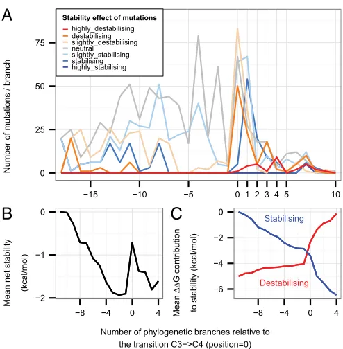
Ances-tral mutations were classified according to their evolutionary
pressures (Fig. 4A), as defined by the TDG09 algorithm (27).
Independently from the distinction between types of selection, mutations were also classified into three groups following the
photosynthetic types of their ancestor and descendant as C3→C3,
C3→C4, and C4→C4(the change C4→C3has not been seen and
detailed comparative analyses show that, if it has occurred, it must be very rare) (28).
On C3→C3branches, the distribution of stability effects
fol-lows a normal distribution, with a peak of stability-neutral
mutations (Fig. 4B,Left) that preserve the global stability of the
structure. In contrast, the C3→C4branches present significantly
more destabilizing mutations (permutation test,P=0.0080; Fig.
4B,Center), which correspond to the second peak (+0.88 kcal/mol) in the global distribution (Fig. 3). This tendency for
destabi-lizing mutations to occur at the C3→C4 transition is also
ap-parent in a timeline of cumulative mutational stability changes
in the ancestral sequences (Fig. 5). In C4→C4branches, a large
fraction of destabilizing mutations is still observed, but there is a significantly greater proportion of mutations with a stabilizing
effect compared with other branches (P < 0.0001; Fig. 4B,
Right). The timeline also shows that there is a large proportion
of stabilizing mutations immediately following the C3→C4
transition (Fig. 5A) and that the preponderance of stabilizing
over destabilizing mutations means that the loss of stability at the transition is largely recovered within the subsequent three
branches (Fig. 5B). Furthermore, considering the cumulative
0 5 10 15
0 2 4 6
50 100 150 200 250 300 350 400 450
Position
Number
Stability
highly_destabilising destabilising slightly_destabilising neutral
slightly_stabilising stabilising highly_stabilising
A
[image:4.585.39.545.52.169.2]B
Fig. 2. Effect of mutations on protein stability. (A) Stability landscape of the large subunit (rbcL). All 19 possible mutations at each position observed in the
O. sativastructure (positions 12–456) are colored on a vertical bar in terms of their stability relative to the native residue. Residues that are part of the active site are indicated by a black bar. The thresholds forΔΔGfoldin kcal/mol are highly stabilizing (<−1.84), stabilizing (−1.84 to−0.92), slightly stabilizing (−0.92 to−0.46), neutral (−0.46 to+0.46), slightly destabilizing (+0.46 to+0.92), destabilizing (+0.92 to+1.84), and highly destabilizing (> +1.84). Positions where the vertical bar is substantially gray or blue are predicted to be tolerant of mutation and where largely red are intolerant. Highly destabilizing mutations are very unlikely to occur in nature. (B) Stability effect of observed mutations at each position, relative to theO. sativa rbcLsequence. Within the monocot species, 105 positions of the 444 aligned residues of the peptide chain have alternate amino acids. The overwhelming majority of observed mutations (79.5%) have modest stability changes in the range of−1.84 to+1.84 kcal/mol.
0.0 0.1 0.2 0.3 0.4 0.5
−3 −2 −1 0 1 2 3 4 5 6 7 8 9 10 ΔΔG (Difference in stability) [kcal mol−1]
Propor
tion
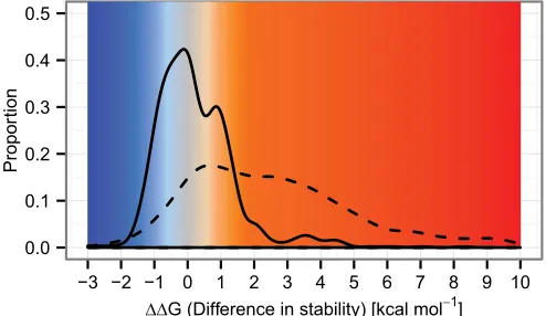
Fig. 3. Distribution of stability effects of possible mutations and those
oc-curring during evolution. The distribution of stability changes arising from mutations observed in the evolutionary history of the reconstructed ances-tral sequences (solid line) stands in contrast to that of all possible simulated mutations (dashed line). Both distributions have their largest peak close to
aΔΔG of zero. The observed mutations have an excess of slightly stabilizing
observed mutations and also a distinct peak of slightly destabilizing and destabilizing values centered at +0.88 kcal/mol. The majority of possible mutations are highly destabilizing and rarely occur during evolution. The probability distributions shown here are obtained by kernel smoothing of the original data (Fig. S1).
Studer et al. PNAS Early Edition | 3 of 6
EVOLUTI
[image:4.585.297.544.478.621.2]contributions to stability of all stabilizing mutations and all destabilizing mutations separately shows that stabilization due to all stabilizing mutations is accumulated more quickly in the
branch following the C3→C4transition than at any other time
(Fig. 5C).
Stability effects and location on the 3D structure.Ancestral mutations were grouped according to their position in the 3D structure of
the hexadecamer (Fig. 4C andD) following the interface
defi-nitions in ref. 29. The stability effects of mutations within the core of the large subunit or the LL1 interfaces within dimers
(e.g., LALB) follow an approximately normal distribution. In
contrast, although small in number, mutations of residues at the
LL2 (e.g., LALH) interface between dimers and at the LS
in-terface between large and small subunits have some tendency to
be highly destabilizing (P=0.0318 andP=0.0053, respectively).
The proportion of mutations at interfaces between large subunits
is significantly greater in the C4→C4 branches (P < 0.0002),
suggesting that the modification of subunit interactions is
im-portant for C4optimization (Fig. 4D).
Positively Selected Sites in the Transition to C4. At the C3→C4
transitions, three positively selected mutations with a destabiliz-ing effect are especially frequent: A328S, A281S, and L270I (Table S1). The A328S mutation and a positively selected, but less frequent, V326I mutation lie on either side of H327, which coordinates the P5 phosphate of the substrate in the closed state of the enzyme. Furthermore, these two residues are at the base
of the active site loop (loop 6 in residues 328–337) that carries
the catalytic lysine K334 and undergoes a disorder-order tran-sition on the binding of both substrates. The replacement of
hydrophobic A328 in the C3form with a polar serine in C4forms
is destabilizing as it disrupts the packing of the base of loop 6
against α-helix 6 (running from residues 338–350). This
de-stabilization could directly alter the catalytic parameters by allowing more flexibility in loop 6, thus affecting the opening and closing of the active site (16). Extensive studies of this loop re-gion in algal and cyanobacterial RubisCOs have shown that catalytic parameters are sensitive to its modification even if the mutated residues have no direct interaction with substrates (30). L270I is located directly beneath H298, which interacts with the P5 phosphate in the preactivated state. Replacement of V326 and L270 will also lead to packing changes that could alter the spatial disposition of the phosphate-binding histidines. Site 281 is
in the core of the C-terminal domain, and its potential to affect activity is not obvious. However, A281 packs against S321 and G322 at the end of the strand, which leads to loop 6, and destabilization Neutral evol./
Neg. selection (N = 385 )
[image:5.585.53.539.51.232.2]Pos. selection [p=1−5%] (N = 109 )
[image:5.585.301.548.370.622.2]Pos. selection [p<1%] (N = 257 )
A
Stability effect of mutations and selective pressurePropor tion 0.0 0 .4 o * Transition C3−>C3 (N = 532 )
Transition C3−>C4 (N = 70 )
Transition C4−>C4 (N = 149 )
B
Stability effect of mutations and photosynthetic branchPropor tion 0.0 0.4 * * o Intra (N = 482 )
LL1 (N = 157 )
LL2 (N = 45 )
LL3 (N = 23 )
LS (N = 44 )
highly_destabilising destabilising slightly_destabilising neutral slightly_stabilising stabilising highly_stabilising
C
Stability effect of mutations at different interfacesPropor tion 0.0 0 .4 0.8 Transition C3−>C3 (N = 532 )
Transition C3−>C4 (N = 70 )
Transition C4−>C4 (N = 149 )
Intra LL1 LL2 LL3 LS
D
Mutation of interfaces in each photosynthetic branchPropor tion 0.0 0 .4 0.8 * o o o * * *
Fig. 4. Stability effect and location of ancestral mutations. The 751 mutations occurring during evolution are separated inAby their selection constraints:
negative selection or neutral evolution (P>0.05 from TDG09 after false discovery rate correction), positive selection (0.01<P<0.05), and strong evidence of positive selection (P<0.01) and binned according to their stability effect. (B) Mutations are separated into their branch type (C3→C3, C3→C4, or C4→C4) and binned by their stability effect. (C) Mutations are classified following the subunit interface definitions in ref. 29 andFig. S2: Intra are in contact only with other residues of the same large subunit, LL1 residues are in contact with the other large subunit of the same dimer (e.g., the LALBinterface), LL2 and LL3 contact a large subunit of another dimer (e.g., LBLCand LBLD, respectively), and LS are all residues in contact only with any of the small subunits. (D) Mutations are separated into their branch type and binned into their contact interfaces. Categories are highlighted by an * when enriched or an“o”when depleted.
A
Number of m
u
tations / br
anch
0 25 50 75
−15 −10 −5 0 1 2 3 4 5 10
Stability effect of mutations highly_destabilising destabilising slightly_destabilising neutral slightly_stabilising stabilising highly_stabilising
B
Mean net stability
(kcal/mol)
0
−1
−2
−8 −4 0 4
C
Mean ΔΔ G contr ib u tionto stability (kcal/mol)
Stabilising Destabilising −6 −4 −2 0
−8 −4 0 4
Number of phylogenetic branches relative to the transition C3−>C4 (position=0)
Fig. 5. Changes in stability through evolution. (A) Frequency of mutations
of this interaction may have a long-range effect on the dynamics at the active site.
Another frequently positively selected mutation, M309I, also identified in some previous phylogenetic studies (20, 24), lies at the interface of the two C-terminal domains within a dimer and also close to the junction between N- and C-terminal domains within each subunit. This mutation has been demonstrated to act
as a catalytic switch between C3-like and C4-like properties (i.e.,
decreasing specificity for CO2over O2and increasing the
turn-over) inFlaveriaspecies and in chimeric enzymes consisting of
large subunits from Flaveria and tobacco small subunits (21).
However, isoleucine is present in only half of all of the C4forms.
Interestingly, sites 309 and 328 are evolutionary coupled (Table
S2). Also under strong positive selection in C3→C4(and C4→C4
branches) is the mutation V101I. The addition of one carbon to
this side chain is anticipated to shift the secondα-helix in the
N-terminal domain toward the active site. Directly on the opposite side of this helix is glutamate-60, which forms a salt bridge with the catalytic K334 in the closed activated state of the enzyme.
Any movement of theα-helix could affect the geometry of the
CO2-bound and transition states of the reaction.
Several of the positively selected mutations found in C3→C4
branches are also present in C4→C4 branches, (i.e., V101I,
L270I, M309I, and A328S). Additionally, three mutations on
α-helix 8, the final element of secondary structure of the
N-ter-minal domain of the large subunit, are positively selected in this type of branch: P142A/T, T143A (also strongly selected in
C3→C4branches), and S145A. This helix forms the symmetric
interface between the N-terminal domains of large subunits on
neighboring dimers (at the LL2 interfaces, e.g., LALH). At each
interface, the threonine and proline from each helix are
in-tercalated (Fig. S3). Structural superposition of the open and
closed forms of rice RubisCO suggests that an asymmetric movement of this helix between open and closed states of the upper active site, such as might occur on ligand binding or product release, will be transmitted to the neighboring active site at its lower left, potentially leading to a preference for the lower site to be closed while the top is open and vice versa.
Discussion
Diversification of RubisCO on an Island of Stability. Throughout their evolutionary histories, RubisCO genes have faced signifi-cant changes, both internal and external to the organism, which have altered the physiologically optimal properties of RubisCO and thus the selective pressures on its evolution (10). In our example of rice RubisCO, residues at nearly all sites contribute favorably to stability, and most putative mutations would lead to
destabilization (Fig. 2A). The change in stability that RubisCO
can withstand without dysfunction has yet to be established ex-perimentally, but the computed stability effects of mutations that have become fixed in some species are largely confined to a narrow range near zero (Fig. 3). This small amplitude of the effects of mutation observed in nature suggests that RubisCO evolves within a small island of stability (3, 5).
The adaptations of RubisCO to C4photosynthesis in
numer-ous plant lineages can be regarded as the result of natural experiments in evolution with a common (or at least similar)
outcome of reduced CO2/O2 specificity and an increase in
turnover (10). The availability of many sequences of closely
re-lated C3and C4species has enabled those branches of the
phy-logenetic tree associated with gain of C4 function, and thus
increased activity, to be identified reliably by parsimony. Ancestral reconstructions of these lineages allow the mutational pathways of evolution to be recovered with high confidence. Because struc-tures of representative proteins are available, the stability effects of mutations at each point on these pathways can also be esti-mated. We found that despite the positively selected mutations forming only a small proportion of the total, overall the changes in stability during evolution display features strongly reminiscent of those previously identified as significant in laboratory experi-ments by site-directed mutagenesis and directed evolution (9).
Destabilizing mutations are more frequently fixed in C4
line-ages. In those evolutionary branches that undergo a functional
change (C3→C4), adaptation is preceded by a long mutational
sequence in which neutral to slightly stabilizing capacitive mutations dominate, i.e., which create the capacity for the pro-tein to tolerate the destabilization required for new function
(Fig. 5B). A variety of often destabilizing mutations occurs
pre-cisely at the transition to C4, and these are immediately followed
by compensatory stabilizing mutations (Fig. 5 andFig. S4).
Except for cases in which folding is coupled to substrate binding, there is no a priori expectation of a direct physical connection between stability and activity. That similar tradeoffs between ac-tivity and stability are consistently found in both directed and natural evolution argues that an indirect connection necessarily arises from the tension between selection for optimal stability and selection for activity from a shared pool of possible mutations.
Modulation of Conformational Change Appears to Be Key to the Adaptation of RubisCO.Adaptive mutations occur in several dis-tinct parts of the RubisCO structure. None are in direct contact with the substrates; however, a small number of second shell mutations (i.e., residues in contact with active site residues) are strongly positively selected. These mutations tend to be destabi-lizing and, on the basis of structural context and earlier mutational studies of algal RubisCOs, are inferred to modify the active site
loop dynamics or position of residues at the P5 and O2/CO2
binding sites. Whereas adaptive mutations 10–20 Å from active
sites have occasionally been identified in other enzymes (31), in RubisCO, these form the majority of positively selected sites that
distinguish C3and C4species. Experiments with RubisCO from the
green alga Chlamydomonas reinhardii previously implicated the
interfaces between large and small subunits in the modulation of catalytic rates (32). The analysis here increases the number of
known functionally significant intersubunit sites (Table S2) and
demonstrates a link with the C3-C4transitions in flowering plants.
Those mutations near the dimer or N- and C-terminal domain interfaces within each large subunit likely affect the substantial relative movements of the domains on substrate binding. Although one of these residue changes (M309I) has previously been shown
to switch the enzyme to C4-like properties in plants (21), it is clear
that this change is not essential, and there are other mutational routes to equivalent functional changes.
Altered Cooperativity May Have an Adaptive Role in Some Species.
Negative cooperativity has been reported for the binding of the transition-state analog 2-carboxyarabinitol bisphosphate to the
active site of the C3RubisCO from spinach (33). Kinetic data fit
a model of rapid binding to one half of the active sites accom-panied by the slower binding to the remainder (34). Although it has proven difficult to generalize these observations to other species (possibly because of the stringent demands for pure and active protein in such experiments and because weak negative cooperativity is also intrinsically difficult to unambiguously identify in standard turnover kinetics), they naturally led to a postulated enzymatic mechanism whereby binding of substrates to one site of each dimer reduces binding at the other (34). Crystallographic studies have not been able to directly address this issue as they produce symmetric structures, either apo or fully saturated (16). The observation of positive selection on mutations in the interface between the N-terminal domains of neighboring dimers suggests a different mechanism of cooperativity. Comparison of hybrid structures of apo and holo forms of RubisCO suggests that con-formational changes at an active site in the ring of active sites at the top of the oligomer are coupled to the lower site in the dimer
to its left. The mutations occurring during the C3to C4transitions
diminish this coupling and would relieve any negative coopera-tivity between the upper and lower sites, thus enhancing turnover. The identified positive selection suggests that these mutations play
a role in the adaptation of some C4species. Consequently, these
mutations and the possibility of a role for cooperativity in RubisCO warrant renewed experimental investigation.
Studer et al. PNAS Early Edition | 5 of 6
EVOLUTI
Conclusions. The mutational landscape of RubisCO is strongly constrained by the need to maintain overall stability. This con-straint limits the adaptation of RubisCO to novel environmental contexts to those amino acid changes that can modify the catalytic efficiency without dramatic effect on the overall folding stability.
Following the repeated origins of C4photosynthesis in flowering
plants, a number of amino acid mutations of RubisCO were preferentially kept by natural selection. These mutations include changes to residues that might modify the geometry of the active site, as well as a substantial number of sites at the interface be-tween domains and subunits, which probably alter the properties of the enzyme via modification of the dynamics of conformational change or alteration of cooperativity between catalytic subunits. It is clear that a substantial proportion of the mutations necessary
for C4adaptation are themselves destabilizing. Evolution
accom-modates such destabilizing functional adaptations thanks to the previous accumulation of stabilizing capacitive mutations and by subsequently fixing stabilizing compensating mutations.
Methods
The multiple sequence alignment of genes for RubisCO large subunit (rbcL) and its associated phylogenetic tree are from Christin et al. (24). The highest-res-olution (1.35 Å) structure of RubisCO currently available, from the C3grass
Oryza sativa(35), was used as the basis for structural analyses. The complete biological unit (L8S8) was directly downloaded from the PDBePISA website (36).
The Protein Data Bank (PDB) structure file for the large subunit contains coordinates for residues 11–475 (465 residues). This structure was used as a template for the homology modeling of 3D octomeric structures (L8) of each ancestralrbcLsequence. The modeling was done with Modeler 9.9 (37). For each sequence, 100 models were built, and the model with the lowest energy (based on its discrete optimized protein energy score) was used in further analyses. Using FoldX 3b5.1 (38), the energies for the WT (ΔGfold,wt) and mu-tant (ΔGfold,mut) protein were computed to give the stability changeΔΔGfold= ΔGfold,mut−ΔGfold,wt. The SD in FoldX is 0.46 kcal/mol (38), and we used this value to bin the ΔΔGfold values into seven categories. Additional FoldX restraints were applied to the conserved active site to avoid the potential for artifacts arising from unparameterised ligands. The inference of ancestral sequences was performed under maximum likelihood as implemented in CodeML (39). Sites under positive selection between C3and C4forms were identified by the TDG09 algorithm (27), which performs a likelihood ratio test to assess if the evolutionary rate at a particular position is similar or different between C3and C4lineages. TheΔΔGfolddue to each mutation on each branch was then mapped onto the phylogenetic tree (Fig. S4). Detailed methods are given inSI Text.
ACKNOWLEDGMENTS.This study benefited from use of the University College
London (UCL)LegionHigh-Performance Computing Facility (Legion@UCL). R.A.S. acknowledges funding from the Fondation du 450ème Anniversaire de l’Université de Lausanne and Swiss National Science Foundation Grants 132476 and 136477. P.-A.C. is funded by Marie Curie International Outgoing Fellowship 252568.
1. Romero PA, Arnold FH (2009) Exploring protein fitness landscapes by directed evo-lution.Nat Rev Mol Cell Biol10(12):866–876.
2. DePristo MA, Weinreich DM, Hartl DL (2005) Missense meanderings in sequence space: A biophysical view of protein evolution.Nat Rev Genet6(9):678–687. 3. Taverna DM, Goldstein RA (2002) Why are proteins marginally stable?Proteins46(1):
105–109.
4. Tokuriki N, Tawfik DS (2009) Stability effects of mutations and protein evolvability.
Curr Opin Struct Biol19(5):596–604.
5. Tokuriki N, Stricher F, Serrano L, Tawfik DS (2008) How protein stability and new functions trade off.PLOS Comput Biol4(2):e1000002.
6. Soskine M, Tawfik DS (2010) Mutational effects and the evolution of new protein functions.Nat Rev Genet11(8):572–582.
7. Wang X, Minasov G, Shoichet BK (2002) Evolution of an antibiotic resistance enzyme constrained by stability and activity trade-offs.J Mol Biol320(1):85–95.
8. Bloom JD, Labthavikul ST, Otey CR, Arnold FH (2006) Protein stability promotes evolvability.Proc Natl Acad Sci USA103(15):5869–5874.
9. Bloom JD, Arnold FH (2009) In the light of directed evolution: Pathways of adaptive protein evolution.Proc Natl Acad Sci USA106(Suppl 1):9995–10000.
10. Tcherkez GG, Farquhar GD, Andrews TJ (2006) Despite slow catalysis and confused substrate specificity, all ribulose bisphosphate carboxylases may be nearly perfectly optimized.Proc Natl Acad Sci USA103(19):7246–7251.
11. Lorimer GH, Andrews TJ (1973) Plant photorespiration—An inevitable consequence of the existence of atmospheric oxygen.Nature243(5406):359–360.
12. Sage RF, Sage TL, Kocacinar F (2012) Photorespiration and the evolution of C4 pho-tosynthesis.Annu Rev Plant Biol63:19–47.
13. Sage RF, Christin PA, Edwards EJ (2011) The C(4) plant lineages of planet Earth.J Exp Bot62(9):3155–3169.
14. Grass Phylogeny Working Group II (2012) New grass phylogeny resolves deep evo-lutionary relationships and discovers C4 origins.New Phytol193(2):304–312. 15. Savir Y, Noor E, Milo R, Tlusty T (2010) Cross-species analysis traces adaptation of
Rubisco toward optimality in a low-dimensional landscape.Proc Natl Acad Sci USA
107(8):3475–3480.
16. Andersson I, Backlund A (2008) Structure and function of Rubisco.Plant Physiol Bio-chem46(3):275–291.
17. Young JN, Rickaby RE, Kapralov MV, Filatov DA (2012) Adaptive signals in algal Ru-bisco reveal a history of ancient atmospheric carbon dioxide.Philos Trans R Soc Lond B Biol Sci367(1588):483–492.
18. Sage RF (2002) Variation in the k(cat) of Rubisco in C(3) and C(4) plants and some implications for photosynthetic performance at high and low temperature.J Exp Bot
53(369):609–620.
19. Hudson GS, et al. (1990) Comparisons of rbcL genes for the large subunit of ribulose-bisphosphate carboxylase from closely related C3 and C4 plant species.J Biol Chem
265(2):808–814.
20. Kapralov MV, Kubien DS, Andersson I, Filatov DA (2011) Changes in Rubisco kinetics during the evolution of C4 photosynthesis in Flaveria (Asteraceae) are associated with positive selection on genes encoding the enzyme.Mol Biol Evol28(4):1491–1503.
21. Whitney SM, et al. (2011) Isoleucine 309 acts as a C4 catalytic switch that increases ribulose-1,5-bisphosphate carboxylase/oxygenase (rubisco) carboxylation rate in Fla-veria.Proc Natl Acad Sci USA108(35):14688–14693.
22. Dessailly BH, Lensink MF, Wodak SJ (2007) Relating destabilizing regions to known functional sites in proteins.BMC Bioinformatics8:141.
23. Beadle BM, Shoichet BK (2002) Structural bases of stability-function tradeoffs in en-zymes.J Mol Biol321(2):285–296.
24. Christin PA, et al. (2008) Evolutionary switch and genetic convergence on rbcL fol-lowing the evolution of C4 photosynthesis.Mol Biol Evol25(11):2361–2368. 25. Wang M, Kapralov MV, Anisimova M (2011) Coevolution of amino acid residues in the
key photosynthetic enzyme Rubisco.BMC Evol Biol11:266.
26. Kapralov MV, Filatov DA (2007) Widespread positive selection in the photosynthetic Rubisco enzyme.BMC Evol Biol7:73.
27. Tamuri AU, Dos Reis M, Hay AJ, Goldstein RA (2009) Identifying changes in selective constraints: Host shifts in influenza.PLOS Comput Biol5(11):e1000564.
28. Christin PA, Freckleton RP, Osborne CP (2010) Can phylogenetics identify C(4) origins and reversals?Trends Ecol Evol25(7):403–409.
29. van Lun M, van der Spoel D, Andersson I (2011) Subunit interface dynamics in hex-adecameric rubisco.J Mol Biol411(5):1083–1098.
30. Parry MA, Andralojc PJ, Mitchell RA, Madgwick PJ, Keys AJ (2003) Manipulation of Rubisco: The amount, activity, function and regulation.J Exp Bot54(386):1321–1333. 31. Thomas VL, McReynolds AC, Shoichet BK (2010) Structural bases for stability-function
tradeoffs in antibiotic resistance.J Mol Biol396(1):47–59.
32. Spreitzer RJ, Peddi SR, Satagopan S (2005) Phylogenetic engineering at an interface between large and small subunits imparts land-plant kinetic properties to algal Ru-bisco.Proc Natl Acad Sci USA102(47):17225–17230.
33. Johal S, Partridge BE, Chollet R (1985) Structural characterization and the de-termination of negative cooperativity in the tight binding of 2-carboxyarabinitol bisphosphate to higher plant ribulose bisphosphate carboxylase.J Biol Chem260(17): 9894–9904.
34. Zhu G, Jensen RG (1990) Status of the substrate binding sites of ribulose bisphosphate carboxylase as determined with 2-C-carboxyarabinitol 1,5-bisphosphate.Plant Physiol
93(1):244–249.
35. Matsumura H, et al. (2012) Crystal structure of rice Rubisco and implications for ac-tivation induced by positive effectors NADPH and 6-phosphogluconate.J Mol Biol
422(1):75–86.
36. Krissinel E, Henrick K (2007) Inference of macromolecular assemblies from crystalline state.J Mol Biol372(3):774–797.
37. Sali A, Blundell TL (1993) Comparative protein modelling by satisfaction of spatial restraints.J Mol Biol234(3):779–815.
38. Schymkowitz J, et al. (2005) The FoldX web server: An online force field.Nucleic Acids Res33(Web Server issue):W382-8.
39. Yang Z (2007) PAML 4: Phylogenetic analysis by maximum likelihood.Mol Biol Evol
Supporting Information
Studer et al. 10.1073/pnas.1310811111
SI Text
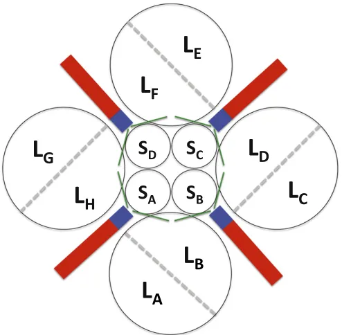
Description of the Biological Unit of Ribulose-1,5-Bisphosphate Carboxylase. In land plants, the ribulose-1,5-bisphosphate car-boxylase (RubisCO) biological unit is an oligomer of 16 subunits.
Two large (L) subunits head-to-tail form a L2 dimer. Four L2
dimers form the main octomeric ring (L8). There are also four
small subunits on the top and four small subunits on the bottom
that together with the large units form the hexadecamer L8S8
complex, which is the biological unit (1, 2). The catalytic activity of RubisCO occurs in the large subunit, and the main component
of this large subunit is the C-terminalα/βbarrel domain
(posi-tions 169–432), in which the catalytic residues lie (3). Various
residues are important for catalysis. Residues 201, 203, and 204 are charged residues and bind a magnesium atom in the catalytic site. Residues 175 and 295 play the role of proton acceptors. Residues 123, 173, 177, 295, 327, and 379 form the binding pocket. Site 334 is a transition state stabilizer. The sixth loop
(328–337) is an important component as it participates in the
opening and closure of the active site (2). We use the nomen-clature of ref. 4 for labeling the different interfaces LL1, LL2, and LL3 (Fig. S2).
Calculation of Stability Effects of Mutation with FoldX.All stability analyses were performed with FoldX 3b5.1 (5, 6), which is one of the best stability predictors and easily implementable in a pipeline (7, 8). No current stability prediction method is very accurate in predicting the effect of individual mutations [evaluations of
FoldX have reported a Matthews correlation coefficient (r) of
0.81 (5) and 0.59 (8) for individual mutations dependent on the protein test set]. However, a number of structure-based methods do predict the trend in average values for binned data very well.
FoldX achieved the highest correlation (r=0.96) for binned data
in a recent evaluation (8). The analysis reported here relies on aggregating results of many mutations in different evolutionary or structural contexts, and it is consequently expected that ob-served trends in stability will be robust. We used the reported accuracy of FoldX of 0.46 kcal/mol (i.e., the SD of the difference
between ΔΔGs calculated by FoldX and the experimental
values) (5) to bin the ΔΔG values into seven categories:
(i) highly stabilizing (ΔΔG< −1.84 kcal/mol); (ii) stabilizing
(−1.84 kcal/mol≤ ΔΔG<−0.92 kcal/mol); (iii) slightly
stabi-lizing (−0.92 kcal/mol≤ ΔΔG<−0.46 kcal/mol); (iv) neutral
(−0.46 kcal/mol<ΔΔG≤+0.46 kcal/mol); (v) slightly
desta-bilizing (+0.46 kcal/mol<ΔΔG≤+0.92 kcal/mol); (vi)
desta-bilizing (+0.92 kcal/mol < ΔΔG≤ +1.84 kcal/mol); and (vii)
highly destabilizing (ΔΔG> +1.84 kcal/mol).
Before the analysis, all of the chains of theOryza sativa
Ru-bisCO structure [Protein Data Bank (PDB) code: 1WDD] were distinctly renamed, and all heteroatoms in the structure that are not well parameterized in FoldX [2-Cabp (CAP), glycerol
(GOL), and Mg2+(MG)] were removed. Although Mg2+is
im-portant to the overall structure of RubisCO, because Mg2+’s
short-range interactions are not well parameterized in FoldX, we
mimic the conformational effects of Mg2+ by constraints that
maintain the binding site conformation very close to that ob-served experimentally. These constraints avoid potential artifacts
due to an inaccurate Mg2+model and are practicable in the case
of RubisCO as all residues surrounding the Mg2+atom are 100%
conserved. None participate in the differences between C3and
C4forms of RubisCO. The closest residue under selection
be-tween C3and C4is 328, but the stability effect of the mutation
A328S is very similar whether the magnesium is present or not.
The binding site restraints are, of course, the result of compro-mise and may themselves be a source of some inaccuracy of pre-dictions for some residues as local structure relaxation of second shell residues bordering the binding site could potentially be im-peded. To perform the stability analyses, we use the standard pipeline for FoldX, which uses two commands: Repair and Build-Model. The Repair command was used to optimize the structure by removing any steric clashes. This optimization improves the global
stability score of the RubisCO L8S8complex, reducing the free
energy (ΔG) from+105.72 kcal/mol (before the repair step)
to−1,506.42 kcal/mol (after the repair step). For the analysis of
all possible mutations, each residue of the O. sativaRubisCO
structure was mutated using the command BuildModel. FoldX
operates in two steps, repeated five times (option <
number-OfRuns>5) to ensure that the minimum energy conformations
of even large residues that possess many rotamers is identified: Step 1, the residue of interest and its neighbors are mutated to themselves and conformationally relaxed to remove any local
clash and the stability of the WT (ΔGwt, in kcal/mol) is
ob-tained by the FoldX energy function; Step 2, the residue is mutated to all 19 other amino acids and all other neighbor
side chains to themselves. The stability of the mutant (ΔGmut, in
kcal/mol) is calculated.
Each mutation is made in all large subunits (LSUs)
simulta-neously, and the reported stability effect of a mutationΔΔG=
ΔGmut −ΔGwt is normalized for the number of large subunits
(by dividing the calculated change by eight). The results of these calculations are presented in Fig. 2 and Table S3.
Whereas calculations were actually carried out for 463 residues
of theO. sativaRubisCO structure (residues 12–475, which are
present in all rbcL subunits less the essential modified residue Lcx201), for the sake of simplifying the presentation (and not affecting any functionally significant residue), the mutational data are presented in text, figures, and tables only for those residues that are common to both reconstructed sequences (i.e., those positions that can be continuously aligned) and structural models, specifically, Gly12 through Ala456 (less the essential Lcx201), i.e., in the case of this stability analysis for 8,436
sim-ulated mutations (19 amino acids×444 residues).
Ancestral Sequence Reconstruction.Recent analyses have success-fully used ancestral sequence reconstruction of extinct proteins to reproduce their evolutionary history in vitro and/or in silico (9, 10). We used the collection of extant large subunit RubisCO protein sequences and the phylogenetic tree for monocot species from the study of Christin et al. (11). The alignment contains 240
sequences [137 of C3, 101 of C4, and 2 of crassulacean acid
metabolism (CAM) plants], all from monocots, one of the major groups of flowering plants. Ancestral sequence reconstruction of extinct RubisCO proteins was performed under maximum like-lihood as implemented in CodeML (PAML package release 4.4d) (12).
Stability Effects of Ancestral Mutations.Homology models of the ancestral octomers were constructed for each branch point of the phylogenetic tree. The stability effect of ancestral mutations occurring in each branch of the tree was estimated using FoldX, as described above, to make the mutations in the appropriate an-cestral octomer structure. Consequently, each mutation in a phy-logenetic branch is evaluated in the context of a structural model containing all of the accumulated mutations up to the branch point,
and many of the possible interactions between mutations that accumulate during evolution are accounted for.
However, in those cases where multiple mutations occur within a branch, the reconstruction procedure cannot tell us the order in which they occur. Consequently, there is a possibility that if mutations within a branch interact, their historical order of
oc-currence could alter theirΔΔG contribution. It is difficult to test
all possible orders (and present the results of that analysis); consequently, in the main results, we assume that there is no interaction between mutations within a branch.
To test the reasonableness of this assumption, we compared,
for all 172 branches with multiple mutations, the sum of theΔΔG
of individual mutations to the ΔΔG of all mutations within
a branch made simultaneously. The differences between these sums of individual and simultaneous mutations are shown in Fig. S5. Because many pairs or triples of ancestral mutations are spatially separated in the structure, the vast majority of groups of mutations in a branch are approximately additive. In only a few cases (15/172) is this difference greater than the FoldX SD of
0.46 kcal/mol, and only one of these lies on a C3→C4branch.
Consequently, it seems reasonable to conclude that any effect of nonadditivity within branches will not significantly impact on the analysis.
The distributions of the stability effects computed with FoldX for all possible mutations and for the ancestral mutations are shown in Fig. 3 and Fig. S1.
Identification of Sites Under Positive Selection.The fixation rate of mutations in the genome depends on selective forces and genetic drift. This rate will be accelerated under positive selection (i.e., to promote a new function) or decelerated under negative selection (i.e., when a mutation has a negative impact on the organismal fitness). Positions under selective constraints are likely to be important in explaining functional changes between subgroups,
such as between C3and C4plants. The TDG09 algorithm (13)
was used to detect sites experiencing changes in selective
con-straints between C3and C4forms. It requires as input a multiple
alignment of homologous sequences, a well-defined phylogenetic tree, and a grouping of these sequences into two categories (in
our cases, C3 and C4). The method estimates parameters for
each site using two assumptions. The first assumption is that evolutionary rates can vary between positions, but these evolu-tionary rates are constant within a given position across species (constant evolutionary rate over time), i.e., the homogeneous model of substitutions (model 2 as described in ref. 13). The second assumption is that evolutionary rates can vary at a given position depending on the species group (shift in evolutionary rate over time), i.e., the nonhomogenous model of substitutions (model 3). For each site, TDG09 computes the likelihood of both model 2 and model 3 and compares them by a likelihood
ratio test (LRT), which will give aPvalue per site. A false
dis-covery rate (FDR) correction is applied. Before the analysis, the lengths of all branches were estimated with CodeML (PAML package release 4.4d) (12) under the Whelan and Goldman
model (14). The two CAM sequences (Ananas andTillandsia)
were omitted for the purpose of this analysis, as they cannot be
clearly assigned to either the C3 or the C4 group and may be
under distinct selective pressures. We used relaxed and stringent FDR thresholds (5% and 1%, respectively) to select sites under functional divergence from the TDG09 output. We identified 18 sites under positive selection at the 5% FDR level, with a subset of 12 sites at the 1% FDR level (Table S1).
In their previous study, using the same dataset, Christin et al. (11) identified 14 sites under codon substitution models (site and branch-site). Codon substitution models use nucleotide sequen-ces to compute the dN/dS ratio of non-synonymous (dN) sub-stitutions over synonymous (dS) subsub-stitutions to infer positive selection. Our analysis of this dataset, analyzed with the TDG09
algorithm (using evolutionary models based on amino acid
se-quences), identified 18 sites under positive selection between C3
and C4forms (Table S1). Nine sites had been detected under the
codon substitution site model M2a in Christin et al. (11). TDG09 detected seven of them with the exception of positions 91 and 265. The branch-site model identified five sites with purifying
selection in C3forms and positive selection in C4(101, 258, 270,
281, and 309). TDG09 identified all of them at the 1% FDR
level. Three sites evolved under neutral evolution in C3 and
under positive selection in C4 (142, 145, and 328). TDG09
identified two of the sites at 1% (145 and 328), whereas position 142 is only detected at the 5% FDR level. In addition, TDG09 identified three new sites at 1% levels (143, 225, and 262) and three at 5% (221, 282, and 326), which were not detected by the codon substitution models used by Christin et al. (11).
Some of the sites under positive selection have been detected in previous studies conducted in conifers (15) and flowering plants (16, 17), such as in monocots (11, 18) and eudicots (19, 20) (Table S1). It is interesting that the codon substitution methods correlate well because codon substitution models can be affected by sat-uration at the dS level (i.e., many synonymous substitutions will be seen only as one substitution), and results can be less reliable as the evolutionary time increases, especially in highly divergent gene families. Evolutionary methods based on amino acids, such as the TDG09 algorithm, can help to override this problem.
Identification of Coevolutionary Sites Under Positive Selection. Co-evolutionary information was detected with EVfold (21) and for
several datasets ofrbcLsequences (Table S2).
Extended Description of Sites Under Positive Selection.In general,
more mutations are observed on average on C3→C4branches
(3.7 per branch) than in C3→C3 branches (2.8) and C4→C4
branches (2.0).
C3→C3 branches. On C3→C3 branches, several sites are under
positive selection. The mutations I225L, A228S, and V262A are found at or near the interface between the C-terminal domain of the large subunit and the small subunit to its right (Fig. S3), and
mutations of I251L and A/V255I/T at the base of theα/β-barrel
are also common. These mutations have a mixed effect on pre-dicted stability, but are usually destabilizing (Table S1).
Despite their being far from the active site, mutations of res-idues in these regions of the structure are positively selected in all branches and have been shown to affect the catalytic properties of
RubisCO from the green alga Chlamydomonas reinhardii.
Ge-netic screening ofC. reinhardiihas shown that A222T or V262L
mutations restore thermal stability and CO2 specificity to a
temperature-sensitive mutant (22). However, inC. reinhardii,the
mutation R258K (which is positively selected in C4→C4branches)
is at the interface of large (LL3) and small (LS1) subunits and is
associated with 23% and 33% increases in maximum reaction rate values for oxygenation (Vo) and carboxylation (Vc), respectively (23). A penta-mutant V221C/V235I/ C256F/K258R/I265V changes
individual Km andkcatvalues for O2and CO2 by factors of 2–3
while leaving specificity unchanged (24). Fig. S3Bshows the close
proximity of the group of positively selected residues, found at the
LBSBinterface (and to a lesser extent the LBLDinterface) and at
the base of theα/βbarrel (in a contiguous stretch of sequence from
221 to 282).
The other positively selected mutation in C3→C3branches is
S328A near the active site (discussed further below). This mu-tation has a slightly stabilizing effect.
loop (loop-6 residues 328–337) that carries the catalytic lysine-334 and makes a disorder-order transition on the binding of both substrates. L270I is located directly beneath H298, which interacts with the P5 phosphate in the preactivated state.
The replacement of hydrophobic alanine in the C3form at 328
with a polar serine in C4forms is destabilizing as it disrupts the
packing of the base of loop 6 against helix 6 (running from residues 338 to 350). Replacement of V326 and L270 will also lead to packing changes that could alter the spatial disposition of the phosphate-binding histidines. The mutation of site 328 from a hydrophobic to a polar residue could also directly alter the catalytic parameters by allowing more flexibility in the sixth loop, altering the kinetics of opening and closing of the active site. Extensive studies of this loop region in algal and cyanobacterial RubisCOs show that catalytic parameters are sensitive to its modification even if the mutated residues have no direct in-teraction with substrates (25). In particular, the mutations
V331A and L326I in the RubisCO inC. reinhardii, which also
modify the loop 6/helix 6 interface, are both known to decrease
CO2specificity (26, 27).
Mutations that have an impact on the active site. Under strong
positive selection in C3→C4and C4→C4is the mutation V101I.
This residue is always valine in C3monocots, and the addition of
one carbon to the side chain could shift the secondα-helix in the
N-terminal domain toward the active site. This movement is sig-nificant as directly on the opposite side of this helix is glutamate-60, which forms a salt bridge with the catalytic K332 in the closed ac-tivated state of the enzyme. Consequently, any movement of the
α-helix could affect the geometry of the CO2-bound and transition
states of the reaction.
Mutations at the interface between subunits.The mutation M309I is under strong positive selection as also identified in some previous studies (11, 19). This mutation has a neutral effect on stability but lies at the interface of the two large subunits that make up the functional dimers and also close to the junction between N- and C-terminal domains. This mutation has been demonstrated to act
as a catalytic switch between C3-like and C4-like behaviors (i.e.,
decreasing specificity for CO2over O2and increasing the
turn-over) inFlaveriaspecies and in chimeric enzymes consisting of
large subunits from Flaveria and tobacco small subunits (28).
However, sequence analysis shows that the isoleucine is only
present in half of all of the monocot C4forms, suggesting that
there are other mechanisms for making similar catalytic changes and/or that the switch may require a particular context in which to function. Interestingly, site 328, which neighbors the P5 binding H327, is coupled to 309 in angiosperm evolution (Table S2), suggesting a coupling of ribulose-1,5-bisphosphate (RBP) binding to conformational change of the dimer involving these residues. [There is also evidence from monocot species that site 326 is coupled to 362 near the interface of N- and C-terminal domains of the large subunit, also suggesting a coupling of binding to conformational change. However, mutations of this latter pair
(I→V) are both positively selected in C3→C3branches, perhaps
indicating an opposite effect on activity.]
A281S is distant from the active site, but has high penetration in
the C4 monocot species in our dataset (67% S) and is also
prevalent in eudicot species (29). It forms part of the well-packed and rigid core of the C-terminal domain, and the sub-stitution is predicted to be destabilizing. This subsub-stitution may result in increased flexibility or conformational changes locally, which could potentially be significant globally as the methyl group
of alanine is in direct contact with S321 and G322 at the end of the strand, which leads to loop 6 and with Q149 at the beginning of the loop that connects the N- and C-terminal domains.
C4→C4branches.Some mutations found in C3→C4branches (i.e.,
V101I, L270I, M309I, and A328S) are also present in C4→C4
branches. Three mutations on α-helix 8, the final element of
secondary structure of the N-terminal domain of the large sub-unit, are positively selected in this branch: P142A/T, T143A (also
strongly positively selected in the C3→C4branches), and S145A.
α-Helix 8 forms the symmetric interface between the N-terminal
domains of large subunits on neighboring dimers. At the terface, the threonine and proline from each helix are
in-tercalated (Fig. S3C). Structural superposition of the open (PDB
code: 3AXM) and closed (PDB code: 1WDD) forms of rice
RubisCO (Fig. S3C) suggests that an asymmetric movement of
this helix between open and closed states of the upper active site, such as might occur on ligand binding or product release, will be transmitted to the neighboring active site at its lower left, leading to a preference for the lower site to be closed while the top is open and vice versa.
In this branch, one mutation, R258K, is positively selected at
the interface of large (LL3) and small (LS1) subunits. In C.
reinhardii, this change is associated with 23% and 33% increases in maximum reaction rate values for Vo and Vc, respectively (23). Furthermore, this residue interacts with the tip of a long
loop in the rbcS subunit (positions 56–58). Residue 57 of the
rbcS has been found to be under weak selective pressure in the
C3→C4transition inFlaveriaspecies (19), and residues 56 and 58
are mutated between C3 rice and C4 corn, supporting a
wide-spread role for interaction with the small subunit in this region
modulating kinetics in the C4transition.
Statistical Analysis and Visualization.Python, Biopython (30), and R (31) were used to prepare the data and perform the computa-tional and statistical analyses. The multiple sequence alignment was visualized with Jalview (32). The phylogenetic tree was an-notated with EvolView (33). The visualizations of sites in 3D were created with Chimera (34).
To test for statistical significance of the observed variations in distributions of stability in different photosynthetic branches, under different selective forces, and in different interfaces (Fig. 4
A–C) or location (Fig. 4D) of mutations, permutation tests were
carried out to investigate the null hypothesis that the distributions in each category are sampled from the same underlying distribu-tion. For example, in comparing the distributions with respect to photosynthetic branch, we randomly permute the branch labels of all mutations 100,000 times and then count the number of times the simulated frequencies for a given stability effect are higher
than the observed sample in each category. The result is aPvalue
for each stability effect, which can be compared using a two-sided
test, where for a single sample, a P < 0.05/2= 0.025 indicates
a significant enrichment for that particular stability effect andP>
0.975 indicates a significant depletion for that particular effect.
ThePvalues for all cases are given in Table S4. The permutation
of the interface mutations takes into account the number of sites in each interface. When attributing significance in this study, we also conservatively corrected for the potential FDR by dividing the
P value threshold by 3 when analyzing the number of selection
pressure categories (Fig. 4A) or branches (Fig. 4BandD) or by
5 when considering interfaces (Fig. 4C).
1. Andersson I, Taylor TC (2003) Structural framework for catalysis and regulation in ribulose-1,5-bisphosphate carboxylase/oxygenase.Arch Biochem Biophys414(2):130–140. 2. Andersson I, Backlund A (2008) Structure and function of Rubisco.Plant Physiol
Biochem46(3):275–291.
3. Chapman MS, et al. (1988) Tertiary structure of plant RuBisCO: Domains and their contacts.Science241(4861):71–74.
4. van Lun M, van der Spoel D, Andersson I (2011) Subunit interface dynamics in hexadecameric rubisco.J Mol Biol411(5):1083–1098.
5. Schymkowitz J, et al. (2005) The FoldX web server: An online force field.Nucleic Acids Res33(Web Server issue):W382-8.
6. Schymkowitz JW, et al. (2005) Prediction of water and metal binding sites and their affinities by using the Fold-X force field.Proc Natl Acad Sci USA102(29):10147–10152.
7. Khan S, Vihinen M (2010) Performance of protein stability predictors.Hum Mutat
31(6):675–684.
8. Potapov V, Cohen M, Schreiber G (2009) Assessing computational methods for predicting protein stability upon mutation: Good on average but not in the details.
Protein Eng Des Sel22(9):553–560.
9. Liberles DA (2007)Ancestral Sequence Reconstruction(Oxford Univ Press, Oxford, UK).
10. Harms MJ, Thornton JW (2010) Analyzing protein structure and function using ancestral gene reconstruction.Curr Opin Struct Biol20(3):360–366.
11. Christin PA, et al. (2008) Evolutionary switch and genetic convergence on rbcL following the evolution of C4 photosynthesis.Mol Biol Evol25(11):2361–2368. 12. Yang Z (2007) PAML 4: Phylogenetic analysis by maximum likelihood.Mol Biol Evol
24(8):1586–1591.
13. Tamuri AU, Dos Reis M, Hay AJ, Goldstein RA (2009) Identifying changes in selective constraints: Host shifts in influenza.PLOS Comput Biol5(11):e1000564.
14. Whelan S, Goldman N (2001) A general empirical model of protein evolution derived from multiple protein families using a maximum-likelihood approach.Mol Biol Evol
18(5):691–699.
15. Sen L, et al. (2011) Molecular evolution of rbcL in three gymnosperm families: Identifying adaptive and coevolutionary patterns.Biol Direct6:29.
16. Kapralov MV, Filatov DA (2007) Widespread positive selection in the photosynthetic Rubisco enzyme.BMC Evol Biol7:73.
17. Wang M, Kapralov MV, Anisimova M (2011) Coevolution of amino acid residues in the key photosynthetic enzyme Rubisco.BMC Evol Biol11:266.
18. Iida S, et al. (2009) Molecular adaptation of rbcL in the heterophyllous aquatic plant Potamogeton.PLoS ONE4(2):e4633.
19. Kapralov MV, Kubien DS, Andersson I, Filatov DA (2011) Changes in Rubisco kinetics during the evolution of C4 photosynthesis in Flaveria (Asteraceae) are associated with positive selection on genes encoding the enzyme.Mol Biol Evol
28(4):1491–1503.
20. Liu L, Zhao B, Zhang Y, Wang J (2012) Adaptive evolution of the rbcL gene in Brassicaceae.Biochem Syst Ecol44(0):13–19.
21. Marks DS, et al. (2011) Protein 3D structure computed from evolutionary sequence variation.PLoS ONE6(12):e28766.
22. Hong S, Spreitzer RJ (1997) Complementing substitutions at the bottom of the barrel influence catalysis and stability of ribulose-bisphosphate carboxylase/oxygenase.
J Biol Chem272(17):11114–11117.
23. Du YC, Peddi SR, Spreitzer RJ (2003) Assessment of structural and functional divergence far from the large subunit active site of ribulose-1,5-bisphosphate carboxylase/oxygenase.J Biol Chem278(49):49401–49405.
24. Spreitzer RJ, Peddi SR, Satagopan S (2005) Phylogenetic engineering at an interface between large and small subunits imparts land-plant kinetic properties to algal Rubisco.Proc Natl Acad Sci USA102(47):17225–17230.
25. Parry MA, Andralojc PJ, Mitchell RA, Madgwick PJ, Keys AJ (2003) Manipulation of Rubisco: The amount, activity, function and regulation.J Exp Bot54(386):1321–1333. 26. Chen ZX, Spreitzer RJ (1989) Chloroplast intragenic suppression enhances the low CO2/O2 specificity of mutant ribulose-bisphosphate carboxylase/oxygenase.J Biol Chem264(6):3051–3053.
27. Zhu G, Spreitzer RJ (1996) Directed mutagenesis of chloroplast ribulose-1,5-bisphosphate carboxylase/oxygenase. Loop 6 substitutions complement for structural stability but decrease catalytic efficiency.J Biol Chem271(31):18494–18498. 28. Whitney SM, et al. (2011) Isoleucine 309 acts as a C4 catalytic switch that increases
ribulose-1,5-bisphosphate carboxylase/oxygenase (rubisco) carboxylation rate in Flaveria.Proc Natl Acad Sci USA108(35):14688–14693.
29. Kapralov MV, Smith JA, Filatov DA (2012) Rubisco evolution in C₄eudicots: An analysis of Amaranthaceae sensu lato.PLoS ONE7(12):e52974.
30. Cock PJ, et al. (2009) Biopython: Freely available Python tools for computational molecular biology and bioinformatics.Bioinformatics25(11):1422–1423.
31. R Development Core Team (2011)R: A Language and Environment for Statistical Computing(R Development Core Team, Vienna).
32. Waterhouse AM, Procter JB, Martin DM, Clamp M, Barton GJ (2009) Jalview Version 2—A multiple sequence alignment editor and analysis workbench.Bioinformatics
25(9):1189–1191.
33. Zhang H, Gao S, Lercher MJ, Hu S, Chen WH (2012) EvolView, an online tool for visualizing, annotating and managing phylogenetic trees.Nucleic Acids Res40(Web Server issue):W569-72.
34. Pettersen EF, et al. (2004) UCSF Chimera—A visualization system for exploratory research and analysis.J Comput Chem25(13):1605–1612.
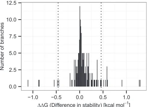
0.0 0.1 0.2 0.3 0.4 0.5
−3 −2 −1 0 1 2 3 4 5 6 7 8 9 10
ΔΔG (Difference in stability) [kcal mol−1]
Propor
[image:11.585.170.419.346.490.2]tion
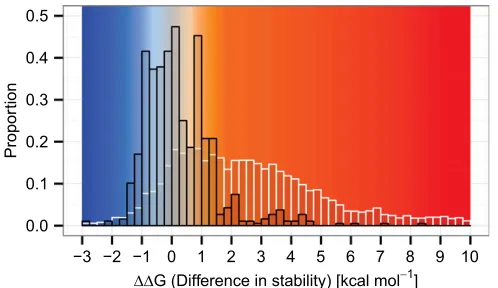
Fig. S1. Distribution of stability effects of ancestral and possible mutations. The distribution of stability effects arising from mutations in the evolutionary
Fig. S2. The RubisCO hexadecamer interfaces. Pairs of large subunits form dimers with an extensive interface; four of these dimers form an octomeric ring. The interdimer interfaces are comparatively small, and the overall structure is stabilized by the binding of eight small subunits that bridge dimers. Each large subunit has interfaces with several neighbors. Interfaces are annotated following van Lun et al. (4): LL1 interfaces within the dimers (LALB, LCLD, LELF, and LGLH) are shown by dashed gray lines, LL2 (LBLC, LDLE, LFLG, and LALH) interfaces are in red, and LL3 (LALC, LBLD, LCLE, LDLF, LELG, LFLH, LALG, and LBLH) interfaces are in blue. LS interfaces of a large subunit with only a small subunit are in green.