REVIEW
Helical growth in plant organs: mechanisms and significance
David R. Smyth*ABSTRACT
Many plants show some form of helical growth, such as the circular searching movements of growing stems and other organs (circumnutation), tendril coiling, leaf and bud reversal (resupination), petal arrangement (contortion) and leaf blade twisting. Recent genetic findings have revealed that such helical growth may be associated with helical arrays of cortical microtubules and of overlying cellulose microfibrils. An alternative mechanism of coiling that is based on differential contraction within a bilayer has also recently been identified and underlies at least some of these growth patterns. Here, I provide an overview of the genes and cellular processes that underlie helical patterning. I also discuss the diversity of helical growth patterns in plants, highlighting their potential adaptive significance and comparing them with helical growth patterns in animals.
KEY WORDS: Cellulose microfibrils, Circumnutation, Handedness, Microtubules, Spiral, Tendrils, G-fibres
Introduction
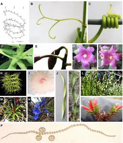
Plant growth is usually linear or circumferential (Steeves and Sussex, 1989) but in some cases it results in twists, spirals or coils; these patterns are generally categorised as helical (see Glossary, Box 1) growth. Well-known examples of plant organs that exhibit helical growth are the tendrils of climbing plants (Jaffe and Galston, 1968), but many other forms have been described (Fig. 1). For example, the tips of growing stems and other organs exhibit circumnutation (see Glossary, Box 1) as they extend and interact with components of the environment, be it light, other vegetation, or soil (Darwin, 1880; Baillaud, 1962a,b). Less well known are the specialised twists of leaf and flower stems that occur to invert their dorsal-ventral orientation (resupination; see Glossary, Box 1) (Hill, 1939), the spiral insertion of petals (contortion; see Glossary, Box 1) in the flower template (Endress, 1999), the coiling (see Glossary, Box 1) of pods and awns during seed dispersal, and the twisting (see Glossary, Box 1) of flat leaves that ensures their rigidity. It should be noted that one common form of helical arrangement, namely the placement of leaves and flowers on stems ( phyllotaxy), represents a different category of helical patterning involving organ initiation rather than subsequent organ growth. The properties and mechanisms of phyllotaxy are relatively well known (Reinhardt et al., 2003; reviewed by Traas, 2013) and will not be discussed here.
In this Review, I first summarise our current knowledge of the genes and cellular processes that underlie helical patterning in plant organs, highlighting the roles of cortical microtubules and cellulose microfibrils. Most of this understanding has come from studies of mutants of Arabidopsis thaliana that exhibit abnormal helical
growth (Hashimoto, 2002, 2013). Although these do not necessarily reflect the patterns associated with normal growth, it seems likely that the processes disrupted in such mutants may sometimes be the same as those recruited to impose helical growth where it has become adaptive and thus now represents the norm. I then discuss the diversity of helical growth patterns observed in plants and consider their possible adaptive significance. Interesting new observations on the presence of gelatinous fibres similar to those in tension wood of trees, and their likely differential extension within a bilayer to generate twists and coils are discussed. The handedness (see Glossary, Box 1) of helical growth in a species can be categorised in two classes–fixed or variable (see Box 2)–and the significance of this is also discussed. Finally, I compare helical growth in plants with that observed in animals and highlight what is still unknown, including the nature of fundamental determinants of the direction of handedness.
The genetic and cellular basis of helical growth Insights fromArabidopsismutants
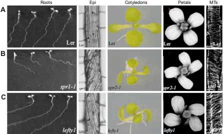
In 1880, Darwin noticed that when primary roots of bean, melon and oak species grew down an inclined plate they oscillated, apparently in a circumnutating helix. Over 100 years later, Okada and Shimura (1990) applied this phenomenon to identify the genes potentially involved in this growth. Using Arabidopsis thaliana, they defined sixWAVY GROWTHgenes that when mutated showed disruptions of this pattern. They attributed the waves to a cyclic response to touch, but Simmons et al. (1995) later provided evidence that they represented a flattened helix, the consequence of interactions between the spiralling circumnutation of the root and the impenetrable plate surface. In some Arabidopsis wild-type strains, roots consistently slant to the left under these growth conditions (as viewed from above; Fig. 2A), possibly as a consequence of right-handed circumnutation (Simmons et al., 1995) or to a left-handed rotation (twisting) of the root itself (Rutherford and Masson, 1996). Either way, changes to this slant angle allowed mutants that disrupted such helical movements to be readily obtained.
Using this approach two genes, namedSPIRAL1andSPIRAL2, were identified from mutant roots that now slanted strongly to the right instead of the left (Fig. 2B) (Furutani et al., 2000). Thespiral1
mutants apparently interfered with circumnutation or twisting by imposing abnormal helical growth within the roots, such that under normal growth conditions the straight cell files now showed a consistent right-handed twist (Fig. 2B). This extended to the hypocotyl and stem, and inspiral2mutants even to the leaf petioles and petals (Fig. 2B). Although the protein products ofSPIRAL1and
SPIRAL2 were unknown at this time, further studies of these mutants strongly implicated microtubules for a number of reasons (Furutani et al., 2000). First, the cortical arrays of microtubules within root epidermal cells in the basal elongation zone were abnormally helical in spiral1 mutants, albeit with a handedness opposite to that of the epidermal cell files (Fig. 2B). Second, low concentrations of propyzamide, a drug that depolymerises
School of Biological Sciences, Monash University, Melbourne, Victoria 3800, Australia.
*Author for correspondence (david.smyth@monash.edu)
D.R.S., 0000-0003-0455-9699
DEVEL
O
microtubules, reversed the root slanting and plant twisting effects observed inspiralmutants. An equivalent effect was obtained with taxol, a microtubule-stabilising drug, suggesting that microtubule depolymerisation alone is not the controlling property. Third, treatment of wild-type roots with the drugs resulted in cortical arrays of microtubules occurring in helices rather than in normal bands, further supporting a role for microtubules.
Understanding the role of microtubules and tubulins
Microtubules are long hollow tubes built from heterodimericα- and β-tubulin subunits. These are arranged in a head-to-tail pattern within longitudinal protofilaments, thirteen of which are arranged side by side in each single microtubule (Nogales, 2015). Microtubules are self-organising and dynamic. They exhibit growth at the plus end through net subunit addition (i.e. when polymerisation exceeds depolymerisation), and shrinkage at the minus end via depolymerisation (Dixit and Cyr, 2004). Individual microtubules can aggregate into arrays of parallel strands and, in elongating plant cells, such arrays are often arranged in lateral hoops or helices around the long axis, lying just underneath the plasma membrane in the cell cortex.
Whereas the initial study ofspiralmutants implicated a role for microtubules in helical growth, a direct involvement of tubulin subunits was only revealed whenArabidopsismutants with roots showing left-handed twisting (i.e. opposite to the right-handed
spiralmutants; Fig. 2C) were found in genes that encodeα-tubulin 6 (lefty1) or α-tubulin 4 (lefty2) (Thitamadee et al., 2002). Again, cortical microtubules now adopted a helical pattern around root epidermal cells, this time right-handed in contrast to the left-handed twists of cell files (Fig. 2C). The mutations were likely to reflect a gain of function as they were semi-dominant, and transgenic plants expressing a mutantα-tubulin in a wild-type background showed the same twisted phenotypes. This is consistent with abnormal
tubulin mutant subunits somehow disrupting the normal microtubule arrays. It should be noted that loss-of-function mutants had no detectable effect on growth or cell file patterns, perhaps because of gene redundancy.
Subsequently, the analysis of a large sample of 32 root-slanting mutations revealed that point mutations in tubulins could result in either left- or right-handed helical phenotypes (Ishida et al., 2007a). Many members of the tubulin family were involved–five of the six α-tubulins and four of the nineβ-tubulins ofArabidopsis–with left-and right-hleft-anded mutants present in each subfamily. The sites of mutational changes within theα/βheterodimer were concentrated in regions of protein-protein interaction, either between heterodimers or between adjacent protofilaments. In addition, there was a strong negative correlation between the slope of the cortical microtubule helices and the magnitude of root slanting for the different mutants, indicating a close functional link.
Microtubule-associated proteins in helical growth
A range of microtubule-associated proteins (MAPs) have also been implicated in helical growth. For example,SPIRAL1 (also known as SKU6) was eventually found to encode a protein localised to the growing ( plus) end of microtubules and the microtubule lattice (Nakajima et al., 2004; Sedbrook et al., 2004). SPIRAL2 (originally named TORTIFOLIA1) also specifically associates with microtubules along their entire lengthin vivo, contains protein-protein interaction domains, and binds directly to microtubules in vitro
(Buschmann et al., 2004). A third gene (GCP2), with a right-handed helical growth mutantspiral3, encodes a component of theγ -tubulin-containing complex GCP2, which is required for microtubule nucleation (Nakamura and Hashimoto, 2009). These and other mutants of MAPs, some with left-handed growth, revealed that disruptions to microtubule dynamics can often lead to twisted growth, although the exact mechanisms are not clear and might be diverse.
Understanding the link between microtubules and cellulose microfibrils
How can the orientation of microtubule arrays lying internal to the plasma membrane be linked with the helical growth of a cell? Classical observations of helical cells in the filamentous green alga
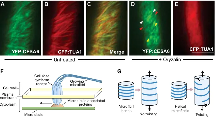
Nitellarevealed that they grow by relatively uniform extension of all parts of the lateral cell wall (Green, 1954). Transverse bands or hoops of cellulose microfibrils occur in plant cell walls and, when cortical microtubules were discovered, a close link between their orientation and that of the overlying cellulose microfibrils was revealed (Ledbetter and Porter, 1963; Baskin, 2001). Microfibrils are now known to be synthesized in a cellulose synthase complex (CSC), a hexagonal rosette embedded in the plasma membrane. During microfibril formation, 18 or 24 newly made cellulose polymer chains are extruded externally, entwine and lie entangled in the cell wall, thus pushing the rosette laterally within the plasma membrane. Thus, the orientation of the new fibre depends on the direction of movement of the rosette (Slabaugh et al., 2014; Kumar and Turner, 2015). The influence of underlying microtubules on this direction of movement was subsequently revealed by live imaging of transgenicArabidopsisplants carrying microtubule and cellulose synthase proteins with different fluorescent tags (Fig. 3A-C) (Paredez et al., 2006). In extending hypocotyl cells, it was shown that microtubules determined the vector adopted by the rosette. Even so, they were not required to maintain its movement, as treatment with the microtubule-destabilising drug oryzalin abolished microtubules but the rosettes continued to move, at least in the short term (Fig. 3D,E).
Box 1. Glossary of terms
Chirality:Existing in two, mirror-image forms (enantiomorphs). Circumnutation:The relatively rapid circular (or elliptical) movement of the ends of growing stems, tendrils, leaves and roots, usually occurring in a fixed rotational direction.
Coiling:The property shown by a rod arranged in a helical conformation (as in a spring).
Contortion:A spiral arrangement of overlapping petals (or sepals) within the flower such that they all overlap with their neighbours in the same way (as in the blades of a propeller).
Handedness:The property shown by two forms of structures that are mirror images of each other. They are often distinguished by the names right (dextral) and left (sinistral) that may be arbitrarily determined. Helical:The property of a line that turns around a rod-shaped space moving continuously from one end to the other. A helix can be defined by its length, radius, and either its pitch (distance between successive intersections of a longitudinal line on the outer edge) or its angle of displacement from the longitudinal axis. Helices can exist in two mirror-image forms (see also chirality, handedness).
Resupination:The twisting of petioles of leaves, or pedicels of flowers, through 180° to reverse the orientation of the upper and lower parts. Spiral:The property of a line that extends outwards from a point in a path of continuously increasing distance from the point; this property can apply to one plane (as in a watch spring), or to a cone-shaped space if the line simultaneously extends at right angles to the plane. The term is often loosely applied to a helix, which, however, is technically not a spiral as it has a fixed radius.
Twining: The process of a helical body coiling around another rod-shaped body.
Twisting:The result of rotating one end of a flexible, rod-shaped body.
DEVEL
O
The subsequent discovery of CELLULOSE SYNTHASE INTERACTIVE PROTEIN 1 (SCI1) – a bridging protein that binds directly to both microtubules and cellulose biosynthetic proteins–revealed a direct mechanical link between these cellular
components (Gu et al., 2010; Li et al., 2012). In mutants of theCSI1
gene (originally calledPOM-POM2), the orientation of cellulose microfibrils and cortical microtubules was uncoupled (Bringmann et al., 2012; Li et al., 2012). Furthermore, elongating roots,
A B C
D E F G
H I J K L
M N O
[image:3.612.108.509.58.522.2]P
Fig. 1. Examples of helical growth forms in plants.(A) Path of circumnutation of the growing apex of the dodderCuscuta gronovii(Convolvulaceae), mapped at 5 min intervals. The apex is moving toward the light (arrows) in wide right-handed circular movements.ωmarks a fixed reference point. Adapted from Baillaud (1962b). (B,C) Tendrils of the grapevineVitis vinifera(Vitaceae) before (B) and after (C) they encounter a support, in this case twining around it in a right-handed helix. (D) Resupination of leaves of the Peruvian lilyAlstroemeria psittacina(Alstroemeriaceae) showing the 180° left-handed twists that bring the abaxial surface uppermost (Ab-Top) with the adaxial surface now below (Ad-Bot). Taken from Chitwood et al. (2012). (E) Resupination of 180° in the pedicel of an orchid bud (Cattleyahybrid, Orchidaceae); the arrow indicates twist. (F,G) Contortion of the petals of the Norfolk Island hibiscusLagunaria Patersonia (Malvaceae), with equal numbers of left-handed (F) and right-handed (G) flowers within an individual plant. (H,I) Seed pod of the legumeMedicago tenoreana (Fabaceae) showing the shallow left-handed helix and surface hooks in side view (H), and a top view (I) following staining with phloroglucinol showing lignin present in the central axis (red) where extension is limited. (H,I) Reproduced with permission from Fourquin et al. (2013). (J,K) Twisted blade-like leaves of the sand lilyPancratium maritimum(Amaryllidaceae; J) and the blood rootHaemodorum venosum(Haemodoraceae; K). These species are unrelated, and the direction of helices is fixed inPancratium(all right-handed) but varies inHaemodorum(50% each). (L) Spiral leaves of the geebung shrubPersoonia helix(Proteaceae) that are uniformly left-handed. Photo courtesy of Sonja Chandler. (M,N) Leaves and flowers of the spiral blue squill (Chamaescilla spiralis, Asparagaceae), showing left-handed helical growth of the blade-like leaves (M), and right-handed contortion of the petals after anthesis (arrow in N). Photos courtesy of Sonja Chandler. (O) Spiral leaves of the rare heathAndersonia grandiflora(Ericaceae), with branches carrying leaves all with left-handed twists (left) or all with right-handed twists (right) within the same individual. Photo courtesy of Sonja Chandler. (P) Elater of the liverwortMarchantia polymorpha(Marchantiaceae) showing double left-handed helices of thickening within the cell that are joined at each end. Three spores are also shown. Adapted from Kny (1890).
DEVEL
O
hypocotyls and leaves now showed left-handed spiralling (Bringmann et al., 2012), although, perhaps unexpectedly, the inflorescence stem incsi1mutants had a slightly right-handed twist (Landrein et al., 2013). The latter study also established the independence of the handedness of helical growth and the direction of phyllotaxy of buds formed on the stem. Thus, our current overview is that the direction of movement of cellulose synthase rosettes through the plasma membrane, leaving a trail of external cellulose microfibrils, is guided by a ‘train-line’ of underlying cytoplasmic microtubules linked together by specific MAPs (Fig. 3F).
The cellular mechanisms involved in helical growth
The microtubule-microfibril link is firmly established, but exactly how it controls helical growth requires explanation. The elongation of cylindrical plant cells usually occurs by longitudinal expansion of side walls while their diameter remains constant. Bands of cellulose microfibrils, especially those in inner, more recently made layers, normally lie transverse to the direction of expansion, and intervening regions apparently loosen evenly as the cell elongates (Fig. 3G). The bands may prevent the cell expanding laterally, while its narrowing is prevented by positive turgor pressure (Frei and Preston, 1961). By contrast, in those cases in which the microfibrils occur in an oblique, helical pattern, lengthwise expansion may be associated with an increase in the pitch of the helices (Fig. 3G).
Thus, to accommodate the increased length of the cell without an increase in the length of the cellulose microfibrils, or a decrease in cell diameter, the cell would necessarily twist (Fig. 3G) (Lloyd and Chan, 2002; Buschmann et al., 2009). This would account for the striking observation that the handedness of abnormal organ twisting among microtubule mutants, and drug-treated cells, is always opposite to the handedness of the distorted underlying cortical microtubule arrays (and thus presumably of the overlying cellulose microfibrils) (Fig. 2B,C). Right-handed organ twisting is always associated with left-handed microtubule arrays, and vice versa. This can be visualised using a coiled spring: if stretched, its diameter will decrease unless one end is rotated in the direction opposite to the coils.
The physical driver of helical growth has been attributed to properties of either individual cells or tissues. Cell interactions are not necessarily involved, as single cultured cells of theArabidopsis tortifolia2mutant (which harbours a point mutation in theα-tubulin gene) nevertheless exhibit helical twisting (Buschmann et al., 2009). Aggregates of such twisted cells, especially in cell files, seem likely to result in twisting of the resulting tissues. However, helical growth is sometimes the result of the regulated, sequential timing of cell divisions, as has been reported inArabidopsisroot meristem cells (Baum and Rost, 1996). A spiralling arrangement of cells also occurs in the root meristem of the floating fernAzollafollowing early cell divisions of defined orientation (Gunning et al., 1978). Proposing another tissue-based mechanism, Furutani et al. (2000) suggested that root twisting inArabidopsis spiralmutants occurs to accommodate the reduced longitudinal extension of internal cells relative to the overlying epidermis.
The question then arises: why left or right? Mutant studies indicate that this depends on an as yet undefined inherent structural property of microtubule arrays. Individual molecular tweaks to microtubules, or to microtubule-associated proteins, can result in spiral changes to their intracellular orientation in one or other direction. Ishida et al. (2007b) proposed that the microtubules of tubulin mutants might be helically twisted by the presence of abnormal amino acids, perhaps because microtubules were now built from abnormal numbers of protofilaments. There is structural evidence that microtubules with more or fewer than the normal 13 protofilaments do twist (Sui and Downing, 2010), but whether such perturbations occur in the microtubules of mutant plants has not yet been investigated.
Exceptions to a strict relationship between spirals of microtubules or microfibrils and cell twisting have been reported. Spiral microtubule arrays can sometimes be observed with a defined chirality (see Glossary, Box 1) in non-twisted wild-type tissues, as in the differentiation zones of roots (Liang et al., 1996), in vessels and tracheids of wood (Barnett and Bonham, 2004) and in immature leaf hairs (Sambade et al., 2014). In these cases, cell expansion may not be occurring. On the other hand, cellulose microfibrils can adopt a defined helical structure even in the likely absence of microtubule function (e.g. Green, 1962; Paredez et al., 2006; Landrein et al., 2013). Furthermore, left-handed twisting is seen in Arabidopsis roots in which microtubules are disrupted by chemicals, or by mutations in the
MICROTUBULE ORGANIZATION1 (MOR1) gene, but the cellulose microfibrils are still arranged in radial bands and are not detectably helical (Sugimoto et al., 2003). It might be that, in many such situations, helical patterning of cellulose microfibrils is overridden by residual transverse bands of cortical microtubule arrays, or some other independent factor. As Landrein et al. (2013) state:‘the main issue with stem twisting is not what is inducing it,
Box 2. Helical forms
Left-handed Sinistral Clockwise
S Negative (–) With sun (NH)
Right-handed Dextral Anti-clockwise
Z Positive (+) Against sun (NH)
In a left-handed (sinistral) helix, as one moves up the helix the line moves to the left across the front face. In a right-handed (dextral) form, the line moves to the right across the front face. Historically, the two forms in plants have also been given other names. Upward movement of the helix can be related to a clock face as viewed from above. The central part of the letters S and Z reflects the slope of the line. The use of + and−is arbitrary. The movement of the ascending line across the back face either matches or countervails the apparent movement of the sun in the southern sky as viewed from the northern hemisphere (NH). Helical handedness in plants is typically either fixed within a species (always left or always right) or variable (50% left, 50% right, usually between shoots within a plant).
DEVEL
O
because stem twisting can occur by default, but what is maintaining the straight stem in the wild type’.
Overall, however, genetic and cellular studies have revealed a strong functional link between microtubule arrangement and the potential twisting of cells and tissues (Fig. 3F,G). This appears to be imparted through the microtubule-mediated guidance of cellulose microfibril patterning in the developing cell wall, although the latter may sometimes exhibit its own independent patterning.
The adaptive roles and significance of helical growth
Having discussed the mechanisms of helical growth, deduced from studies of abnormal plants, I now turn to the likely biological significance of helical growth during normal plant growth and development. Clearly there are many different categories of helical growth, each having arisen sporadically across the family tree, and their functional roles are presumably diverse.
Circumnutation
In 1880, Darwin introduced the word‘circumnutation’to describe plant movements like those of the stem of a climbing plant,‘which bends successively to all points of the compass, so that it revolves’ (Fig. 1A) (Darwin, 1880). Earlier, Darwin had published a typically erudite characterisation of circumnutation in many climbing species (Darwin, 1867). He noted that the growing tip often moved in wide circles (or ellipses) with a period of several hours, and that when an obstacle was encountered the stem commenced to wind around it for support as it continued to grow. He also noted that the stem was usually twisted before it encountered a support, possibly‘to gain rigidity…so as either to be enabled to pass over inequalities in its spiral ascent, or to carry its own weight when allowed to revolve
freely’, and that the twisting is associated with increased
‘turgescence’ and extensibility of cells in the part of the stem lying on the outside of the helix (Darwin, 1880). Interestingly, in the great majority of species, including members of the legume family (Fabaceae), bindweeds and dodders (Convolvulaceae) (Fig. 1A), and the dogbane family (Apocynaceae), the helix movement was fixed and right-handed, although some adopted only left-handed spiralling (hops, Moraceae), and yet others exhibited mixed spiralling, even in individual plants (e.g. in the yam family, Diosceraceae) (see also Baillaud, 1962b).
Darwin (1880) further observed that circumnutation applied more broadly to the newly growing stems of many non-climbing species, as well as to roots and leaves. Indeed, he claimed that:‘the capacity of acquiring the revolving power in which most climbers depend is inherent, though underdeveloped, in almost every plant in the vegetable kingdom’(Darwin, 1867). Generally, non-twining shoots and leaves might circumnutate (with positive feedback) in their search for optimum light conditions, whereas circumnutation in roots might facilitate movement around obstacles in the soil.
Models of circumnutation have been characterised as involving
‘internal oscillators’ or ‘geotropic feedback’ (Johnsson and Heathcote, 1973). The fixed handedness of circumnutation led Darwin (1880) to favour an internal control mechanism; based on current data, this is perhaps linked to a fixed helical direction of microtubule cortical arrays. On the other hand, the role of an external influence, gravity, in moderating circumnutation has been studied experimentally, for example inArabidopsisplants grown in space (e.g. Johnsson et al., 2009). The conclusion in this case was that circumnutation still occurred under conditions of micro-gravity, but that its amplitude was much larger when significant gravity-like
A
B
C
Ler
spr1-1
lefty1
[image:5.612.83.529.58.326.2]Roots Epi Cotyledons Petals MTs
Fig. 2.Arabidopsismicrotubule mutants.(A) Wild-type Lerseedlings; (B)spiral1orspiral2mutant seedlings; (C)lefty1mutant seedlings. Wild-type (Ler) seedling roots grow with a leftward slant on tilted plates, whereasspiralmutants show a rightward slant, andleftymutants a more extreme leftward slant. Epidermal cell files (Epi) in the differentiated zone of roots are straight in wild type, twisted in a right-handed helix inspiral1-1mutants, or in a left-handed helix in lefty1mutants. Cotyledons and petals can also follow this twisting pattern. Cortical arrays of microtubules (MTs) in the root elongation zone are radially organised in wild-type cells, but are in a left-handed helix inspiral1-1mutants, and a right-handed inlefty1mutants. Scale bars: 10μm. Reproduced with permission from Hashimoto (2002), Furutani et al. (2000) and Thitamadee et al. (2002).
DEVEL
O
conditions (centrifugal force equivalent to 0.8 g) were applied. Other results have indicated that gravity is essential for stem circumnutation inArabidopsis, as it does not occur in scarecrow
mutants that lack gravity-sensing endodermal cells (Kitazawa et al., 2005). Furthermore, circumnutation can be restored to these mutants by adding the wild-type version of the SCARECROWgene from morning glory (Ipomoea nil, Convolvulaceae), but not the version found in a mutant that has lost stem circumnutation and coiling. Clearly, internal properties and gravity interact, although the underlying mechanisms have not yet been defined.
Tendrils
In climbing plants, more specialised helical growth occurs in tendrils (Darwin, 1867; Baillaud, 1962a; Jaffe and Galston, 1968). Tendrils are thin, rod-shaped structures that circumnutate as they extend, searching from the growing plant (Fig. 1B, Fig. 4A). They variously develop as modifications of: (1) axillary shoots, for example in the melon family (Cucurbitaceae); (2) inflorescences, as in grape (Vitaceae) and passionfruit vines (Passifloraceae); or (3) leaflets, as in some beans and peas (Fabaceae) andBignoniavines (Bignoniaceae). The study of mutants with modified tendrils has revealed that their identity can be controlled by transcription factors. In pea (Pisum sativum), a class I HD-Zip protein controls tendril identity (Hofer et al., 2009), whereas in melon (Cucumis melo) and cucumber (C. sativus), a TCP family member is involved (Mizuno et al., 2015; Wang et al., 2015). In grapevines, gibberellins have been implicated in controlling tendril identity; tendrils revert to an inflorescence identity in plants with disrupted GA response (Boss and Thomas, 2002).
Tendrils carry a touch-sensitive region close to their tip, and if this contacts a suitable object the tendril rapidly twists around it, usually commencing within seconds or minutes (Fig. 1C). In many species
the tendril end has a hook, within which the touch-sensitive region is located. In others, the full circumference is sensitive, and coiling is triggered in the direction of the initial contact. The basis of touch sensitivity is not yet known, although physiological experiments have been extensively reported (Jaffe and Galston, 1968). The handedness of the coiling is apparently not predetermined by pre-existing twists in the tendril itself, unlike stems of twining (see Glossary, Box 1) plants, but depends on the orientation of the initial contact.
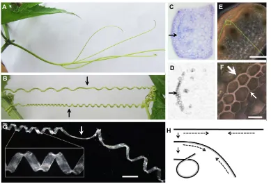
A second property of many shoot- or inflorescence-based tendrils is a subsequent, slower coiling between the anchored tip and the plant itself, resulting in a spring-like structure (Fig. 4B). The outcome is a reduction in this distance and a consequent lifting of the plant body, as well as providing increased flexibility in response to wind and other stresses (Darwin, 1867). Darwin also noticed that coiling reversed its handedness in the centre of the tendril at a point now called a‘perversion’(Darwin, 1867). He deduced that this was a mechanical consequence of the coiling of a flexible rod with fixed (i.e. non-rotating) ends.
The likely cellular basis for coiling in tendrils and twining stems was recently discovered (Meloche et al., 2007; Bowling and Vaughn, 2009). It involves bands or cylinders of specialised fibre cells, each containing an inner gelatinous cell wall layer that is rich in longitudinal cellulose microfibrils and associated arabinogalactans and pectins (G-fibres) (Fig. 4C,D). The outer wall of these cells progressively lignifies as helical growth occurs. Such fibres had been characterised in tension wood of flowering plants, where their contraction serves to straighten and lift growing branches (Goswami et al., 2008; Mellerowicz and Gorshkova, 2012). More recently, it was shown that in cucumber tendrils the gelatinous G-fibres occur as a bilayer of cells (Gerbode et al., 2012), with the layer closer to the inner, concave edge of the coil being YFP:CESA6
Untreated
CFP:TUA1 YFP:CESA6
Merge CFP:TUA1
A B C D E
+ Oryzalin
Cell wall
Plasma membrane
Cytoplasm
Growing microfibril
Microtubule
Microtubule-associated proteins Cellulose
synthase rosette
Microfibril bands
Helical microfibrils
No twisting Twisting
[image:6.612.130.484.58.253.2]G F
Fig. 3. The role of microtubules and cellulose microfibrils in helical growth.(A-E) Confocal fluorescence images of cells from etiolated hypocotyls of Arabidopsisplants carrying YFP-tagged cellulose synthase A6 (CESA6) and CFP-taggedα-tubulin 1 (TUA1). There is a close correspondence between the orientation of cellulose microfibrils (green) and that of the microtubule arrays (red) in untreated cells (A-C). Following treatment with the microtubule-destabilising drug oryzalin (D,E) microtubule structure is lost, although microfibrils may continue to show oblique growth (arrowheads). The average of 30 frames collected at 10 s intervals is shown. Reproduced with permission from Paredez et al. (2006). Scale bar: 10μm. (F) A simplified model illustrating the dependence of the orientation of growing cellulose microfibrils within the developing cell wall on the pattern of underlying cortical microtubules. As multiple cellulose polymers are synthesised by the cellulose synthase rosette in the plasma membrane, they entwine in a microfibril that is embedded in the external cell wall. The rosette may thus be pushed sideways, with its direction of movement being determined by the cytoplasmic microtubule array to which it is attached via microtubule-associated proteins. (G) Comparison of the longitudinal expansion of cells in which cellulose microfibrils occur in parallel bands or hoops (left) with those that occur in a helical pattern (right). In each case, the diameter of the cell is held constant due to osmotic pressure, but the length increases due to loosening of the cell wall matrix without lengthening of the microfibrils. Individual bands of microfibrils simply move further apart (left). However, if they occur in a helical pattern, the cell wall undergoes a twist with the opposite handedness to accommodate the expansion (right, large arrowheads).
DEVEL
O
more heavily lignified (Fig. 4E,F). Physical models implicated the differential contraction of this bilayer in coiling, with the inner lignified cells being more compressible and shrinking more than the outer layer, perhaps losing more water because of their increased hydrophobicity (Fig. 4G,H). As in nature, the result was equal numbers of coils of opposite handedness separated by a perversion point, as well as the overwinding of the coils when the two ends were pulled further apart (Gerbode et al., 2012).
Resupination
Some plants have leaf petioles that are also touch sensitive and wind around a support in a manner analogous to tendrils (Darwin, 1867). However, petioles in other species undergo a different form of helical growth called resupination (Hill, 1939). Here they do not coil upon contact, but all twist through 180° as they approach maturity so that the originally lower surface of the leaf is now uppermost (Fig. 1D). The adaptive significance of this is not clear, but the anatomy of the mature leaf shows some reversal of normal tissue distribution. A recent study of a well-known example, the Peruvian lilyAlstroemeria(Alstroemeriaceae), revealed that the orientation of the twist is always left-handed (Fig. 1D), independent of the variable left- or right-handed phyllotaxis of the leaf insertion pattern (Chitwood et al., 2012).
Resupination may also occur in flower pedicels (Hill, 1939); for example, in many orchids (Orchidaceae) where flowers are twisted around by 180° at maturity so that the labellum, a modified adaxial
petal, is now abaxially located, i.e. at the flower’s base (Fig. 1E). In this position, the labellum provides a convenient landing platform for fertilising insects (Darwin, 1862). Resupination also occurs in other species sporadically across the flowering plant phylogeny, more frequently in flowers with bilateral symmetry that are often animal pollinated (Dworaczek and Classen-Bockhoff, 2015). For example, it occurs in legume species in which the flowering stem is pendulous rather than upright (Wisteria,Robinia, Fabaceae) and thus locates the specialised keel to its usual position at the base of hanging flowers (Hill, 1939).
The twisting observed in resupination might be provoked by differential expansion (or shrinkage) of subregions of the petiole or pedicel. For example, long, raphide-bearing cells occur in three ribs in the pedicel and ovary of the orchidLemboglossum bictoniense. To induce resupination, these raphides apparently extend more than the intervening tissues, thus generating a twist so that their relative length remains the same (Dines and Bell, 1994). The direction of the twist might sometimes be controlled by the developmental context of the leaf or bud within the plant. For example, in orchids with an alternating (distichous) arrangement of buds along the flowering stem, the direction of the twist also alternates between left- and right-handedness (Nyman et al., 1984; Dines and Bell, 1994). In some cases, however, the mechanism presumably has an internal genetic component, as the handedness is fixed (Hill, 1939; Chitwood et al., 2012; Dworaczek and Classen-Bockhoff, 2015). In either case, gravity sensing is also likely to be involved and B
G H
A C E
[image:7.612.114.503.55.320.2]D F
Fig. 4. Gelatinous fibres in tendrils.(A,B) Tendrils of the loofah melon (Luffaspecies, Cucurbitaceae) before (A) and after (B) they encounter a support, when tendrils have contracted by internal coiling around a point of perversion (arrows), with equal numbers of coils of opposite handedness on each side. (C,D) Sections through a coiled tendril of the wild cucumberEchynocistus lobata(Cucurbitaceae) stained with Toluidine Blue (C), or with an anti-xylan antibody (D) that detects gelatinous G-fibres (arrow) on the inner concave side; the other reactive cells within the section are xylem elements. C,D reproduced with permission from Bowling and Vaughn (2009). (E,F) Unstained section of a coiled cucumber tendril, showing a lignified band on the inner concave side (E). Higher magnification of the boxed region (F) reveals that this band has two layers, with heavier lignin thickening on the inner side (large arrow) than the outer side (small arrow). (G) A G-fibre ribbon extracted from a cucumber tendril retains its coiled state, including a perversion point (arrow). E-G reproduced with permission from Gerbode et al. (2012). (H) Proposed mechanism of coiling of a tendril by preferential contraction (dashed arrows) of the inner, more heavily lignified layer ( pale shading) of a gelatinous fibre ribbon in side view. The outer, less lignified layer (dark shading) resists compression and together this results in curving and then coiling of the tendril. In this diagram only one end (the left end) is fixed, and coils of only one handedness arise (see Fig. 1C). If both ends are anchored, the tendril coils with different handedness on each side of a perversion point (B,G). Scale bars: 100μm in E; 10μm in F; 1 mm in G.
DEVEL
O
somehow limits the twist to 180°. Consistent with this, the petioles (or pedicels) in some cases may retwist if the leaf (or flower) is artificially inverted.
Contortion
Asymmetric, slightly helical growth can also occur in petals. It is usually associated with contortion – the spiral overlapping arrangement of all petals in a flower (Endress, 1999). Here, each petal overlaps its neighbour on the same side, so that the pattern is propeller-like when viewed from above (Fig. 1F,G). Contorted petals occur sporadically across the angiosperms, but especially in species with multi-symmetric (i.e. non-bilateral) flowers with five petals. Two major occurrences are in malvids (e.g. Malvaceae, including
HibiscusandLagunaria; Fig. 1F,G) (Endress and Matthews, 2006) and in lamiids (e.g. Apocynaceae, including periwinkle, Vinca; Endress, 2010). Interestingly, it was noted that the direction of spiralling is usually fixed in asterids (mostly lamiids), although it changed from left to right during evolution of the Apocynaceae, whereas in the rosids (including malvids) there are also examples in which both left- and right-handed contortion occur in the same individual (Endress, 1999). Thus, the mechanism establishing the fixed helical pattern in asterids and some rosids is apparently very different from that in other rosids with a variable pattern.
Seed dispersal
Roles for helical patterning in seed dispersal exemplify the diverse functions of helical growth that have arisen. The seed pods of some species of the pasture legume genusMedicagocoil, whereas others are straight. In the case of coiled, compact pods with Velcro-like hooks on the outer surface, attachment to passing animals may aid seed dispersal (Fig. 1H). Legume pods develop from a single carpel, with one side of the pod opening preferentially at maturity. The comparison of 11 coiled and 6 uncoiledMedicagospecies revealed that coiling is associated with lignified thickening along the opening side, which presumably retards growth, resulting in a tight coil (Fig. 1I) (Fourquin et al., 2013). In addition, coiling was shown to be correlated with a polymorphism in the single Medicago SHATTERPROOF transcription factor gene known to define a lignin band in equivalent regions ofArabidopsisseed pods. This gene might thus play a role in imposing helical growth inMedicago, although another gene must be responsible for defining chirality. Notably, chirality is usually fixed and left-handed in Medicago
species, but the genetic study of species with both left- and right-handed plants showed that a single dominant gene can impose right-handedness (Lilienfeld and Kihara, 1956).
A second example of helical patterning in seed dispersal is the corkscrew coiling of the long awn attached to seeds ofGeraniumand other Geraniaceae that drive them into the soil. These awn structures contain a longitudinal bilayer of hygroscopically sensitive inner cells and a more rigid outer layer (Abraham and Elbaum, 2013). Shrinkage of the inner layer upon drying results in coiling of the awn, but this only occurs in species with an unusual tilted helix of cellulose microfibrils in the inner layer, and not in those with a regular helix. This tilt results in a coiling motion of the drying awn without inducing any twists. As predicted by modelling, both the cellulose microfibrils and the coils run in the same direction, in this case right-handed (Abraham et al., 2012). This example demonstrates that tissues do not have to be alive to demonstrate coiling.
Wood
Wood is mostly made up of lignified tracheids, vessels and fibre cells generated from the cambium, a cylindrical secondary meristem
on the outside of the stem. In many trees, files of wood cells appear to spiral weakly around the trunk, especially in younger-formed wood (Harris, 1989). This helical pattern appears to be the result of the plane of ancestral cell divisions in the cambium being slightly offset from radial. The direction of the spiral varies but is predictable depending on the species, indicating some inherent determinative process. In this regard, wood exhibits a near-universal right-handed arrangement of cellulose microfibrils in the main secondary (S2) cell wall layer (Barnett and Bonham, 2004). However, this is unlikely to impact on growth as the cells have already fully extended when the microfibrils are laid down, and the significance of this arrangement is unknown.
Flat organs
The twisting or coiling of long, flat leaves has also been frequently observed (e.g. Fig. 1J,K). Its significance has been analysed, for example in bulrush/cat-tail leaves (Typhaspecies, Typhaceae) where twisting increases their rigidity, allowing increased height without bending (Schulgasser and Witztum, 2004a). Such twisting is also likely to limit wind drag by minimising the surface area exposed to wind from any direction (Rowlatt and Morshead, 1992). The same phenomenon could apply to sun exposure where minimising this might be adaptive in warm climates, for example in the highly helical leaves of the Australian shrubPersoonia helix(Proteaceae) (Fig. 1L). The control of twisting can be genetic where its direction is fixed, as observed inTypha(left-handed),Pancratium maritimum
(Amaryllidaceae, right-handed; Fig. 1J) (Schulgasser and Witztum, 2004b), Persoonia (right-handed; Fig. 1L), and in leaves of the rosette species Chamaescilla spiralis, Asparagaceae (left-handed; Fig. 1M). In the latter case, the petals are also contorted, but always in the opposite direction, i.e. they are right-handed (Fig. 1N). Alternatively, in species where handedness is not fixed between shoots of the same plant (e.g. Andersonia grandiflora, Ericaceae; Fig. 1O), twisting presumably represents a self-perpetuating response of the leaf primordium to some early stochastic asymmetry in the shoot meristem. It will be interesting to test if this is related to the left- or right-handed direction of helical phyllotaxy which, characteristically, can also vary between shoots.
Elaters
As in the angiosperms, sporadic examples of helical growth occur throughout other green plants, including algae (Green, 1954; Frei and Preston, 1961). In liverworts (basal land plants) such as
Marchantia polymorpha, spores are expelled from the parent plant by elaters that, like awns, contract and coil in response to reduced humidity (Fig. 1P) (Ingold, 1939). Marchantiaelaters are single elongated cells with twin spirals of secondarily thickened material that are uniquely attached at their ends to make a continuous loop. This means that as the pitch of the spirals decrease, the elaters coil (as in a twisted rubberband) and interact with other elaters to disrupt the spore mass. If the link between the twin helices is cut at both ends, the elaters do not coil (Ingold, 1939). Primary and secondary cell wall layers have different polysaccharide compositions in the liverwort Radula buccinifera (Kremer et al., 2004), and their differential contraction during drying might be the basis of coiling.
Helical growth in metazoans
Metazoans, too, can show spiral growth. Indeed, early cell divisions in most lophotrochozoans occur in a spiral arrangement (Hejnol, 2010). A well-known example is shell coiling in gastropods such as snails. This coiling can be traced back to the third cell division of the
DEVEL
O
new embryo, in which the four spindles are uniformly offset from a perfectly radial orientation (Freeman and Lundelius, 1982). Four smaller daughter cells (micromeres) move rightwards into the grooves between the four larger daughter cells (macromeres). Subsequent cell divisions maintain a right-handed spiral outcome, eventually including the linear mantle that develops and secretes the shell. Spiral growth of the shell occurs when material is produced more abundantly in a gradient from one end of the mantle to the other (Shimizu et al., 2013). It is remarkable that almost all extant gastropods coil in a dextral, right-handed helix, implying an internal determinant (Palmer, 1996). This is consistent with a single dominant Dlocus controlling right-handedness, with its function occurring in maternal cells rather than the zygote (Sturtevant, 1923; Freeman and Lundelius, 1982). Cellular and cytoplasmic manipulations, and the disruption of spindle microtubules and the actin cytoskeleton, all point to a localised determinant of the asymmetric spindle orientation (Shibazaki et al., 2004; Kuroda et al., 2009). This has been confirmed recently by the cloning of the
Dlocus of the pond snailLimnaea stagnalis, which revealed that it encodes a formin (Davison et al., 2016). Formins interact with the actin and microtubule cytoskeleton, and in snails maternal transcripts are already localised to only one of the two cells of the first zygotic division. This finding parallels known cellular determinants of helical growth in plants in that the cytoskeleton with its inherent chiral properties is involved, but differs because the animal determinant controls asymmetric cell division rather than twisted cell expansion as in plants.
A closer analogy with twisted growth in plants is revealed by roller mutants of the nematode Caenorhabditis elegans. In these mutants, the body is twisted in either a left-handed or right-handed helix, causing them to roll as they move forward. Severalroland other genes encode collagens that assemble into trimeric polymers in the cuticle (Kramer et al., 1990), and studies have shown that the regular helical structure of collagens is disrupted in rol mutants (Bergmann et al., 1998), paralleling the changes in microtubules in
Arabidopsis spiralandleftymutants, and is the likely cause of body twists.
Finally, it should be noted that left-right handedness in some vertebrates is conferred by the directed movement of an unidentified signal by beating cilia, which are offset from the vertical towards the posterior pole and all rotate clockwise (Nonaka et al., 1998, 2005). However, such a process cannot be involved in imposing helical handedness in flowering plants because their ability to generate cilia has been lost (Merchant et al., 2007).
Conclusions
Helical growth in plants involves many different patterns, ranging from the exploratory looping of extending stems and other organs, to the coiling of tendrils to support the climbing plant body, the twisting of supporting stems through 180° to reverse the orientation of leaves and flowers, the contortion of petals, and the twisting of flat organs to increase their rigidity. Helical rotation of growing organ tips may be universal, but other cases might reflect adaptive specialisations that have arisen independently and sporadically during the evolution of flowering plants.
At the molecular and cellular level, at least some cases of helical growth have been firmly linked with the helical arrangement of arrays of microtubules in the cellular cortex, and the related coiling of cellulose microfibrils around the cell (Fig. 3F,G). A further generalisation is that some helical coils owe their origin to differential contraction within a longitudinal bilayer of specialised cells (Fig. 4H).
Another important fact highlighted throughout this Review is that the handedness of a helical pattern comes in two forms: either fixed in the same direction in all individuals; or variable (50:50), usually between organs of the same plant. This reflects the likely mechanism of twisting. A fixed orientation implies that it is controlled by an internal defining property, which is ultimately likely to involve dependence on a chemical polymer that exists in a single chiral form. Variable handedness indicates that the signal that sets the direction is a chance event with two alternative outcomes that self-perpetuate once established. The latter might depend on anisotropic cell interactions, possibly related to an early developmental asymmetry in the newly arising meristem that generates the plant stem or organs involved.
The main unresolved question is what determines the handedness of helical growth, be it a specific chemical structure, a polar physical influence within self-organising plant tissue, or something unknown. The basis of rapid circumnutation movements and the nature of touch-sensitive zones in tendrils and climbing stems are also, at present, ripe for investigation. It will also be interesting to determine how frequently bilayers form the basis of twisting and coiling. Another perspective worth considering is the possibility that plant growth has evolved to avoid underlying helical forces, and that some examples discussed here represent reversions to this pattern. Finally, the adaptive significance of some helical growth patterns is understood, but for others remains obscure, and imagination and further experimental testing will be required to provide an agreed interpretation.
Acknowledgements
I thank Peter and Sonja Chandler for stimulating interaction during a survey of helical growth in the flora of south-western Western Australia, and the WA Department of Parks and Wildlife and the WA Herbarium for permits, hospitality and help. John Alvarez, Peter Chandler, Chihiro Furumizu and Staffan Persson kindly commented on the text, Ros Gleadow led me to twisted leaf studies, and John Bowman provided relevant liverwort folklore.
Competing interests
The author declares no competing or financial interests.
References
Abraham, Y. and Elbaum, R.(2013). Hygroscopic movements in Geraniaceae: the
structural variations that are responsible for coiling or bending.New Phytol.199,
584-594.
Abraham, Y., Tamburu, C., Klein, E., Dunlop, J. W. C., Fratzl, P., Raviv, U. and Elbaum, R.(2012). Tilted cellulose arrangement as a novel mechanism for
hygroscopic coiling in the stork’s bill awn.J. R. Soc. Interface9, 640-647.
Baillaud, L.(1962a). Mouvements autonomes des tiges, vrilles, et autres organes à
l’exception des organes volubiles at des feuilles. In Handbuch der
Pflanzenphysiologie, Vol. 17 (ed. H. Ruhland), pp. 562-634. Berlin:
Springer-Verlag.
Baillaud, L.(1962b). Les mouvements d’exploration et d’enroulement des plantes
volubiles. InHandbuch der Pflanzenphysiologie, Vol. 17 (ed. H. Ruhland), pp.
637-715. Berlin: Springer-Verlag.
Barnett, J. R. and Bonham, V. A.(2004). Cellulose microfibril angle in the cell wall
of wood fibres.Biol. Rev.79, 461-472.
Baskin, T. I. (2001). On the alignment of cellulose microfibrils by cortical
microtubules: a review and a model.Protoplasma215, 150-171.
Baum, S. F. and Rost, T. L.(1996). Root apical organization inArabidopsis thaliana.
1. Root cap and protoderm.Protoplasma192, 178-188.
Bergmann, D. C., Crew, J. R., Kramer, J. M. and Wood, W. B.(1998). Cuticle
chirality and body handedness inCaenorhabditis elegans. Dev. Genet. 23,
164-174.
Boss, P. K. and Thomas, M. R.(2002). Association of dwarfism and floral induction
with a grape‘green revolution’mutation.Nature416, 847-850.
Bowling, A. J. and Vaughn, K. C.(2009). Gelatinous fibers are widespread in
coiling tendrils and twining vines.Am. J. Bot.96, 719-727.
Bringmann, M., Li, E., Sampathkumar, A., Kocabek, T., Hauser, M.-T. and Persson, S.(2012). POM-POM2/CELLULOSE SYNTHASE INTERACTING1 is essential for the functional association of cellulose synthase and microtubules in
Arabidopsis.Plant Cell24, 163-177.
DEVEL
O
Buschmann, H., Fabri, C. O., Hauptmann, M., Hutzler, P., Laux, T., Lloyd, C. W. and Schäffner, A. R.(2004). Helical growth of theArabidopsismutanttortifolia1
reveals a plant-specific microtubule-associated protein.Curr. Biol.14, 1515-1521.
Buschmann, H., Hauptmann, M., Niessing, D., Lloyd, C. W. and Schäffner, A. R.
(2009). Helical growth of theArabidopsismutanttortifolia2does not depend on
cell division patterns but involves handed twisting of isolated cells.Plant Cell21,
2090-2106.
Chitwood, D. H., Naylor, D. T., Thammapichai, P., Weeger, A. C. S., Headland, L. R. and Sinha, N. R.(2012). Conflict between intrinsic leaf asymmetry and
phyllotaxis in the resupinate leaves ofAlstroemeria psittacina.Front. Plant Sci.3,
182.
Darwin, C.(1862).On the Various Contrivances by which British and Foreign
Orchids are Fertilised by Insects and the Good Effects of Inter-Crossing. London:
John Murray.
Darwin, C.(1867). On the movements and habits of climbing plants.J. Linn. Soc.
Lond. Bot.9, 1-118.
Darwin, C.(1880).The Power of Movements in Plants. London: John Murray.
Davison, A., McDowell, G. S., Holden, J. M., Johnson, H. F., Koutsovoulos, G. D., Liu, M. M., Hulpiau, P., Van Roy, F., Wade, C. M., Banerjee, R. et al.
(2016). Formin is associated with left-right asymmetry in the pond snail and the
frog.Curr. Biol.26, 654-660.
Dines, T. D. and Bell, A. D.(1994). Differential cell enlargement and its possible
implication for resupination in Lemboglossum bictoniense (Orchidaceae).
Bot. J. Linn. Soc.114, 67-79.
Dixit, R. and Cyr, R.(2004). The cortical microtubule array: from dynamics to
organization.Plant Cell16, 2546-2552.
Dworaczek, E. and Classen-Bockhoff, R.(2015). ‘False resupination’ in the
flower-pairs ofThalia(Marantaceae).Flora221, 65-74.
Endress, P. K.(1999). Symmetry in flowers: diversity and evolution.Int. J. Plant Sci.
160Suppl., S2-S23.
Endress, P. K.(2010). Flower structure and trends of evolution in eudicots and their
major subclades.Ann. Missouri Bot. Gard.97, 541-583.
Endress, P. K. and Matthews, M. L.(2006). First steps towards a floral structural
characterization of the major rosid subclades.Plant Syst. Evol.260, 223-251.
Fourquin, C., del Cerro, C., Victoria, F. C., Vialette-Guiraud, A., de Oliveira, A. C. and Ferrándiz, C.(2013). A change in SHATTERPROOF protein lies at the origin
of a fruit morphological novelty and a new strategy for seed dispersal inMedicago
genus.Plant Physiol.162, 907-917.
Freeman, G. and Lundelius, J. W.(1982). The developmental genetics of dextrality
and sinistrality in the gastropodLymnaea peregra.Roux’s Arch. Dev. Biol.191,
69-83.
Frei, E. and Preston, R. D.(1961). Cell wall organization and wall growth in the
filamentous green algaeCladophoraandChaetomorpha. II. Spiral structure and
spiral growth.Proc. R. Soc. B Biol. Sci.155, 55-77.
Furutani, I., Watanabe, Y., Prieto, R., Masukawa, M., Suzuki, K., Naoi, K., Thitamadee, S., Shikanai, T. and Hashimoto, T.(2000). TheSPIRALgenes are
required for directional control of cell elongation in Arabidopsis thaliana.
Development127, 4443-4453.
Gerbode, S. J., Puzey, J. R., McCormick, A. G. and Mahadevan, L.(2012). How
the cucumber tendril coils and overwinds.Science337, 1087-1091.
Goswami, L., Dunlop, J. W. C., Jungnikl, K., Eder, M., Gierlinger, N., Coutand, C., Jeronimidis, G., Fratzl, P. and Burgert, I.(2008). Stress generation in the
tension wood of poplar is based on the lateral swelling power of the G-layer.Plant
J.56, 531-538.
Green, P. B.(1954). The spiral growth pattern of the cell wall inNitella axillaris.
Am. J. Bot.41, 403-409.
Green, P. B.(1962). Mechanism for plant cellular morphogenesis.Science138, 1404-1405.
Gu, Y., Kaplinsky, N., Bringmann, M., Cobb, A., Carroll, A., Sampathkumar, A., Baskin, T. I., Persson, S. and Somerville, C. R. (2010). Identification of a
cellulose synthase-associated protein required for cellulose biosynthesis.Proc.
Natl. Acad. Sci. USA107, 12866-12871.
Gunning, B. E. S., Hughes, J. E. and Hardhan, A. R.(1978). Formative and proliferative cell divisions, cell differentiation, and developmental changes in the
meristem ofAzollaroots.Planta143, 121-144.
Harris, J. M.(1989).Spiral Grain and Wave Phenomena in Wood Formation. Berlin: Springer-Verlag.
Hashimoto, T.(2002). Molecular genetic analysis of left-right handedness in plants.
Philos. Trans. R. Soc. B Biol. Sci.357, 799-808.
Hashimoto, T.(2013). Dissecting the cellular functions of plant microtubules using
mutant tubulins.Cytoskeleton70, 191-200.
Hejnol, A.(2010). A twist in time–the evolution of spiral cleavage in the light of
animal phylogeny.Integr. Comp. Biol.50, 695-706.
Hill, A. W.(1939). Resupination studies in flowers and leaves.Ann. Bot.3, 871-887.
Hofer, J., Turner, L., Moreau, C., Ambrose, M., Isaac, P., Butcher, S., Weller, J., Dupin, A., Dalmais, M., Le Signor, C. et al.(2009). Tendril-less regulates tendril
formation in pea leaves.Plant Cell21, 420-428.
Ingold, C. T.(1939).Spore Discharge in Land Plants. Oxford: Oxford UP.
Ishida, T., Kaneko, Y., Iwano, M. and Hashimoto, T.(2007a). Helical microtubule
arrays in a collection of twisting tubulin mutants ofArabidopsis thaliana.Proc. Natl.
Acad. Sci. USA104, 8544-8549.
Ishida, T., Thitamadee, S. and Hashimoto, T. (2007b). Twisted growth and
organization of cortical microtubules.J. Plant Res.120, 61-70.
Jaffe, M. J. and Galston, A. W.(1968). The physiology of tendrils.Annu. Rev. Plant
Physiol.19, 417-434.
Johnsson, A. and Heathcote, D.(1973). Experimental evidence and models on
circumnutations.Z. Pflanzenphysiol.70, 371-405.
Johnsson, A., Solheim, B. G. B. and Iversen, T.-H.(2009). Gravity amplifies and
microgravity decreases circumnutations inArabidopsis thalianastems: results
from a space experiment.New Phytol.182, 621-629.
Kitazawa, D., Hatakeda, Y., Kamada, M., Fujii, N., Miyazawa, Y., Hoshino, A., Iida, S., Fukaki, H., Morita, M. T., Tasaka, M. et al.(2005). Shoot circumnutation
and winding movements require gravisensing cells.Proc. Natl. Acad. Sci. USA
102, 18742-18747.
Kny, L. (1890). Bau und Entwickelung von Marchantia polymorpha
L. Sonderabdruck aus dem Text der VIII. Abtheilung der “Botanischen
Wandtaflen”. Berlin: Paul Parey.
Kramer, J. M., French, R. P., Park, E.-C. and Johnson, J. J. (1990). The
Caenorhabditis elegans rol-6gene, which interacts with thesqt-1collagen gene to
determine organismal morphology, encodes a collagen.Mol. Cell. Biol. 10,
2081-2089.
Kremer, C., Pettolino, F., Bacic, A. and Drinnan, A.(2004). Distribution of cell wall
components inSphagnumhyaline cells and in liverwort and hornwort elaters.
Planta219, 1023-1035.
Kumar, M. and Turner, S. (2105). Plant cellulose synthesis: CESA proteins
crossing kingdoms.Phytochemistry112, 91-99.
Kuroda, R., Endo, B., Abe, M. and Shimizu, M. (2009). Chiral blastomere
arrangement dictates zygotic left–right asymmetry pathway in snails.Nature462,
790-794.
Landrein, B., Lathe, R., Bringmann, M., Vouillot, C., Ivakov, A., Boudaoud, A., Persson, S. and Hamant, O.(2013). Impaired cellulose synthase guidance leads
to stem torsion and twists phyllotactic patterns inArabidopsis.Curr. Biol.23,
895-900.
Ledbetter, M. C. and Porter, K. R.(1963). A“microtubule”in plant cell fine structure.
J. Cell Biol.19, 239-250.
Li, S., Lei, L., Somerville, C. S. and Gu, Y.(2012). Cellulose synthase interactive
protein 1 (CSI1) links microtubules and cellulose synthase complexes.Proc. Natl.
Acad. Sci. USA109, 185-190.
Liang, B. M., Dennings, A. M., Sharp, R. E. and Baskin, T. I.(1996). Consistent
handedness of microtubule helical arrays in maize andArabidopsisprimary roots.
Protoplasma190, 8-15.
Lilienfeld, F. A. and Kihara, H. (1956). Dextrality and sinistrality in plants. I
Medicago tuberculata.Proc. Japan Acad.32, 620-625.
Lloyd, C. and Chan, J.(2002). Helical microtubule arrays and spiral growth.Plant
Cell14, 2319-2324.
Mellerowicz, E. J. and Gorshkova, T. A.(2012). Tensional stress generation in gelatinous fibres: a review and possible mechanism based on cell-wall structure
and composition.J. Exp. Bot.63, 551-565.
Meloche, C. G., Knox, J. P. and Vaughn, K. C.(2007). A cortical band of gelatinous fibers causes the coiling of redvine tendrils: a model based upon cytochemical
and immunocytochemical studies.Planta225, 485-498.
Merchant, S. S., Prochnik, S. E., Vallon, O., Harris, E. H., Karpowicz, S. J., Witman, G. B., Terry, A., Salamov, A., Fritz-Laylin, L. K., Marécahl-Drouard, L. et al.(2007). TheChlamydomonasgenome reveals the evolution of key animal
and plant functions.Science318, 245-250.
Mizuno, S., Sonoda, M., Tamura, Y., Nishino, E., Suzuki, H., Sato, T. and Oizumi,
T.(2015). Chiba Tendril-Less locus determines tendril organ identity in melon
(Cucumis melo L.) and potentially encodes a tendril-specific TCP homolog.
J. Plant Res.128, 941-951.
Nakajima, K., Furutani, I., Tachimoto, H., Matsubara, H. and Hashimoto, T.
(2004). SPIRAL1 encodes a plant-specific microtubule-localization protein
required for directional control of rapidly expanding Arabidopsis cells.Plant Cell
16, 1178-1190.
Nakamura, M. and Hashimoto, T.(2009). A mutation in theArabidopsisγ– tubulin-containing complex causes helical growth and abnormal microtubule branching.
J. Cell Sci.122, 2208-2217.
Nogales, E.(2015). An electron microscopy journey in the study of microtubule
structure and dynamics.Protein Sci.24, 1912-1919.
Nonaka, S., Tanaka, Y., Okada, Y., Takeda, S., Harada, A., Kanai, Y., Kido, M. and Hirokawa, N.(1998). Randomization of left–right asymmetry due to loss of nodal cilia generating leftward flow of extraembryonic fluid in mice lacking KIF3B
motor protein.Cell95, 829-837.
Nonaka, S., Yoshiba, S., Watanabe, D., Ikeuchi, S., Goto, T., Marshall, W. F. and Hamada, H.(2005). De novo formation of left–right asymmetry by posterior tilt of
nodal cilia.PLoS Biol.3, e268.
Nyman, L. P., Soediono, N. and Arditti, J.(1984). Opening and resupination in
buds and flowers ofDendrobium(Orchidaceae) hybrids.Bot. Gaz.145, 215-221.
DEVEL
O
Okada, K. and Shimura, Y.(1990). Reversible root tip rotation inArabidopsis
seedlings induced by obstacle-touching stimulus.Science250, 274-276.
Palmer, A. R. (1996). From symmetry to asymmetry: phylogenetic patterns of
asymmetry variation in animals and their evolutionary significance.Proc. Natl.
Acad. Sci. USA93, 14279-14286.
Paredez, A. R., Somerville, C. R. and Ehrhardt, D. W.(2006). Visualization of cellulose synthase demonstrates functional association with microtubules.
Science312, 1491-1495.
Reinhardt, D., Pesce, E.-R., Stieger, P., Mandel, T., Baltensperger, K., Bennett, M., Traas, J., Friml, J. and Kuhlemeier, C.(2003). Regulation of phyllotaxis by
polar auxin transport.Nature426, 255-260.
Rowlatt, U. and Morshead, H.(1992). Architecture of the leaf of the greater reed
mace,Typha latifoliaL.Bot. J. Linn. Soc.110, 161-170.
Rutherford, R. and Masson, P. H. (1996). Arabidopsis thaliana sku mutant seedlings show exaggerated surface-dependent alteration in root growth vector.
Plant Physiol.111, 987-998.
Sambade, A., Findlay, K., Schäffner, A. R., Lloyd, C. W. and Buschmann, H.
(2014). Actin-dependent and -independent functions of cortical microtubules in
the differentiation ofArabidopsisleaf trichomes.Plant Cell26, 1629-1644.
Schulgasser, K. and Witztum, A.(2004a). Spiralling upward.J. Theor. Biol.230, 275-280.
Schulgasser, K. and Witztum, A.(2004b). The hierarchy of chirality.J. Theor. Biol.
230, 281-288.
Sedbrook, J. C., Ehrhardt, D. W., Fisher, S. E., Scheible, W.-R. and Somerville, C. R.(2004). The ArabidopsisSKU6/SPIRAL1gene encodes a plus end-localized
microtubule-interacting protein involved in directional cell expansion.Plant Cell
16, 1506-1520.
Shibazaki, Y., Shimizu, M. and Kuroda, R.(2004). Body handedness is directed by
genetically determined cytoskeletal dynamics in the early embryo.Curr. Biol.14,
1462-1467.
Shimizu, K., Iijima, M., Setiamarga, D. H. E., Sarashina, I., Kudoh, T., Asami, T., Gittenberger, E. and Endo, K.(2013). Left-right asymmetric expression ofdppin
the mantle of gastropods correlates with asymmetric shell coiling.EvoDevo4, 15.
Simmons, C., Söll, D. and Migliaccio, F.(1995). Circumnutation and gravitropism
cause root waving inArabidopsis thaliana.J. Exp. Bot.46, 143-150.
Slabaugh, E., Davis, J. K., Haigler, C. H., Yingling, Y. G. and Zimmer, J.(2014).
Cellulose synthases: new insights from crystallography and modeling.Trends
Plant Sci.19, 99-106.
Steeves, T. A. and Sussex, I. M.(1989).Patterns in Plant Development, 2nd edn. Cambridge: Cambridge University Press.
Sturtevant, A. H.(1923). Inheritance of direction of coiling inLimnaea.Science58, 269-270.
Sugimoto, K., Himmelspach, R., Williamson, R. E. and Wasteneys, G. O.(2003). Mutation or drug-dependent microtubule disruption causes radial swelling without
altering parallel cellulose microfibril deposition inArabidopsisroot cells.Plant Cell
15, 1414-1429.
Sui, H. and Downing, K. H.(2010). Structural basis of interprotofilament interaction
and lateral deformation of microtubules.Structure18, 1022-1031.
Thitamadee, S., Tuchihara, K. and Hashimoto, T.(2002). Microtubule basis for
left-handed helical growth inArabidopsis.Nature417, 193-196.
Traas, J.(2013). Phyllotaxis.Development140, 249-253.
Wang, S., Yang, X., Xu, M., Lin, X., Lin, T., Qi, J., Shao, G., Tian, N., Yang, Q., Zhang, Z. et al.(2015). A rare SNP identified a TCP transcription factor essential
for tendril development in cucumber.Mol. Plant8, 1795-1808.


