Jie Min, Ping Yu, Zhou Xu, Zhi Li, Qianqian Zhang, Haiyang Yu and Shanshan Gao
Abstract
This study aims to investigate the gradient nanomechanical behavior of dental fluorosis enamel and provide appropriate selection criteria for restorative materials. The nanomechanical properties of the outer, middle, and inner layers of normal tooth enamel, mild dental fluorosis enamel, and severe dental fluorosis enamel were tested by nanoindentation under an applied load of 2000μN and holding time of 30 s. The nanotribological properties were then evaluated through nanoscratch tests under an applied load of 1000μN. In addition, the nanotribological property of the outer layer of dental fluorosis enamel was compared with that of four restorative materials, namely, lithium disilicate glass-ceramic (IPS e.max CAD), polymer-infiltrated-ceramic network (PICN), composite resin block (Lava™ultimate), and conventional composite resin (Fltek™Z350XT). The nanohardness and elastic modulus of mild dental fluorosis enamel increased from the outer to the middle layers and then decreased from the middle to the inner layers. By contrast, the changed displacement, friction coefficient, and nanoscratch depth and width decreased from the outer to the middle layers and then increased from the middle to the inner layers. In severe dental fluorosis enamel, nanohardness and elastic modulus increased from the outer to the inner layers, but the changed displacement, friction coefficient, and nanoscratch depth and width decreased from the outer to the inner layers. The nanoscratch depth and width of Lava™ultimate were similar to those of the outer layer of the mild dental fluorosis enamel. The gradient nanomechanical behavior of dental fluorosis enamel significantly differed from that of normal tooth enamel. Dental materials with a wear resistance similar to that of the opposing enamel are a good choice for restoring dental fluorosis (trial registration: WCHSIRB-D-2014-126, registered 25 December 2014).
Keywords:Dental fluorosis, Enamel, Microstructure, Nanoindentation, Nanoscratch
Introduction
Dental fluorosis is a tooth malformation caused by the intake of excess fluoride from various sources, such as water, food, and air, during tooth development and mineralization [1, 2]. The regional concentration of fluoride and extensive application of fluoride to prevent dental caries have resulted in the high incidence of this malformation. The incidence of dental fluorosis reaches 80–90% in some high-fluoride areas [3,4]. Dental fluor-osis is characterized by the presence of chalky, opaque patches or tooth defects that affect the appearance and function of the teeth (Fig. 1a). This condition may
further result in serious mental burden and socialization barrier [5]. Patients with dental fluorosis often require restoration to recover their dental appearance and func-tion [6, 7]. Matching of the mechanical and tribological properties of the dental restoration to those of the op-posing tooth enamel is very important to achieve good clinical outcomes [8, 9]. Mismatches between material properties can cause the excessive wear of the opposing natural tooth or the restoration itself [10, 11]. Thus, a thorough investigation of the microstructure, nanome-chanical properties, and nanotribological properties of dental fluorosis enamel is necessary to select the appro-priate restorative materials [12].
The outermost layer of the enamel protects the dentin and vital pulp from the oral environment. The dental en-amel should be able to withstand the mastication forces
* Correspondence:876439179@qq.com
State Key Laboratory of Oral Diseases, National Clinical Research Center for Oral Diseases, West China Hospital of Stomatology, Sichuan University, Chengdu, China
over millions of cycles throughout the entire lifetime of an individual [13–15]. It must exhibit superior mechan-ical properties to dissipate stress in the tooth and to pre-vent crack initiation [12]. Given that the microstructure and composition of enamel change from the outer enamel toward the enamel-dentin junction (EDJ), the natural tooth enamel exhibits gradient mechanical behaviors [15–18]. Chronic exposure to high fluoride levels results in struc-tural changes in dental enamel and leads to dental fluorosis [19–21]. These changes are often accompanied by alter-ations in the mechanical behavior of the enamel [22–24]. Shearer et al. [22] and Suckling et al. [23] used an animal model to study the mechanical behavior of the dental fluorosis enamel. Fan et al. [24] investigated the mechanical behavior of human mild dental fluorosis enamel. To date, however, the gradient nanomechanical behavior of the den-tal fluorosis enamel remains unclear. In addition, the selec-tion criteria for restorative materials for dental fluorosis are also ambiguous. Therefore, this study investigates the gra-dient nanomechanical behavior of the mild dental fluorosis enamel and severe dental fluorosis enamel. The nanotribo-logical properties of four different restorative materials are compared with those of the outer layer of the dental fluor-osis enamel. The results of this study will guide the clinical selection and development of restorative materials for den-tal fluorosis.
Materials and Methods
A total of 30 caries-free premolars (10 normal teeth, 10 mild dental fluorosis teeth showing chalky, opaque patches [Fig. 1b] and 10 severe dental fluorosis teeth showing chalky, opaque patches and tooth defects [Fig. 1c]) were collected. The ages of the donors ranged from 19 to 25 years. All donors with dental fluorosis had lived in areas with high fluorine concentration. The study protocol was approved by the Ethics Committee of West China Hospital. After extraction, the teeth were stored in Hank’s balanced salt solution (HBSS, Solarbio, Beijing, China) at 4 °C to
prevent dehydration and demineralization prior to sample preparation. All samples were tested within 1 week after extraction.
Sample Preparation
Tooth crowns were separated from the roots by using a high-speed cutting machine (Struers Minitom, Struers, Denmark) with a diamond abrasive cutoff wheel (Struers, Denmark) operating at 300 rpm under water irrigation. The crowns were then cut into two halves and embedded in epoxy resin (EpoFix, Struers, Denmark) with their lon-gitudinal sections exposed. One half of the crown was used for the nanoindentation tests, and the other half was used for the nanoscratch tests. Five specimens (4 mm × 4 mm × 2 mm) for each restorative material [lithium disili-cate glass-ceramic (IPS e.max CAD) (Ivoclar Vivadent AG), polymer-infiltrated-ceramic networks (PICN) (Vita Zahnfabrik, Bad Sackingen, Germany), composite resin block (Lava™ultimate) (3M ESPE, Seefeld, Germany), and conventional composite resin (Fltek™Z350XT) (3M ESPE, MN, USA)] were also prepared. The specimens were se-quentially polished, beginning with #800 mesh SiC paper (silicon carbide paper, Struers) and then with increasingly finer abrasives until #4000 mesh. Thereafter, the speci-mens were polished with 3μm and 0.04μm abrasive par-ticle solutions (OP-S NonDry, Struers, Denmark) with a water base. Finally, the specimens were ultrasonically cleaned for 15 s. In this study, the enamel was divided into three layers, namely, the outer enamel, which has a max-imum distance of at most 100μm from the occlusal sur-face; the middle enamel, which is located midway between the occlusal surface and the EDJ (middle enamel); and the inner enamel, which has a maximum distance of at most 100μm from the EDJ (inner enamel) [25].
Nanoindentation Tests
[image:2.595.61.539.89.219.2]nanohardness were measured through the conventional Oliver and Pharr approach [26,27]. The contact displace-ments before and after the holding time were recorded. Then, the changed displacement was calculated by subtracting the initial depth at the beginning of the holding time from the penetration depth at the end of the holding period under the maximum load. The changed displacement was used to assess the nanoin-dentation creep response.
Nanoscratch Tests
Nanoscratch tests were performed using a nanoscratch device (Triboindenter TI950, Hysitron, USA), with a conical diamond indenter (nominal radius of ~ 1 μm) (Hysitron Triboscope, MN, USA). The scratches were applied under a load of 1000 μN at a rate of 0.5 μm/s and scratch length of 10μm. Fifty scratches were applied in each enamel layer of normal tooth enamel, mild den-tal fluorosis enamel, and severe denden-tal fluorosis enamel, as well as the restorative materials. The distance be-tween scratches was set to over 5 μm. After the nano-scratch tests, the friction coefficient and nanonano-scratch depth and width were recorded by the system.
Statistical Analysis
Statistical analyses were performed using SPSS 18.0. One-way ANOVA and Student’st tests were performed to analyze the data. Apvalue of less than 0.05 was con-sidered statistically significant.
SEM Observation
The microstructures of the three enamel layers of the normal tooth, mild dental fluorosis, and severe dental fluorosis were investigated by field emission gun scanning electron microscopy (SEM) (INSPECT F, Czech Republic).
Results and Discussion
Microstructure and Gradient Nanomechanical Behavior of Dental Fluorosis Enamel
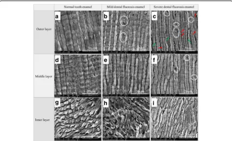
The microstructures of the three enamel layers of the normal tooth, mild dental fluorosis, and severe dental fluorosis are shown in Fig.2. The outer and middle en-amel rods of the normal teeth exhibited uniform diame-ters and were arranged in an upright manner (Fig.2a,d),
increasing crystal clearance and micropores (red arrow in Fig. 2c). A small number of pores (white circles in Fig. 2f) were also found in the middle layer. The struc-ture of the inner enamel of severe dental fluorosis was similar to that of the normal teeth (Fig. 2i). Compared with those of the normal teeth, the microstructures of the outer enamel of mild dental fluorosis and the outer and middle enamel of severe dental fluorosis showed marked differences, which could be attributed to two factors [28–31]. One factor is the interference of exces-sive fluoride intake during the normal formation of tooth enamel at puberty. This process results in excessive matrix protein retention, enamel rod hypomineralization, and a loose crystalline arrangement of the enamel rods [28–30]. The other factor is the chemical change in hydroxyapatite crystals caused by excessive fluoride intake. Fluoride apatite is formed when fluoride element displaces the hy-droxyl in hydroxyapatite crystals [31].
dental fluorosis enamel increased from the outer to the middle layers and then decreased from the middle to the inner layers (Fig. 3). The changed displacement (7.70 ± 2.71 nm) of the outer layer of the mild dental fluorosis enamel was significantly larger than that of the normal tooth enamel (p< 0.05), and the changed displacement decreased from the outer to the middle layers and then slightly increased from the middle to the inner layers
(Fig.4). For the severe dental fluorosis enamel, the nano-hardness and elastic modulus increased from the outer to the inner layers. The nanohardness (2.04 ± 0.89 GPa) and elastic modulus (46.63 ± 11.19 GPa) of the outer layer of the severe dental fluorosis enamel were lower than those of its middle layer, and the inner layer showed the highest values among those layers (p< 0.05) (Fig. 3). The changed displacement of severe dental
Fig. 3Nanomechanical properties of the enamel layers of the normal tooth, mild dental fluorosis, and severe dental fluorosis.aNanohardness. bElastic modulus. Identical symbols denote no significant difference in nanohardnesses and elastic moduli among the corresponding layers of the normal tooth enamel, mild dental fluorosis enamel, and severe dental fluorosis enamel
[image:4.595.58.537.87.379.2] [image:4.595.58.540.554.695.2]fluorosis enamel decreased from the outer to the inner layers, and the changed displacement (11.50 ± 3.77 nm) of the outer layer was larger than that of the middle layer (8.79 ± 2.24 nm). Among the layers, the inner layer showed the lowest displacement (p< 0.05) (Fig.4).
The friction coefficients of the three enamel layers of the normal tooth, mild dental fluorosis, and severe den-tal fluorosis are shown in Fig. 5. The friction coefficient of the normal tooth enamel increased from the outer to the inner layers. In mild dental fluorosis enamel, the Fig. 4Nanoindentation creep behavior of the enamel layers of the normal tooth, mild dental fluorosis, and severe dental fluorosis. Identical symbols denote no significant difference in nanoindentation creep behavior among the corresponding layers of the normal tooth enamel, mild dental fluorosis enamel, and severe dental fluorosis enamel
[image:5.595.58.537.88.336.2] [image:5.595.57.538.472.713.2]friction coefficient decreased from the outer to the mid-dle layers and then increased from the midmid-dle to the inner layers. In severe dental fluorosis enamel, the fric-tion coefficients of the outer (0.25 ± 0.044) and middle (0.18 ± 0.025) layers were significantly larger than those of the mild dental fluorosis enamel and normal tooth enamel (p< 0.05). In addition, the friction coefficient of the severe dental fluorosis enamel decreased from the outer to the inner layers (p< 0.05).
The nanoscratch depths and widths of the three en-amel layers of the normal tooth, mild dental fluorosis, and severe dental fluorosis are shown in Fig. 6. Normal tooth enamel showed a nanoscratch depth and width that increased from the outer to the inner layers (Fig.6a), While the mild dental fluorosis enamel revealed a nano-scratch depth and width that decreased from the outer to the middle layers and then increased from the middle to the inner layers (Fig. 6b). Variations in the nano-scratch depth and width of the severe dental fluorosis enamel were significantly different from those of the normal tooth enamel. Specifically, nanoscratch depths and widths decreased from the outer to the inner layers of severe dental fluorosis enamel (Fig.6c).
The wear resistance of the normal tooth enamel de-creased from the outer to the inner layers, and this
behavior corresponds with that observed in previous studies [42–44]. Excess fluoride can form fluoride-like deposits on the enamel surface and reduce wear resist-ance [3,45,46]. In this study, the wear resistances of the outer and middle layers of severe dental fluorosis enamel and the outer layer of mild dental fluorosis enamel were remarkably lower than those of the normal tooth en-amel. The inter-rod enamel contains more protein than the enamel rod, acts as a buffer layer that absorbs and disperses the pressure on the tooth, and affects the wear resistance of the tooth enamel [43]. Excessive fluoride intake leads to hypomineralized enamel rod formation and excessive matrix protein retention in the inter-rod enamel of dental fluorosis [28–31], both of which dra-matically affect the wear resistance of the dental fluor-osis enamel.
An understanding of the nanomechanical and nanotri-bological properties of different layers of dental fluorosis is an important contribution of this investigation, as knowledge of such properties can help guide the selec-tion of the appropriate restorative materials to use in clinical practice and promote the development of dental restorative materials. Dental fluorosis enamel presents a distinct gradient nanomechanical behavior that differs from that of the normal tooth enamel. Therefore, the
[image:6.595.57.539.416.701.2]layers, whereas the changed displacement decreased from the outer to the inner layers. In-depth analysis was subsequently performed to address the large standard deviation of the nanohardness and elastic moduli ob-served in severe dental fluorosis enamel. The outer and middle enamel layers of severe dental fluorosis can be divided into two types according to the features of their enamel rods, namely, normal and abnormal enamel rods (Fig.7). Certain enamel rods (i.e., normal enamel rods in severe dental fluorosis) appear complete but exhibit loosely arranged crystal structures and numerous micro-pores (Fig.7). Another portion of the enamel rods (i.e., the abnormal enamel rods in severe dental fluorosis) is characterized by numerous pores (white circles in Fig.7). In this study, the outer and middle layers of se-vere dental fluorosis enamel presented lower nanohard-ness and elastic moduli and higher creep deformation than the corresponding layers of the natural tooth en-amel, especially in the outer layer. In the outer layer of severe dental fluorosis enamel, normal and abnormal enamel rods showed low nanohardness and elastic moduli and high changed displacement; by contrast, the corresponding features in the abnormal enamel rods were greater (Fig. 8). Studies have suggested that
Appropriate Dental Materials for the Clinical Restoration of Dental Fluorosis
The nanoscratch depths and widths of the outer layers of the normal tooth, mild dental fluorosis, and severe dental fluorosis were compared with those of four re-storative materials (Fig.9). While IPS e.max CAD pre-sented the lowest nanoscratch depth and width, Vita Enamic, polymer-infiltrated-ceramic network (PICN), revealed a nanoscratch depth and width similar to those of the outer layer of the normal tooth enamel. The nanoscratch depth and width of the composite resin block Lava™ultimate (LUV) were similar to those of the outer layer of the mild dental fluorosis enamel, while the nanoscratch depth and width of the conventional com-posite resin Fltek™Z350XT (Z350) were higher than those of the outer layer of the mild dental fluorosis enamel. Among the samples tested, the outer layer of severe dental fluorosis enamel presented the largest nanoscratch depth and width.
Dental fluorosis in the anterior teeth affects the tooth appearance, and severe dental fluorosis with tooth de-fects in posterior teeth negatively influences mastication [5]. Restorations, such as crowns, inlays, or onlays, are often required to restore the teeth damaged by dental
[image:7.595.57.540.504.693.2]fluorosis [6, 7]. Matching of the mechanical behavior of the restorative material to that of the opposing tooth en-amel is especially important to prevent excessive wear of the natural tooth enamel or the applied material itself [8–11]. Ceramics are widely used as restorative mate-rials because of their high biocompatibility and similar esthetics to the natural tooth enamel [47]. However, ceramics present high wear resistance, which causes excessive wear of the opposing natural tooth enamel [47, 48]. Materials with low wear resistance, such as PICN and composite resin block, have been developed as alternatives to ceramics [48, 49]. PICN exhibits a wear resistance similar to that of the outer layer of the normal tooth enamel. Thus, when the opposing tooth is a normal tooth, PICN is the proper material for restor-ation. However, the opposing tooth in dental fluorosis re-quiring restoration likely presents mild dental fluorosis. In this case, materials with nanotribological properties simi-lar to those of the mild dental fluorosis enamel are neces-sary to restore dental fluorosis. Conventional composite resins, such as Z350, reveal a wear resistance lower than that of the outer layer of mild dental fluorosis; such a characteristic may lead to increased wear of the restorative materials. Composite resin block, such as LUV, is fabri-cated under high temperatures and high pressures and possesses mechanical properties superior to those of the conventional composite resins [50]. In the present study,
composite resin block showed a wear resistance similar to that of the outer layer of the mild dental fluorosis enamel. This characteristic implies that this material is appropriate for use as restorative material for dental fluorosis. As the nanomechanical behavior of dental fluorosis enamel deter-mines the selection of the restorative material, the appro-priate material should be applied for dental fluorosis to achieve better clinical outcomes. Thus, additional studies on the nanomechanical behavior of dental fluorosis en-amel should be conducted, and novel restorative materials should be further developed.
Conclusion
Based on the results of our analysis, the following con-clusions can be drawn:
1. The microstructure and gradient nanomechanical behavior of dental fluorosis enamel were
drastically different from those of the normal tooth enamel. The differences were observed in the outer layer of mild dental fluorosis enamel and the outer and middle layers of severe dental fluorosis enamel.
2. Normal and abnormal enamel rods could be observed in dental fluorosis enamel. In particular, the microstructures of abnormal enamel rods in dental fluorosis enamel
[image:8.595.58.537.88.357.2]drastically differed from those of the normal enamel rods. Specifically, abnormal enamel rods displayed lower nanohardness and elastic modulus but higher creep deformation than those of the normal enamel rods.
3. The wear resistance of the composite resin block was similar to that of the outer layer of mild dental fluorosis enamel. Thus, compared with ceramics, composite resin block is a more appropriate restorative material for dental fluorosis.
Abbreviations
EDJ:Enamel-dentin junction; IPS: IPS e.max CAD; LVU: Lava™ultimate; MFE: Mild dental fluorosis enamel; NTE: Normal tooth enamel; PICN: Polymer-infiltrated-ceramic network; SEM: Scanning electron microscopy; SFE: Severe dental fluorosis enamel; SPM: Scanning probe microscope; Z350: Fltek™Z350XT
Acknowledgements
The authors acknowledge Mr. Chaoliang Zhang for the technical support of SEM and People’s Hospital of Gulin County for assisting in the sample collection.
Funding
This work was supported by the Sichuan Province Science and Technology Support Program (0040305302015).
Availability of Data and Materials
The datasets supporting the conclusions of this article are included within the article.
Authors’Contributions
JM, HYY, and SSG conceived and designed the study. JM prepared the manuscript and performed the experiments. JM, PY, ZX, ZL, and QQZ analyzed the data. SSG reviewed and edited the manuscript. All authors read and approved the final manuscript.
Competing Interests
The authors declare that they have no competing interests.
Publisher’s Note
Springer Nature remains neutral with regard to jurisdictional claims in published maps and institutional affiliations.
Received: 7 June 2018 Accepted: 18 October 2018
References
1. Mochizuki K, Fujii H, Mizuguchi Y et al (2006) Tetrcycline-tooth interaction: an elemental analysis from prenatal period to early childhood. Pediatr. Dent. J. 16(1):43–49
2. Carvalho CAP, Nicodemo CAZ, Mercadante DCF et al (2013) Dental fluorosis in the primary dentition and intake of manufactured soy-based foods with fluoride. Clin Nutr 32:432–437
3. Jeng YR, Lin TT, Shieh DB (2009) Nanotribological characterization of tooth enamel rod affected by surface treatment. J Biomech 42:2249–2254 4. Chen HF, Yan M, Yang XF et al (2012) Spatial distribution and temporal
variation of high fluoride contents in groundwater and prevalence of fluorosis in humans in Yuanmou County, Southwest China. J Hazard Mater 235-236:201–209
5. Chankanka O, Levy SM, Warren JJ et al (2010) A literature review of aesthetic perceptions of dental fluorosis and relationships with psychosocial aspects/oral health-related quality of life. Community Dent Oral Epidemiol 38(2):97–109
[image:9.595.58.537.86.350.2]6. Petridis HP, Zekeridou A, Malliari M et al (2012) Survival of ceramic veneers made of different materials after a minimum follow-up period of five years: a systematic review and meta-analysis. Eur J Esthet Dent 7(2):138–152 7. Mazurek K, Mierzwińska-Nastalska E, Molak R et al (2012) Strength and
thickness of the layer of materials used for ceramic veneers bonding. Acta Bioeng Biomech 14(3):75–78
8. Wang L, Liu YH, Si WJ et al (2012) Friction and wear behaviors of dental ceramics against natural tooth enamel. J Eur Ceram Soc 32:2599–2606 9. Wiley MG (1989) Effects of porcelain on occluding surfaces of restored
teeth. J Prosthet Dent 61:133–137
10. Chen H, Tang Z, Liu J et al (2006) Acellular synthesis of a human enamel-like microstructure. Adv Mater 18:1846–1851
11. Zhang JL, Jiang DL, Zhang JX et al (2010) Synthesis of dental enamel-like hydroxyapaptite through a solution mediated solid-state conversion. Langmuir 26(5):2989–2994
12. Habelitz S, Marshall SJ, Marshall GW Jr et al (2001) Mechanical properties of human dental enamel on the nanometre scale. Arch Oral Biol 46:173–183 13. Elfrink MEC, Cate JM, Ruijven LJ et al (2013) Mineral content in teeth with
deciduous molar hypomineralisation (DMH). J Dent 41:974–978 14. Kirkham J, Robinson C, Strafford SM (2000) The chemical composition of
tooth enamel in junctional epidermolysis bullosa. Arch Oral Biol 45:377–386 15. He LH, Yin ZH, Vuuren LJ et al (2013) A natural functionally graded
biocomposite coating-human enamel. Acta Biomater 9:6330–6337 16. Park S, Quinn JB, Romberg E et al (2008) On the brittleness of enamel and
selected dental materials. Dent Mater 24:1477–1485
17. Barani A, Bush MB, Lawn BR (2012) Effect of property gradients on enamel fracture in human molar teeth. J Mech Behav Biomed 15:121–130 18. An B, Wang R, Arola D et al (2012) The role of property gradients on the
mechanical behavior of human enamel. J Mech Behav Biomed 9:63–72 19. Colaco MV, Barroso RC, Porto IM et al (2012) Synchrotron X-ray diffraction
characterization of healthy and fluorotic human dental enamel. Radiat Phys Chem 81:1578–1585
20. Al-Jawada M, Steuwerb A, Kilcoynec SH et al (2007) 2D mapping of texture and lattice parameters of dental enamel. Biomaterials 28:2908–2914 21. DenBesten PK, Yan Y, Featherstone JDB et al (2002) Effects of fluoride on rat
dental enamel matrix proteinases. Arch Oral Biol 47:763–770
22. Shearer TR, Britton JL, Desart DJ et al (1980) Microhardness of molar teeth in cattle with fluorosis. Am J Veterinary Res 41(9):1543–1545
23. Suckling G, Thurley DC, Nelson DG (1988) The macroscopic and scanning electron-microscopic appearance and microhardness of the enamel, and the related histological changes in the enamel organ of erupting sheep incisors resulting from prolonged low daily dose of fluoride. Arch Oral Biol 33(5):361–373
24. Fan H, Gao S, Liu Y et al (2014) The micromechanical and tribological feature of mild mottled enamel. J Mech Med Biol 14(4):1450050–1–14 25. Zheng Q, Xu H, Song F et al (2013) Spatial distribution of the human
enamel fracture toughness with aging. J Mech Behav Biomed 26:148–154 26. Oliver WC, Pharr GM (1992) An improved technique for determining
hardness and elastic modulus using load and displacement sensing indentation experiments. J Mater Res 7:1564–1583
27. He LH, Swain MV (2007) Nanoindentation derived stress–strain properties of dental materials. Dent Mater 23:814–821
28. Robinson C, Connell S, Kirkham J et al (2004) The effect of fluoride on the developing tooth. Caries Res 38(3):268–276
29. Fejerskov O, Larsen MJ, Richards A et al (1994) Dental tissue effects of fluoride. Adv Dent Res 8(1):15–31
30. Fejerskov O, Yanagisawa T, Tohda H et al (1991) Posteruptive changes in human dental fluorosis-a histological and ultrastructural study. Proc Finn Dent Soc 87(4):607–619
31. Aoba T (1997) The effect of fluoride on apatite structure and growth. Crit Rev Oral Biol Med 8(2):136–153
32. Braly A, Darnell LA, Mann AB et al (2007) The effect of prism orientation on the indentation testing of human molar enamel. Arch Oral Bio 52:856–860 33. Ge J, Cui FZ, Wang XM et al (2005) Property variations in the prism and the
organic sheath within enamel by nanoindentation. Biomaterials 26:3333–3339 34. IL J¨g (2005) Viscoelastic behavior of organic materials: consequences of a
logarithmic dependence of force on strain rate. J Biomech 38:1451–1458 35. Bechtle S, Ang SF, Schneider GA (2010) On the mechanical properties of hierarchically structured biological materials. Biomaterials 31:6378–6385
36. Yahyazadehfar M, Bajaj D, Arola D (2013) Hidden contributions of the enamel rods on the fracture resistance of human teeth. Acta Biomater 9: 4806–4814
37. Yahyazadehfar M, Arola D (2015) The role of organic proteins on the crack growth resistance of human enamel. Acta Biomater 19:33–45
38. Ji B, Gao H (2004) Mechanical properties of nanostructure of biological materials. J Mech Phys Solids 52:1963–1990
39. He LH, Swain MV (2007) Enamel-A“metallic-like”deformable biocomposite. J Dent 35:431–437
40. Bajaj D, Arola D (2009) Role of prism decussation on fatigue crack growth and fracture of human enamel. Acta Biomater 5:3045–3056
41. Bronckers AL, Lyaruu DM, Bervoets TJ et al (2002) Fluoride enhances intracellular degradation of amelogenins during secretory phase of amelogenesis of hamster teeth in organ culture. Connect Tissue Res 43(2–3):456–465
42. Zheng J, Li Y, Shi MY et al (2013) Microtribological behaviour of human tooth enamel and artificial hydroxyapatite. Tribol Int 63:177–185 43. Zheng SY, Zheng J, Gao SS et al (2011) Investigation on the
microtribological behaviour of human tooth enamel by nanoscratch. Wear 271:2290–2296
44. Jeng YR, Lin TT, Hsu HM et al (2011) Human enamel rod presents anisotropic nanotribological properties. J Mech Behav Biomed 4:515–522 45. Garrido MA, Ceballos IGL, Río MTG et al (2011) Nanotribological behaviour
of tooth enamel rod affected by bleaching treatment. Wear 271:2334–2339 46. Gao SS, Huang SB, Qian LM ea (2009) Wear behavior of early carious
enamel before and after remineralization. Wear 267:726–733
47. Goujat A, Abouelleil MH, Colon P et al (2018) Mechanical properties and internal fit of 4 CAD-CAM block materials. J Prosthet Dent 119:384–389 48. Coldea A, Swain MV, Thiel N (2013) In-vitro strength degradation of dental
ceramics and novel PICN material by sharp indentation. J Mech Behav Biomed Mater 26:34–42
49. Min J, Arola D, Yu DD et al (2016) Comparison of human enamel and polymer-infiltrated-ceramic-network material“ENAMIC”through micro- and nano-mechanical testing. Ceram Int 42:10631–10637