0095-1137/06/$08.00
⫹
0
doi:10.1128/JCM.44.3.970–975.2006
Copyright © 2006, American Society for Microbiology. All Rights Reserved.
Rapid Screening of Fluoroquinolone Resistance Determinants in
Streptococcus pneumoniae
by PCR-Restriction Fragment Length
Polymorphism and Single-Strand Conformational Polymorphism
Margaret Ip,* Shirley S. L. Chau, Fang Chi, Amy Qi, and Raymond W. M. Lai
Department of Microbiology, The Chinese University of Hong Kong, Prince of Wales Hospital, Shatin, Hong Kong
Received 10 November 2005/Returned for modification 23 December 2005/Accepted 6 January 2006
A rapid method, using PCR-restriction fragment length and single-strand conformation polymorphism
(SSCP), was applied to screen for mutations of the fluoroquinolone resistance determinants in
Streptococcus
pneumoniae
. One hundred nonduplicate
Streptococcus pneumoniae
isolates with ciprofloxacin MICs of
>
4.0
g/ml from the Prince of Wales Hospital, Hong Kong, years 2000 to 2003, were examined. For each isolate, PCR
amplicons of quinolone resistance-determining regions (QRDRs) of
gyrA
,
gyrB
,
parC
, and
parE
genes were
digested with AluI, HinfI, Sau3AI, and MspI, respectively, and analyzed by SSCP. Each SSCP pattern was
given a number, and each isolate obtained a four-digit code, e.g., 1111, that represented the SSCP profile. The
SSCP patterns were correlated to mutations characterized from sequence analyses of PCR amplicons. The
most common SSCP profile obtained was no. 5232 (40%), which included strains with two amino acid
substitutions in the ParC (Lys-137-Asn) and ParE (Ile-460-Val) genes, followed by the SSCP profile 5223
(17%), which included strains with amino acid substitutions in the ParE (Ile-460-Val) gene only. Ten isolates
(10%) with amino acid substitutions at GyrA and ParE (
ⴞ
ParC) genes were resistant to levofloxacin with a
MIC of
>
16
g/ml. Other SSCP profiles were unique in distinguishing the common amino acid substitutions
in GyrA (Ser-81-Phe) and ParC (Lys-137-Asn, Ser-79-Phe plus Lys-137-Asn, Asp-83-Asn plus Lys-137-Asn,
Ser-79-Phe, and Glu-96-Asp). SSCP analysis of restricted fragments generated patterns that were highly
discriminative for mutations present in the QRDRs of
gyrA
,
gyrB
,
parC
, and
parE
. This method provides a
database of high resolution profiles on these mutations and allows rapid screening for new mutations of the
fluoroquinolone resistance genes.
Antibiotic-resistant
Streptococcus pneumoniae
has evolved to
be a worldwide problem in the last decade. Fluoroquinolones
are an important class of antibiotics, and agents such as
levo-floxacin or moxilevo-floxacin are incorporated in guidelines for the
empirical treatment of community-acquired pneumonia (17,
18). The prevalence of levofloxacin nonsusceptibility (MIC of
ⱖ
4.0
g/ml) varies by region and has remained low, ranging
from 0% in some European countries (24) to 1.3% in the
United States (25). Higher rates have been documented in
some countries, e.g., South Korea (2.9%) (4), and in Hong
Kong, a rate of 13% has been reported in 2001 (9) and the
spread hypothesized to be a result of clonal dissemination of
the Spanish 23F strain of
S. pneumoniae
(10). However, there
is increasing evidence that interspecies transfer of the
parE
-parC
gene region arising from other viridans group
strepto-cocci occurs (7, 28). It is thus important to identify and monitor
the spread of fluoroquinolone resistance determinants among
S. pneumoniae
strains.
Fluoroquinolone resistance in
S. pneumoniae
is primarily
due to mutations in the quinolone resistance-determining
re-gions (QRDRs) of the genes encoding the A and B subunits of
DNA gyrase and topoisomerase IV, in particular, the
parC
and
gyrA
genes (3, 6, 14). Often, the level of resistance increases as
more amino acids are substituted for by additional mutations.
Mutations in
parE
and
gyrB
have also been reported, but to a
lesser extent (2, 23, 30). Reduced susceptibility to
fluoroquin-olone may also be due to altered accumulation of the drug or
efflux, but this plays a less significant role (31).
Screening for mutations in the QRDRs is performed by
sequencing of the
gyrA
,
gyrB
,
parC
, and
parE
genes of the
fluoroquinolone-resistant strains. Methods for detection of
known mutations in the QRDRs have included
PCR-restric-tion fragment length polymorphism (PCR-RF) (1),
oligonucle-otide probe assay (6), TaqMan assay (8), and single-strand
conformational polymorphism (SSCP) (27). Often, these
methods are not designed to have, or do not have, a sufficiently
high sensitivity or resolution to identify new mutations. Tawata
et al. (29) introduced a modified method of using a
combina-tion of PCR-restriccombina-tion fragment length polymorphism and
[image:1.585.302.541.613.714.2]* Corresponding author. Mailing address: Dept. of Microbiology,
Chinese University of Hong Kong, The Prince of Wales Hospital,
Shatin, Hong Kong. Phone: (852) 2632 1265. Fax: (852) 2647 3227.
E-mail: [email protected].
TABLE 1. PCR primers for QRDRs of the fluoroquinolone
resistance genes
Gene Primer (5⬘to 3⬘)a Amplicon
size (bp) Reference
gyrA
CCG TCG CAT TCT TTA CG
382
22
AGT TGC TCC ATT AAC CA
gyrB
TTC TCC GAT TTC CTC ATG
457
19
AGA AGG GTA CGA ATG TGG
parC
TGG GTT GAA GCC GGT TCA
366
20
TGC TGG CAA GAC CGT TGG
parE
AAG GCG CGT GAT GAG AGC
289
22
TCT GCT CCA ACA CCC GCA
aFor each gene, the first primer listed is forward, and the second is reverse.
970
on May 16, 2020 by guest
http://jcm.asm.org/
single-strand conformational polymorphism (PCR-RF–SSCP)
as a tool for mass screening of the genome. We thus sought to
identify and monitor the spread of fluoroquinolone resistance
determinants in
S. pneumoniae
using a modified method of
SSCP to examine and screen for new mutations at the QRDRs
of the respective fluoroquinolone resistance genes in
S.
pneu-moniae.
MATERIALS AND METHODS
[image:2.585.48.540.71.376.2]Bacterial isolates and susceptibility tests.One thousand eighty-one nondu-plicateS. pneumoniaeisolates from patients admitted to the Prince of Wales Hospital, a 1,350-bed teaching and tertiary hospital in Hong Kong, from 2000 to 2003 were screened for fluoroquinolone nonsusceptibility. The MICs of cipro-floxacin, levocipro-floxacin, moxicipro-floxacin, and gatifloxacin were determined by the microdilution broth method as described by NCCLS (21). One hundred isolates that were nonsusceptible to ciprofloxacin (CIP) with MICs ofⱖ4g/ml were
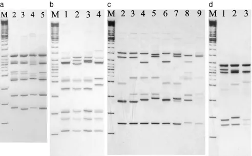
FIG. 1. SSCP patterns of DNA amplicons of
gyrA
(a),
gyrB
(b),
parC
(c), and
parE
(d) after restriction digestion with AluI, HinfI, Sau3AI, and
MspI, respectively. (a) SSCP patterns 2 to 5 of the
gyrA
gene after digestion with AluI restriction enzyme. Lane M represents a 100-bp DNA ladder.
(b) SSCP patterns 1 to 4 of the
gyrB
gene after restriction digestion using HinfI. Lane M represents a 100-bp DNA ladder. (c) SSCP patterns 2
to 9 of the
parC
gene after restriction digestion using Sau3AI. Lane M represents a 100-bp DNA ladder. (d) SSCP patterns 1 to 3 of the
parE
gene
after restriction digestion using MspI. Lane M represents a 100-bp DNA ladder.
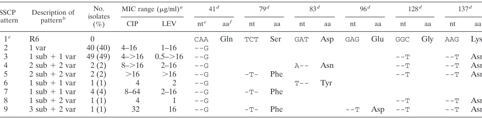
TABLE 2. Correlation of SSCP patterns with nucleotide sequences and amino acid substitutions in
gyrA
gene,
compared to that of
S. pneumoniae
R6
SSCP pattern
Description of
patternb No. isolates
(%)
MIC range (g/ml)a 75d 81d 83d
CIP LEV nte
aaf
nt aa nt aa
1
cR6
0
TAC
Tyr
TCC
Ser
ATT
Ile
2
1 sub
⫹
1 var
9 (9)
16–64
⬎
16
--T
-T-
Phe
3
1 sub
1 (1)
8
16
-T-
Phe
4
1 var
1 (1)
4
2
--C
5
1 var
89 (89)
4–
⬎
16
0.5–16
--T
aCIP, ciprofloxacin; LEV, levofloxacin.
bsub, no. of amino acid substitution; var, no. of base pair variation compared to R6.
cS. pneumoniaeR6 strain.
dNumeral indicates amino acid position. nt, nucleotide; aa, amino acid. eNucleotide sequence changes relative toS. pneumoniaeR6. fAmino acid substitution corresponding to nt change.
on May 16, 2020 by guest
http://jcm.asm.org/
[image:2.585.43.540.595.676.2]further examined on the mutations and amino acid substitutions at the QRDRs of the respective fluoroquinolone resistance genes using PCR-RF–SSCP.
Analysis by PCR-RF–SSCP.Chromosomal DNA from the isolates was ob-tained by melting a small piece of DNA plug in 150l double-distilled H2O at
65°C. DNA plug was prepared for pulsed-field gel electrophoresis according to the method previously described (11, 12). For each isolate, the QRDRs ofgyrA,
gyrB,parC, andparEgenes were amplified by PCR using the primers listed in Table 1 and conditions as previously described (19, 20, 22). For the restriction digestion, the PCR amplicons ofgyrA,gyrB,parC, andparEgenes were digested with AluI, HinfI, Sau3AI, and MspI enzymes (Amersham Biosciences), respec-tively, each in a 10-l reaction mixture containing reaction buffer and incubated at 37°C overnight as recommended by the manufacturer. For example, AluI was the restriction enzyme used for the digestion of the PCR amplicons of thegyrA
gene, HinfI forgyrB, and so forth. A quantity of 3.5l (approximately 100 to 150 ng) of the digested product was mixed with an equal volume of denaturing solution (94% formamide, 0.05% xylene cyanol solution, 0.4 mg/ml bromophenol blue) according to the manufacturer’s instructions (ExcelGel DNA Analysis Kit; Amersham Biosciences), the mixture was denatured at 95°C for 8 min in a thermocycler, and the mixture was placed on ice immediately. The denatured mixture was electrophoresed on precast gels for SSCP (ExcelGel DNA Analysis Kit;, Amersham Biosciences) at 600 V, with a current of 50 mA and 30 W power for 90 min at 4°C using the Multiphor II electrophoresis unit (Amersham Bio-sciences). The precast gels were 12.5% acrylamide gels and contained 48 wells for sample loading. The DNA gel was stained by silver stain according to the manufacturer’s instructions (DNA silver staining kit; Amersham Biosciences) to visualize and permanently stain the discrete DNA bands.
Analysis of SSCP patterns.Each SSCP pattern was given a number and each isolate obtained a four-digit code that represented the SSCP profile. For exam-ple, 1234 represented the SSCP profile of pattern 1 forgyrA, pattern 2 forgyrB, pattern 3 forparC, and pattern 4 for theparEgene. The SSCP patterns were
correlated to the mutations characterized from the sequence analyses of the respective PCR amplicons, and any amino acid substitutions were noted. At least three pairs of forward and reverse sequences of the PCR amplicons, if available, for each corresponding SSCP pattern were sequenced for confirmation. Se-quencing was performed with an ABI 310 sequencer and an ABI Prism dRhodamine terminator cycle sequencing kit (Applied Biosystems, Foster City, Calif.). The sequences were aligned and compared with the corresponding QRDR regions of the four fluoroquinolone resistance genes of theS.
pneu-moniaeR6 strain from the GenBank.
RESULTS
The different SSCP patterns obtained from PCR-RF–SSCP
of the QRDRs of the
gyrA
,
gyrB
,
parC
, and
parE
genes are
shown in Fig. 1a to d, respectively. Four SSCP patterns were
observed for
gyrA
, four patterns for
gyrB
, eight for
parC
, and
three for
parE
. The corresponding nucleotide change and
amino acid substitution for each of the SSCP patterns are
shown in Tables 2 to 5. The sequencing results confirmed that
the same SSCP pattern represented the same sequence of the
QRDR of the PCR amplicons of the specific
fluoroquinolone-resistant genes.
Four SSCP patterns were obtained for
gyrA
(Fig. 1a). The
majority of isolates (90%) belonged to pattern 5 and were
found to have one base pair difference from
S. pneumoniae
R6
[image:3.585.45.543.89.165.2](Table 2). Only one amino acid substitution (Ser-81-Phe) was
TABLE 4. Correlation of SSCP patterns with nucleotide sequences and amino acid substitutions in
parC
compared to that of
S. pneumoniae
R6
SSCP pattern
Description of patternb
No. isolates
(%)
MIC range (g/ml)a 41d 79d 83d 96d 128d 137d
CIP LEV nte
aaf
nt aa nt aa nt aa nt aa nt aa
1
cR6
0
CAA
Gln
TCT
Ser
GAT
Asp
GAG
Glu
GGC
Gly
AAG
Lys
2
1 var
40 (40)
4–16
1–16
--G
3
1 sub
⫹
1 var
49 (49)
4–
⬎
16
0.5–
⬎
16
--G
--T
--T
Asn
4
2 sub
⫹
2 var
2 (2)
8–
⬎
16
2–16
--G
A--
Asn
--T
--T
Asn
5
2 sub
⫹
2 var
2 (2)
⬎
16
⬎
16
--G
-T-
Phe
--T
--T
Asn
6
1 sub
⫹
1 var
1 (1)
4
2
--G
T--
Tyr
7
1 sub
⫹
1 var
4 (4)
8–64
2–16
--G
-T-
Phe
8
1 sub
⫹
2 var
1 (1)
4
1
--G
--T
--T
Asn
9
3 sub
⫹
2 var
1 (1)
32
16
--G
-T-
Phe
--T
Asp
--T
--T
Asn
aCIP, ciprofloxacin; LEV, levofloxacin.
bsub, no. of amino acid substitution; var, no. of base pair variation compared to R6.
cS. pneumoniaeR6 strain.
dNumeral indicates amino acid position. nt, nucleotide; aa, amino acid. eNucleotide sequence change relative toS. pneumoniaeR6.
fAmino acid substitution corresponding to nt change.
TABLE 3. Correlation of SSCP patterns with nucleotide sequences and amino acid substitutions in
gyrB
compared to that of
S. pneumoniae
R6
SSCP pattern
Description of patternb
No. isolates
(%)
MIC range (g/ml)a 381d 384d 386d 461d 472d
CIP LEV nte
aaf
nt aa nt aa nt aa nt aa
1
cR6
14 (14)
4
1–2
GTA
Vla
GGA
Gly
TTG
Leu
AAC
Asn
GCT
Ala
2
3 var
80 (80)
4–64
0.5–
⬎
16
--G
--G
--A
3
2 var
5 (5)
4–16
1–4
--G
--A
4
2 var
1 (1)
4
2
--T
--C
a
CIP, ciprofloxacin; LEV, levofloxacin.
b
sub, no. of amino acid substitution; var, no. of base pair variation compared to R6.
c
S. pneumoniaeR6 strain.
d
Numeral indicates amino acid position. nt, nucleotide; aa, amino acid.
e
Nucleotide sequence changes relative toS. pneumoniaeR6.
f
Amino acid substitution corresponding to nt change.
on May 16, 2020 by guest
http://jcm.asm.org/
[image:3.585.47.538.555.676.2]found, represented in both SSCP patterns 2 and 3, and was
present in 10 (10%) of the isolates. The CIP and levofloxacin
(LEV) MICs of these isolates were
ⱖ
8
g/ml and
ⱖ
16
g/ml,
respectively. Four SSCP patterns were obtained for
gyrB
(Fig.
1b), and these included a number of silent mutations, but there
was no amino acid substitution. Fourteen (14%) of the
se-quences were identical to that of the wild-type strain, R6, while
86% had two or three nucleotide changes (patterns 2 to 4)
(Table 3). Nine SSCP patterns were obtained for
parC
(Fig.
1c). SSCP pattern 2 included 40 (40%) isolates that differed
from the R6 strain by a base pair, while the other 60 (60%)
isolates had one, two, or three amino acid substitutions, as
shown in SSCP patterns 3 to 9. These amino acid substitutions
included Ser-79-Phe, Asp-83-Asn/Tyr, Glu-96-Asp, and
Lys-137-Asn. The most common substitution is represented by
SSCP pattern 3, with 49 (49%) of the isolates with an amino
acid substitution of Lys-137-Asn (Table 4). Ninety-six (96%)
isolates had an amino acid substitution of Ile-460-Val in
parE
,
as represented by SSCP patterns 3 and 4 (Table 5). These
isolates have various MICs ranging 4 to 64
g/ml and 0.5 to
ⱖ
16
g/ml for CIP and LEV, respectively.
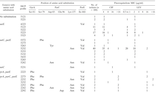
The correlation of the different SSCP profiles with the
amino acid substitutions at GyrA, ParC, and ParE and the
fluoroquinolone MICs are summarized in Table 6. The most
common SSCP profile was 5232 (40%), which included strains
with two amino acid substitutions in the ParC (Lys-137-Asn)
and ParE (Ile-460-Val) genes, followed by the SSCP profile of
5223 (17%), which included strains with amino acid
substitu-tions in the ParE (Ile-460-Val) gene only. Ten isolates (10%)
[image:4.585.39.542.90.154.2]with amino acid substitutions at the GyrA and ParE (
⫾
ParC)
TABLE 5. Correlation of SSCP patterns with nucleotide sequences and amino acid substitutions in
parE
compared to that of
S. pneumoniae
R6
SSPC pattern
Description of patternb
No. isolates (%)
MIC range (g/ml)a 460d 476d
CIP LEV nte
aaf
nt aa
1
cR6
4 (4)
4–16
1–2
ATC
Ile
ATC
Ile
2
1 sub
⫹
1 var
54 (54)
4–32
0.5–
⬎
16
G--
Val
--T
3
1 sub
42 (42)
4–64
1–16
G--
Val
a
CIP, ciprofloxacin; LEV, levofloxacin.
b
sub, no. of amino acid substitution; var, no. of base pair variation compared to R6.
c
S. pneumoniaeR6 strain.
d
Numeral indicates amino acid position. nt, nucleotide; aa, amino acid.
e
Nucleotide sequence change relative toS. pneumoniaeR6.
f
Amino acid substitution corresponding to nt change.
TABLE 6. Correlation of different SSCP profiles with amino acid substitutions of GyrA, ParC, and ParE and fluoroquinolone MICs
Gene(s) with amino acid substitution
SSCP profile
Position of amino acid substitution
No. of isolates (n
⫽100)
Fluoroquinolone MIC (g/ml)
GyrA ParC ParE CIP LEV
Ser-81- Ser-79- Asp-83- Glu-96- Lys-137- Ile-460- 4 8 16 ⬎16 0.5 to 1 2 4 8 16 ⬎16
No substitution
5121
1
1
1
5221
2
2
1
1
parE
4223
Val
1
1
1
5123
11
11
4
7
5222
3
3
1
2
5223
17
16
1
8
8
1
5323
4
3
1
1
2
1
parC
,
parE
5372
Phe
Val
1
1
1
5273
1
1
1
5263
Tyr
Val
1
1
1
5232
Asn
Val
40
35
4
1
20
18
2
5233
2
2
2
5132
2
2
2
5433
1
1
1
5283
1
1
1
5242
Asn
Asn
Val
1
1
1
parC
5231
Asn
1
1
1
gyrA
,
parE
2223
Phe
Val
1
1
1
gyrA
,
parC
,
parE
2273
Phe
Phe
Val
2
2
2
2232
Phe
Asn
Val
2
2
2
3232
1
1
1
2252
Phe
Phe
Asn
Val
2
2
1
1
2242
Phe
Asn
Asn
Val
1
1
1
2292
Phe
Asp
Asn
Val
1
1
1
on May 16, 2020 by guest
http://jcm.asm.org/
[image:4.585.45.544.426.724.2]genes are clearly resistant to levofloxacin with MICs of
ⱖ
16
g/ml.
DISCUSSION
PCR-RF–SSCP has been described as a useful tool for mass
screening for DNA polymorphism analysis on other genes (15,
26) and is able to detect a nucleotide substitution, deletion, or
insertion in up to a 22,000-base-pair amplicon (29). We applied
this method for the analysis of the QRDRs of the
gyrA
,
gyrB
,
parC
, and
parE
genes of
S. pneumoniae
. The restriction
diges-tion of the PCR amplicons generates a higher degree of
poly-morphism that could be readily detected by SSCP. Each
re-striction enzyme was chosen to cut the respective PCR
amplicons in regions to produce fragments in the range of 100
to 200 bp. Up to nine bands in the SSCP pattern were observed
by using this method. However, using PCR-SSCP alone, only
two to three bands were produced, and the method was unable
to discriminate single nucleotide changes (results not shown).
The SSCP patterns were able to detect a single base difference
(for example, patterns 3, 4, and 5 in
gyrA
); therefore, the
patterns produced from PCR-RF–SSCP were highly
discrimi-native for mutations present in
gyrA
,
gyrB
,
parC
, and
parE
.
Among the hundred isolates screened, the SSCP profiles
dis-tinguished the common amino acid substitutions in GyrA
(Ser-81-Phe), ParC (Ser-79-Phe, Asp-83-Asn/Tyr, and Lys-137-Asn)
and ParE (Ile-460-Val) (5, 6, 14). Of the 36 isolates that possessed
an amino acid substitution (Ile-460-Val) at ParE alone, 34 (94%)
strains had a CIP MIC of 4
g/ml and a LEV MIC of 0.5 to 2
g/ml, similar to isolates that had no amino acid substitution
detected, supporting that the ParE substitution did not contribute
to significant fluoroquinolone resistance (14). However, two
iso-lates had higher fluoroquinolone MICs (LEV MIC of 4 or 8
g/ml) that may be attributed to other mutations in regions that
were not studied here or some other mechanism, such as those
that alter the drug permeation.
ParC substitution has been reported as contributing to
low-level fluoroquinolone resistance (13, 16, 20). Although CIP
MICs may be increased, the LEV MIC generally remained
low, in the range of 0.5 to 2
g/ml. Many reports showed that
ParC and GyrA substitutions are responsible for high-level
fluoroquinolone resistance and ParC substitution was a
first-step prerequisite in the development of high-level resistance
(13, 16, 20). Our findings supported that all nine of our isolates
with ParC and GyrA substitutions had LEV MICs of
ⱖ
16
g/ml. These amino acid substitutions have previously been
described to be responsible for high levels of resistance (13, 16,
20). An exception, Glu-96-Asp, is an unusual substitution
found in ParC, and its importance remains to be determined.
Our data are similar to the previous findings from Hong
Kong in which Ser-81-Phe (9) was the substitution found in
GyrA that contributed to high-level resistance to
fluoroquino-lones. However, an additional number of new substitutions in
ParC (Asp-83-Tyr/Asn and Glu-96-Asp) were identified in the
present study.
PCR-RF–SSCP provides a database of high-resolution
pro-files on these mutations and allows rapid screening for any new
mutation. The analysis and comparison of the pattern with the
known sequences provide an alternative method for detecting
mutation in the gene studied other than direct nucleotide
se-quencing. The four-digit code that represents the SSCP profile
for an isolate also provides an objective way of recognizing the
QRDR mutations associated with that strain. If the SSCP
patterns for a fluoroquinolone-resistant gene, e.g.,
gyrA
, exceed
10, then the single-digit number could be replaced by a single
alphabet letter, e.g., A to indicate the eleventh pattern, B for
the twelfth pattern, etc. The method is sensitive and cheaper
and easier to perform compared to direct nucleotide
sequenc-ing of all sequences. In addition, this method is rapid, and up
to 48 samples could be run per gel. The method could readily
be adaptable to a clinical laboratory if the laboratory has a
PCR facility. The only additional requirement is an
electro-phoresis system for SSCP and the gels and reagents are readily
available commercially. It is discriminatory and could readily
be applied as a screening method to examine for known and
new mutations of the fluoroquinolone resistance genes.
ACKNOWLEDGMENT
The work described in this paper was supported by an earmarked
grant from the Research Grants Council of the Hong Kong Special
Administrative Region (Project No. CUHK 4432/03 M).
REFERENCES
1.Alonso, R., M. Galimand, and P. Courvalin.2004. An extended PCR-RFLP assay for detection ofparC,parEandgyrAmutations in fluoroquinolone-resistantStreptococcus pneumoniae. J. Antimicrob. Chemother.53:682–683. 2.Bast, D. J., D. E. Low, C. L. Duncan, L. Kilburn, L. A. Mandell, R. J. Davidson, J. C. de Azavedo, et al.2000. Fluoroquinolone resistance in clin-ical isolates ofStreptococcus pneumoniae: contributions of type II topoisom-erase mutations and efflux to levels of resistance. Antimicrob. Agents Che-mother.44:3049–3054.
3.Brueggemann, A. B., S. L. Coffman, P. Rhomberg, H. Huynh, L. Almer, A. Nilius, R. Flamm, and G. V. Doern.2002. Fluoroquinolone resistance in
Streptococcus pneumoniaein United States since 1994–1995. Antimicrob.
Agents Chemother.46:680–688.
4.Canton, R., M. Morosini, M. C. Enright, and I. Morrissey.2003. Worldwide incidence, molecular epidemiology and mutations implicated in fluoroquin-olone-resistantStreptococcus pneumoniae: data from the global PROTEKT surveillance programme. J. Antimicrob. Chemother.52:944–952. 5.Davies, T. A., A. Evangelista, S. Pfleger, K. Bush, D. F. Sahm, and R.
Goldschmidt.2002. Prevalence of single mutations in topoisomerase type II genes among levofloxacin-susceptible clinical strains ofStreptococcus
pneu-moniaeisolated in the United States in 1992 to 1996 and 1999 to 2000.
Antimicrob. Agents Chemother.46:119–124.
6.Davies, T. A., and R. Goldschmidt.2002. Screening of large numbers of
Streptococcus pneuomoniaeisolates for mutations associated with
fluoro-quinolone resistance using an oligonucleotide probe assay. FEMS Microbiol. Lett.17:219–224.
7.de la Campa, A. G., L. Balsalobre, C. Ardanuy, A. Fenoll, E. Perez-Trallero, J. Linares, et al. 2004. Fluoroquinolone resistance in penicillin-resistant
Streptococcus pneumoniaeclones, Spain. Emerg. Infect. Dis.10:1751–1759.
8.Giles, J., J. Hardick, J. Yuenger, M. Dan, K. Reich, and J. Zenilman.2004. Use of Applied Biosystems 7900HT sequence detection system and Taqman assay for detection of quinolone-resistantNeisseria gonorrhoae. J. Clin. Mi-crobiol.42:3281–3283.
9.Ho, P. L., R. W. Yung, D. N. Tsang, T. L. Que, M. Ho, W. H. Seto, T. K. Ng, W. C. Yam, and W. W. Ng.2001. Increasing resistance ofStreptococcus
pneumoniaeto fluoroquinolones: results of a Hong Kong multicentre study
in 2000. J. Antimicrob. Chemother.48:659–665.
10.Ho, P. L., T. L. Que, S. S. Chiu, R. W. H. Yung, T. K. Ng, D. N. C. Tsang, W. H. Seto, and Y. L. Lau.2004. Fluoroquinolone and other antimicrobial resistance in invasive pneumococci, Hong Kong, 1995–2001. Emerg. Infect. Dis.10:1250–1257.
11.Ip, M., D. J. Lyon, R. W. H. Yung, C. Chan, and A. F. Cheng.1999. Evidence of clonal dissemination of multidrug-resistantStreptococcus pneumoniaein Hong Kong. J. Clin. Microbiol.37:2834–2839.
12.Ip, M., D. J. Lyon, R. W. H. Yung, L. Tsang, and A. F. Cheng.2002. Introduction of new clones of penicillin-nonsusceptibleStreptococcus
pneu-moniaein Hong Kong. J. Clin. Microbiol.40:1522–1525.
13.Janoir, C., V. Zeller, M.-D. Kitzis, N. J. Moreau, and L. Gutmann.1996. High-level fluoroquinolone resistance inStreptococcus pneumoniaerequires mutations inparCandgyrA. Antimicrob. Agents Chemother.40:2760–2764. 14.Jones, M. E., D. F. Sahm, N. Martin, S. Scheuring, P. Heisig, C. Thorns-berry, K. Ko¨hrer, and F. J. Schmitz.2000. Prevalence ofgyrA,gyrB,parC,
on May 16, 2020 by guest
http://jcm.asm.org/
andparEmutations in clinical isolates ofStreptococcus pneumoniaewith decreased susceptibilities to different fluoroquinolones and originating from worldwide surveillance studies during the 1997–1998 respiratory season. Antimicrob. Agents Chemother.44:462–466.
15.Kurihara, A., M. Tawata, Y. Ikegishi, K. Aida, and T. Onaya.1999. The procedure of polymerase chain reaction-restriction fragment-single strand conformation polymorphism analysis by Hha I/Hinc II to detect mitochon-drial DNA mutations. Life Sci.64:1223–1230.
16.Lim, S., D. Bast, A. McGeer, J. de Azavedo, and D. E. Low.2003. Antimi-crobial susceptibility breakpoints and first-stepparCmutations in
Strepto-coccus pneumoniae: redefining fluoroquinolone resistance. Emerg. Infect.
Dis.9:833–837.
17.Mandell, L. A., J. G. Bartlett, S. F. Dowell, T. M. File, Jr., D. M. Musher, and C. Whitney.2003. Update of practice guidelines for the management of community-acquired pneumonia in immunocompetent adults. Clin. Infect. Dis.37:1405–1433.
18.Mandell, L. A., T. J. Marrie, R. F. Grossman, A. W. Chow, R. H. Hyland, Canadian Infectious Disease Society, and Canadian Thoracic Society.2000. Summary of Canadian guidelines for the initial management of community-acquired pneumonia: an evidence-based update by the Canadian Infectious Disease Society and the Canadian Thoracic Society. Can. Respir. J.7:371– 382.
19.Mun˜oz, R., M. Bustamante, and A. G. de la Campa.1995. Ser-127-to-Leu substitution in the DNA gyrase B subunit ofStreptococcus pneumoniaeis implicated in novobiocin resistance. J. Bacteriol.177:4166–4170. 20.Mun˜oz, R., and A. G. de la Campa.1996. ParC subunit of DNA
topoisom-erase IV ofStreptococcus pneumoniaeis a primary target of fluoroquinolones and cooperates with DNA gyrase A subunit in forming resistance phenotype. Antimicrob. Agents Chemother.40:2252–2257.
21.National Committee for Clinical Laboratory Standards.2004. Performance standards for antimicrobial susceptibility testing. M100-S14. National Com-mittee for Clinical Laboratory Standards, Wayne, Pa.
22.Pan, X. S., and L. M. Fisher.1999.Streptococcus pneumoniaeDNA gyrase
and topoisomerase IV: overexpression, purification, and differential inhibi-tion by fluoroquinolone. Antimicrob. Agents Chemother.43:1129–1136. 23.Perichon, B., J. Tankovic, and P. Courvalin.1997. Characterization of a
mutation in theparEgene that confers fluoroquinolone resistance in
Strep-tococcus pneumoniae. Antimicrob. Agents Chemother.41:1166–1167.
24.Reinert, R. R., S. Reinert, M. van der Linden, M. Y. Cil, A. Al-Lahham, and P. Appelbaum. 2005. Antimicrobial susceptibility of Streptococcus
pneu-moniaein eight European countries from 2001 to 2003. Antimicrob. Agents
Chemother.49:2903–2913.
25.Richter, S. S., K. P. Heilmann, S. E. Beekmann, N. J. Miller, C. L. Rice, and G. V. Doern.2005. The molecular epidemiology ofStreptococcus pneumoniae
with quinolone resistance mutations. Clin. Infect. Dis.15:225–235. 26.Sato, Y., and T. Nishio.2003. Mutation detection in rice waxy mutants by
PCR-RF-SSCP. Theor. Appl. Genet.107:560–567.
27.Sougakoff, W., N. Lemaitre, E. Cambau, M. Szpytma, V. Revel, and V. Jarlier. 1997. Nonradioactive single-strand conformation polymorphism analysis for detection of fluoroquinolone resistance in mycobacteria. Eur. J. Clin. Microbiol. Infect. Dis.16:395–398.
28.Stanhope, M. J., S. L. Walsh, J. A. Becker, M. J. Italia, K. A. Ingraham, M. N. Gwynn, T. Mathie, J. A. Poupard, L. A. Miller, J. R. Brown, and H. Amrine-Madsen. 2005. Molecular evolution perspectives on intraspecific lateral DNA transfer of topoisomerase and gyrase loci inStreptococcus
pneumoniae, with implications for fluoroquinolone resistance development
and spread. Antimicrob. Agents Chemother.49:4315–4326.
29.Tawata, M., E. Iwase, K. Aida, and T. Onaya.1996. A mass screening device of genome by polymerase chain reaction-restriction fragment-single strand conformation polymorphism analysis. Genet. Anal.12:125–127.
30.Weigel, L. M., G. J. Anderson, R. R. Facklam, and F. C. Tenover.2001. Genetic analyses of mutations contributing to fluoroquinolone resistance in clinical isolates of Streptococcus pneumoniae. Antimicrob. Agents Che-mother.45:3517–3523.
31.Zeller, V., C. Janoir, M. D. Kitzis, L. Gutmann, and N. J. Moreau.1997. Active efflux as a mechanism of resistance to ciprofloxacin inStreptococcus
pneumoniae. Antimicrob. Agents Chemother.41:1973–1978.