Copyright © 1999, American Society for Microbiology. All Rights Reserved.
Evaluation of Accuracy and Repeatability of Identification
of Food-Borne Pathogens by Automated
Bacterial Identification Systems
JOSEPH A. ODUMERU,
1* MARINA STEELE,
1LYNNE FRUHNER,
1CAROLYN LARKIN,
1JIANGDONG JIANG,
1ELROY MANN,
3AND
W. BRUCE M
CNAB
2Laboratory Services Division, University of Guelph, Guelph, Ontario, Canada N1H 8J7
1;
Ontario Ministry of Agriculture, Food and Rural Affairs, Guelph, Ontario, Canada,
N1G 4Y2
2; and Health Canada, Health of Animals Laboratory, Guelph,
Ontario, Canada N1G 3W4
3Received 17 August 1998/Returned for modification 24 September 1998/Accepted 15 December 1998
The performances of five automated microbial identification systems, relative to that of a reference
identi-fication system, for their ability to accurately and repeatedly identify six common food-borne pathogens were
assessed. The systems assessed were the MicroLog system (Biolog Inc., Hayward, Calif.), the Microbial
Identification System (MIS; MIDI Inc., Newark, Del.), the VITEK system (bioMe´rieux Vitek, Hazelwood, Mo.),
the MicroScan WalkAway 40 system (Dade-MicroScan International, West Sacramento, Calif.), and the
Replianalyzer system (Oxoid Inc., Nepean, Ontario, Canada). The sensitivities and specificities of these
systems for the identification of food-borne isolates of
Bacillus cereus
,
Campylobacter jejuni
,
Listeria
monocyto-genes
,
Staphylococcus aureus
,
Salmonella
spp., and verotoxigenic
Escherichia coli
were determined with 40
reference positive isolates and 40 reference negative isolates for each pathogen. The sensitivities of these
systems for the identification of these pathogens ranged from 42.5 to 100%, and the specificities of these
systems for the identification of these pathogens ranged from 32.5 to 100%. Some of the systems had difficulty
correctly identifying the reference isolates when the results were compared to those from the reference
identification tests. The sensitivity of MIS for the identification of
S. aureus
,
B. cereus
,
E. coli
, and
C. jejuni
, for
example, ranged from 47.5 to 72.5%. The sensitivity of the Microlog system for the identification of
E. coli
was
72.5%, and the sensitivity of the VITEK system for the identification of
B. cereus
was 42.5%. The specificities
of four of the five systems for the identification of all of the species tested with the available databases were
greater than or equal to 97.5%; the exception was MIS for the identification of
C. jejuni
, which displayed a
specificity of 32.5% when it was tested with reference negative isolates including
Campylobacter coli
and other
Campylobacter
species. All systems had >80% sensitivities for the identification of
Salmonella
species and
Listeria
species at the genus level. The repeatability of these systems for the identification of test isolates ranged
from 30 to 100%. Not all systems included all six pathogens in their databases; thus, some species could not
be tested with all systems. The choice of automated microbial identification system for the identification of a
food-borne pathogen would depend on the availability of identification libraries within the systems and the
performance of the systems for the identification of the pathogen.
Bacterial food-borne pathogens are an important food safety
issue worldwide. Rapid and accurate identification of bacterial
pathogens isolated from food samples is important both for
food quality assurance and for the tracing of outbreaks of
bacterial pathogens within the food supply. Automated
micro-bial identification systems have become widely used in both
clinical and food microbiology laboratories. These systems
of-fer some important advantages over conventional methods,
including reduced labor, reduced human error, increased
sam-ple throughput, and faster turnaround times for test results.
Some examples of automated microbial identification systems
currently on the market include the Microbial Identification
System (MIS; MIDI Inc., Newark, Del.), the MicroScan
Walk-Away 40 system (Dade Diagnostics Corp., Mississauga,
On-tario, Canada), the MicroLog system (Biolog Inc., Hayward,
Calif.), the VITEK system (bioMe´rieux Vitek, Hazelwood,
Mo.), and the Replianalyzer system (Oxoid Inc., Nepean,
On-tario, Canada).
While several studies have examined the performances of
automated microbial identification systems for the
examina-tion of clinical isolates (11, 18, 21–27, 29), little work has been
done to study the sensitivities and specificities of these systems
for the testing of pathogens isolated from food samples. Many
of the previous studies of clinical isolates were performed with
versions of these automated microbial identification systems
that contained now obsolete databases (11, 14, 18, 25, 28), and
many of the studies did not include species that are often
detected as pathogens in food samples. Therefore, it is
essen-tial to determine the sensitivities, specificities, and
repeatabili-ties of these systems for the identification of important
patho-gens isolated from food samples.
The objectives of the study were (i) to determine the
sensi-tivities and specificities of the MicroLog system, MIS, the
VITEK system, the WalkAway 40 system, and the
Repliana-lyzer system for the identification of food isolates of
Bacillus
cereus
,
Campylobacter jejuni
,
Listeria monocytogenes
,
Staphylo-coccus aureus
,
Salmonella
spp., and verotoxigenic
Escherichia
* Corresponding author. Mailing address: Laboratory Services
Di-vision, University of Guelph, 95 Stone Road West, Guelph, Ontario,
Canada N1H 8J7. Phone: (519) 767-6243. Fax: (519) 767-6240. E-mail:
[email protected].
944
on May 15, 2020 by guest
http://jcm.asm.org/
coli
and (ii) to determine the repeatabilities of these systems
for the identification of these food isolates.
MATERIALS AND METHODS
Bacterial isolates.The bacterial isolates used in this study included 40
refer-ence positive isolates and 40 referrefer-ence negative isolates each ofB. cereus,C. jejuni,L. monocytogenes,S. aureus,Salmonellaspecies, and verotoxigenicE. coli
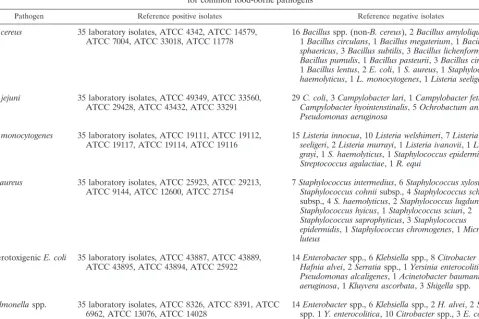
(Table 1). All of the reference positive isolates were obtained from food samples and included five American Type Culture Collection (ATCC) strains and 35 laboratory isolates from a wide variety of food sources. The reference negative isolates were cultured from food, clinical, or environmental samples and included five ATCC strains and 35 laboratory isolates which were related to but not identical to the pathogen of interest. All isolates were confirmed as either reference positive isolates or reference negative isolates by using reference identification tests, and only those isolates which were correctly identified by the reference identification tests at a good confidence level were included in the study.
Preparation of study isolates.Stock cultures of reference positive isolates and
reference negative isolates were subcultured from a frozen state onto Trypticase soy agar (TSA) with 5% sheep blood (BBL, Cockeysville, Md.) and were incu-bated aerobically at 35°C for 24 h; however,C. jejunireference positive isolates and reference negative isolates were incubated microaerophilically for 48 to 72 h. Second and third subcultures were performed with the media and incubation conditions recommended by the manufacturer of each identification system. Isolates from the third subculture was used to test each system.
Reference identification tests.The identities of the reference positive isolates
and the reference negative isolates of verotoxigenicE. coliandSalmonellaspp. were confirmed with the API 20E identification kit (bioMe´rieux Vitek) according to directions that accompanied the product. The identities of the reference positive isolates and the reference negative isolates ofB. cereuswere confirmed by a biochemical testing scheme consisting of Gram staining, catalase reaction, hemolysin production on TSA plus 5% sheep blood (BBL), and lecithinase production onB. cereusselective agar (Oxoid) supplemented with polymyxin B (Oxoid) and egg yolk emulsion (Oxoid). The identities of theB. cereusisolates were further confirmed by using the API 50 CHB (bioMe´rieux Vitek) test kit. The identities of theC. jejunireference positive isolates and reference negative
isolates were confirmed by using a biochemical identification scheme consisting of Gram staining, catalase reaction, oxidase reaction, the presence of corkscrew-like motility as seen under a dark-field microscope, hippurate hydrolysis, and sensitivity to nalidixic acid and resistance to cephalothin (20). The reference method used in this study to confirm the identities of the L. monocytogenes
reference positive isolates and reference negative isolates was a biochemical scheme, which included Gram staining, catalase reaction, esculin hydrolysis on Oxford agar, hemolysis on horse blood agar, CAMP reaction, fermentation of rhamnose, xylose, and mannitol, and the presence of tumbling motility. A mod-ified version of the simplmod-ified scheme of Kloos and Schleifer (13) was used to confirm the identities of theS. aureusreference positive isolates and reference negative isolates. This scheme consisted of Gram staining, hemolysin production on TSA plus 5% sheep blood (BBL), colony pigment production, catalase reac-tion, tube coagulase test, urea and nitrate reactions, and fermentation of arab-inose, lactose, maltose, mannitol, sucrose, trehalose, and xylose. These reference identification tests were designed to identify the reference positive isolates to the species level and to confirm that the reference negative isolates were species other than that of the reference positive isolate to which they were being com-pared.
Bacterial identification systems.The automated microbial identification
[image:2.612.65.545.88.407.2]sys-tems included in this study are listed in Table 2. The WalkAway 40 and the VITEK systems both entail inoculation of a microbial suspension into prepared microwell plates for the WalkAway 40 system or test cards for the VITEK system. These microwell plates and test cards contain a variety of conventional and proprietary biochemical substrates and antibiotics. Growth of bacteria within the microwells (WalkAway 40 system) or test card wells (VITEK system) results in biochemical substrate changes which can be interpreted by a specialized plate reader (WalkAway 40 system) or automated test card reader (VITEK system) to produce a biochemical profile. This profile can be compared to the profiles of known microorganisms to generate an identification. Operation of the MicroLog system also involves inoculation of a microbial suspension into specialized mi-crowell plates. The wells of these plates contain buffered media with different carbon sources and an indicator dye, tetrazolium violet. The dye is reduced when different carbon sources are utilized, resulting in a biochemical profile which can be compared to the profiles of known microorganisms to generate an identifi-cation. The Replianalyzer is similar to the WalkAway 40 and VITEK systems in that a profile of biochemical reactions is generated and compared to those of
TABLE 1. Reference isolates used to determine specificities of five automated microbial identification systems
for common food-borne pathogens
Pathogen Reference positive isolates Reference negative isolates
B. cereus
35 laboratory isolates, ATCC 4342, ATCC 14579,
ATCC 7004, ATCC 33018, ATCC 11778
16
1
Bacillus
Bacillus circulans
spp. (non-
, 1
B. cereus
Bacillus megaterium
), 2
Bacillus amyloliquifaciens
, 1
Bacillus
,
sphaericus
, 3
Bacillus subtilis
, 3
Bacillus lichenformis
, 2
Bacillus pumulis
, 1
Bacillus pasteurii
, 3
Bacillus circulans
,
1
Bacillus lentus
, 2
E. coli
, 1
S. aureus
, 1
Staphylococcus
haemolyticus
, 1
L. monocytogenes
, 1
Listeria seeligeri
C. jejuni
35 laboratory isolates, ATCC 49349, ATCC 33560,
ATCC 29428, ATCC 43432, ATCC 33291
29
Campylobacter hyointenstinalis
C. coli
, 3
Campylobacter lari
, 1
, 5
Campylobacter fetus
Ochrobactum anthropi
, 1
, 1
Pseudomonas aeruginosa
L. monocytogenes
35 laboratory isolates, ATCC 19111, ATCC 19112,
ATCC 19117, ATCC 19114, ATCC 19116
15
seeligeri
Listeria innocua
, 2
Listeria murrayi
, 10
Listeria welshimeri
, 1
Listeria ivanovii
, 7
Listeria
, 1
Listeria
grayi
, 1
S. haemolyticus
, 1
Staphylococcus epidermidis
, 1
Streptococcus agalactiae
, 1
R. equi
S. aureus
35 laboratory isolates, ATCC 25923, ATCC 29213,
ATCC 9144, ATCC 12600, ATCC 27154
7
Staphylococcus intermedius
Staphylococcus cohnii
subsp., 4
, 6
Staphylococcus xylosus
Staphylococcus schleiferi
, 5
,
subsp., 4
S. haemolyticus
, 2
Staphylococcus lugdunensis
, 4
Staphylococcus hyicus
, 1
Staphylococcus sciuri
, 2
Staphylococcus saprophyticus
, 3
Staphylococcus
epidermidis
, 1
Staphylococcus chromogenes
, 1
Micrococcus
luteus
Verotoxigenic
E. coli
35 laboratory isolates, ATCC 43887, ATCC 43889,
ATCC 43895, ATCC 43894, ATCC 25922
14
Hafnia alvei
Enterobacter
, 2
spp., 6
Serratia
Klebsiella
spp., 1
Yersinia enterocolitica
spp., 8
Citrobacter
spp., 2
, 1
Pseudomonas alcaligenes
, 1
Acinetobacter baumanii
, 1
P.
aeruginosa
, 1
Kluyvera ascorbata
, 3
Shigella
spp.
Salmonella
spp.
35 laboratory isolates, ATCC 8326, ATCC 8391, ATCC
6962, ATCC 13076, ATCC 14028
14
spp. 1
Enterobacter
Y. enterocolitica
spp., 6
Klebsiella
, 10
Citrobacter
spp., 2
spp., 3
H. alvei
E. coli
, 2
Serratia
, 1
Edwardsiella tarda
, 1
S. sonnei
on May 15, 2020 by guest
http://jcm.asm.org/
known microorganisms, but this system uses agar plates rather than microwell plates. MIS is significantly different from the other systems because it is based on a comparison of the fatty acid methyl ester profiles for unknown microorganisms to those for known microorganisms to generate an identification.
Isolates to be identified with MIS were grown and processed as described in the MIS operating manual (17) for the CLIN library and were tested with the Sherlock, version 1.06, CLIN library database, version 3.8. Isolates to be identi-fied with the Microscan WalkAway 40 system were grown and processed as described in the WalkAway 40 system operating manual (5), inoculated into Dried Overnight Gram Positive ID panels (L. monocytogenesandS. aureus) or Dried Overnight Gram Negative ID panels (Salmonellaspp. and verotoxigenicE. coli), and evaluated with the Microscan Data Management System, version 20.57, database. Isolates to be identified with the Replianalyzer system were grown and processed as described in the Replianalyzer operating manuals (3) and were evaluated with the Replianalyzer, version 2.11, database. Isolates to be identified with the MicroLog system were grown and processed as described in the Biolog system manual (6), inoculated into GP MicroPlates (L. monocytogenes) or GN MicroPlates (Salmonellaspp. and verotoxigenicE. coli), and analyzed with the version DE, release 3.5, database. Isolates to be identified with the VITEK system were grown and processed as described in the VITEK operator’s manual (7), inoculated into the GPI card (L. monocytogenesandS. aureus), the GNI card (Salmonellaspp. and verotoxigenicE. coli), or the BAC card (B. cereus), and evaluated with the VITEK, version DSAMSO-R10.3, database. Reference neg-ative isolates and reference positive isolates of each pathogen were examined with the same systems under the same conditions.
Analysis of results.The different systems generated identification results in
different formats. Most of the automated systems had a specific minimum level of probability that was required for the identification of an unknown organism to be interpreted with good confidence. The MicroLog system required a minimum similarity index of 0.500. MIS required a minimum similarity index of 0.300 with a minimum separation of 0.100 between the first identification and any secondary identifications. The WalkAway 40 system required a probability of greater than or equal to 85%. The Replianalyzer system required a probability of 95%. The VITEK system did not have a specific probability cutoff level for acceptable identifications. A probability of greater than or equal to 95% was used so that the results obtained with the VITEK system could be directly compared with those obtained with the other systems.
The sensitivities of the test systems were determined by testing each system with 40 known reference positive isolates of each of the six pathogens. Sensitivity in this study was defined as the proportion of the reference positive isolates which were correctly identified with the automated microbial identification tems with an acceptable identification confidence level, as specified by the sys-tem’s manufacturer. The proportion of reference positive isolates which were correctly identified with the automated microbial identification systems but with unacceptably low confidence levels was also examined.
The specificities of the test systems were determined by testing each system with 40 isolates of bacteria which were not the pathogen of interest but which showed similarities in terms of their biochemical reactions and Gram staining results to those of the pathogen of interest. Specificity in this study was defined as the proportion of reference negative isolates which were not identified as the pathogen of interest.
The repeatabilities of the test systems were determined by performing repeat analyses for 20 randomly selected ATCC strains and laboratory isolates from among both the reference positive isolates and the reference negative isolates of each pathogen. The second subculture of the first analysis was used as a starting point for the replicate analysis of each isolate, and the replicate analysis was performed once for each isolate on days different from the days of performance of the initial analysis. The identification results and the confidence levels of the identifications were examined. Repeatability of identification was defined as the proportion of repeat analyses which generated the same identification with similar confidence levels, i.e., having either acceptable or unacceptable
confi-dence levels in both analyses. The proportion of repeat analyses which generated the same identification but at different confidence levels was also examined.
RESULTS
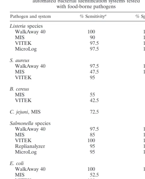
The sensitivities and specificities of the automated microbial
identification systems for each of the pathogens of interest are
summarized in Table 3. The data indicate the sensitivity and
specificity of each system for the pathogens of interest when
identification results with acceptable confidence levels were
analyzed. The repeatabilities of the different automated
micro-TABLE 2. Summary of automated bacterial identification systems included in the study and the food-borne
bacterial pathogens used to test these systems
aSpecies Reference test System used
WalkAway 40 MIS VITEK Replianalyzer MicroLog
L. monocytogenes
Biochemical scheme
Yes
Yes
Yes
No
Yes
S. aureus
bBiochemical scheme
Yes
Yes
Yes
No
Yes
B. cereus
bBiochemical scheme, API 50 CHB
No
Yes
Yes
No
Yes
C. jejuni
Biochemical scheme
No
Yes
No
No
No
Salmonella
spp.
API 20E
Yes
Yes
Yes
Yes
Yes
E. coli
API 20E
Yes
Yes
Yes
Yes
Yes
aThe following database versions of the automated microbial identification systems were used in this study: WalkAway 40 system, version 20.37; MIS, version 3.8;
VITEK system, version DSAMSO-R10.3; Replianalyzer system, version 2.11; MicroLog system, version 3.5.
[image:3.612.52.553.92.183.2]bS. aureusandB. cereuscultures were not tested with the Biolog databases because these databases were in the process of being revised at the time of the study.
TABLE 3. Summary of sensitivities and specificities of several
automated bacterial identification systems tested
with food-borne pathogens
Pathogen and system % Sensitivitya % Specificityb
Listeria
species
WalkAway 40
100
100
MIS
90
100
VITEK
97.5
100
MicroLog
97.5
100
S. aureus
WalkAway 40
97.5
100
MIS
47.5
100
VITEK
95
95
B. cereus
MIS
55
97.5
VITEK
42.5
97.5
C. jejuni
, MIS
72.5
32.5
Salmonella
species
WalkAway 40
97.5
100
MIS
85
100
VITEK
100
100
Replianalyzer
95
100
MicroLog
95
100
E. coli
WalkAway 40
100
100
MIS
52.5
97.5
VITEK
100
97.5
Replianalyzer
90
100
MicroLog
72.5
100
aProportion of reference positive strains which were correctly identified with
an acceptable confidence rating.
bProportion of reference negative strains which were not identified as the
pathogen of concern with an acceptable confidence rating.
on May 15, 2020 by guest
http://jcm.asm.org/
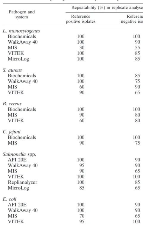
[image:3.612.309.547.402.696.2]bial identification systems for reference positive isolates and
reference negative isolates for the pathogens of interest are
presented in Table 4. The values represent the proportions of
repeat analyses which generated the same identification with
similar confidence levels.
DISCUSSION
There are a number of factors to be considered when
deter-mining which automated microbial identification system is
most appropriate for the detection of food-borne pathogens
within a particular laboratory. These factors include the initial
investment that is required, operating costs, technician time,
range of organisms within the system’s database, and the ability
of the system to correctly identify food pathogens of interest.
The last two variables were examined in this study. The
auto-mated microbial identification systems evaluated included
MIS, the WalkAway 40 system, the MicroLog system, the
VITEK system, and the Replianalyzer system.
The WalkAway 40 system, the VITEK system, and the
Mi-croLog system all contained databases for the identification of
Listeria
spp. MIS contained a database for the identification of
L. monocytogenes
specifically. To compare the systems fairly,
only identifications to the genus level are reported in Table 3.
All four of the systems performed well in identifying
L.
mono-cytogenes
to the genus level, with sensitivities of 90 to 100% and
specificities of 100%. A much lower specificity was observed
when MIS was used to identify
L. monocytogenes
to the species
level because
Listeria innocua
isolates were frequently
identi-fied as
L. monocytogenes
. The repeatabilities observed for the
different systems were also good, with the exception of the
MIS, which showed a lower repeatability than the other
sys-tems. The repeatability of the MIS was increased to 70% when
species identification, but not confidence rating, was
consid-ered. While all of these systems could be used reliably as a
screening method for the detection of
Listeria
spp. to the genus
level, further biochemical tests would be required to determine
whether the isolate was a potentially pathogenic
L.
monocyto-genes
isolate or a nonpathogenic
Listeria
species. Presumptive
identification of
Listeria
species from colonies on selective
me-dia, such as Oxford agar, requires only a few simple tests, so
there is at present little advantage in using these automated
identification systems for the identification of
L.
monocyto-genes
.
The WalkAway 40 system, the VITEK system, and MIS all
contained databases for the identification of
S. aureus
. The
MicroLog and the Replianalyzer system databases for the
identification of
Staphylococcus
and related species were being
developed or upgraded, and they were not included in this
study. The WalkAway 40 and the VITEK systems both showed
good sensitivities and specificities for the identification of
S.
aureus
. MIS showed 100% specificity for the identification of
S.
aureus
but a sensitivity of only 47.5%. This sensitivity of MIS
for the identification of
S. aureus
increased to 87.5% if correct
identifications with low confidence levels were included,
sug-gesting that the fatty acid compositions of the food isolates
examined were slightly different from those of the isolates
within the database. The specificity of the system was reduced
to 90% when results with low confidence levels were included
in the analysis. Repeatability levels for all three systems were
slightly higher for reference positive isolates than for reference
negative isolates, ranging from 60 to 100% when both
identi-fication and confidence level were considered. Presumptive
identification of
S. aureus
requires only a few biochemical tests
which are available in most nonautomated methods; hence,
there is little advantage in using these automated identification
systems for the identification of this organism.
[image:4.612.51.291.101.478.2]Both MIS and the VITEK system contained databases for
the identification of
B. cereus
isolates. At the time of this study,
the MicroLog system database for
B. cereus
was being
up-graded; therefore, this system was not evaluated for
B. cereus
.
Neither MIS nor the VITEK system was able to identify
B.
cereus
with a high sensitivity, even if correct identifications with
low confidence levels were included. Some investigators have
proposed that
B. cereus
,
Bacillus mycoides
, and
Bacillus
thurin-giensis
are very closely related and should be merged into a
single species (2, 8). When the results were reanalyzed with the
definition of a correct identification expanded to include
B.
mycoides
and
B. thuringiensis
species, the sensitivities of MIS
and the VITEK system increased to 82.5 and 67.5%,
respec-tively. The two systems showed a high specificity (97.5%) for
B.
cereus
. The repeatability of the VITEK system for this
organ-ism was somewhat low at 60%, while MIS showed a
repeat-ability of 90%. The results obtained by the API 50 CHB test kit
corresponded well with those obtained by the biochemical
ref-erence scheme when tests were performed with
B. cereus
iso-lates.
TABLE 4. Repeatabilities of identifications generated
by automated bacterial identification systems against
food-borne pathogens and closely related species
Pathogen and system
Repeatability (%) in replicate analysesa
Reference
positive isolates negative isolatesReference
L. monocytogenes
Biochemicals
100
100
WalkAway 40
100
90
MIS
30
55
VITEK
100
85
MicroLog
100
85
S. aureus
Biochemicals
100
85
WalkAway 40
100
75
MIS
60
90
VITEK
90
65
B. cereus
Biochemicals
100
100
MIS
90
80
VITEK
60
80
C. jejuni
Biochemicals
100
100
MIS
90
75
Salmonella
spp.
API 20E
100
90
WalkAway 40
95
90
MIS
90
65
VITEK
100
100
Replianalyzer
100
85
MicroLog
85
65
E. coli
API 20E
100
90
WalkAway 40
100
90
MIS
70
65
VITEK
95
100
Replianalyzer
100
85
MicroLog
70
65
aProportion of replicate analyses which generated the same identification with
similar confidence rating.
on May 15, 2020 by guest
http://jcm.asm.org/
MIS was the only system with a database for the
identifica-tion of
Campylobacter
species. The sensitivity and specificity of
this system for the identification of
C. jejuni
were 72.5 and
32.5%, respectively. The sensitivity increased to 92.5% if
cor-rect identifications with low confidence levels were included.
However, the specificity of the system declined to 30% when
other
Campylobacter
species misidentified as
C. jejuni
with low
confidence levels were included in the estimation of specificity.
The low specificity of MIS for
C. jejuni
may be due to the fact
that most of the reference negative isolates used in the study
were
Campylobacter coli
and the system appears to have
diffi-culty distinguishing
C. coli
from the closely related species
C.
jejuni
. Both
C. jejuni
and
C. coli
are common food-borne
pathogens; thus, MIS may be useful as a screening method for
the identification of pathogenic
Campylobacter
isolates. The
repeatability of MIS was found to be 90% for
C. jejuni
and 75%
for related species when the same identification and
confi-dence levels were considered in the analysis. The identification
of
C. jejuni
is similar to that of
L. monocytogenes
in that
pre-sumptive identification from selective media requires only a
few simple laboratory tests; hence, there is little advantage in
the use of automated systems for the identification of
Campy-lobacter
to the genus level unless these systems are able to
differentiate pathogenic and nonpathogenic
Campylobacter
species with high specificities and sensitivities.
All of the systems included in this study had developed
databases for the identification of
Salmonella
spp., and all of
the systems showed good sensitivities for
Salmonella
identifi-cation, with values ranging from 85 to 100%. Specificities of
100% were observed for all of the systems tested, suggesting
that false-positive identifications would not be a problem for
Salmonella
isolates. The repeatabilities of the different systems
when they were tested with
Salmonella
spp. and related isolates
were highest with the VITEK, the WalkAway 40, and the
Replianalyzer systems and were somewhat higher for reference
positive isolates than for reference negative isolates.
While all of the systems included in this study had databases
for the identification of
E. coli
, none of these databases
distin-guished between verotoxigenic
E. coli
and other
E. coli
strains.
One study, however, reported that a limited number of
bio-chemical profiles were generated when
E. coli
O157:H7 strains
were screened with the MicroScan Overnight Panel System (1).
The Replianalyzer database may also detect
E. coli
O157:H7
on the basis of a sorbitol-negative reaction. The WalkAway 40,
the VITEK, and the Replianalyzer systems appeared to be well
adapted for the identification of
E. coli
isolates, with
sensitiv-ities of 90 to 100% and specificsensitiv-ities of 97.5 to 100%. Both the
MicroLog system and MIS reported somewhat lower
sensitiv-ities; however, these sensitivities increased to acceptable levels
if correct identifications with low confidence levels were
in-cluded in the analysis. Inclusion of results with lower
confi-dence levels in the analysis resulted in a slightly lower
speci-ficity of MIS for this organism. The WalkAway 40, VITEK, and
Replianalyzer systems appeared to be the most repeatable for
verotoxigenic
E. coli
and related isolates, while MIS and the
MicroLog system had somewhat lower repeatabilities.
In summary, none of the systems appeared to offer any
advantage over biochemical identification schemes for
L.
monocytogenes
identification; however, very good results
(
.
90% sensitivity) were obtained for the identification of
S.
aureus
,
Salmonella
spp., and verotoxigenic
E. coli
by the
Walk-Away 40 and the VITEK systems. The Replianalyzer system
had a
$
90% sensitivity for the identification of
Salmonella
and
verotoxigenic
E. coli
. The MicroLog system had
.
90%
sensi-tivity for the identification of
Salmonella
spp. and
.
90%
sen-sitivity for the identification of verotoxigenic
E. coli
when
correct identification results with or without acceptable
confi-dence ratings were included in the results. Similarly, MIS had
.
90% sensitivity for the identification of the
B. cereus
group,
C. jejuni
, and
Salmonella
spp. when correct identification
re-sults with or without acceptable confidence ratings were
in-cluded in the analysis. The lower sensitivities observed when
some of these pathogens were tested with MIS may be
influ-enced by the fact that the reference systems for these species
were based on biochemical reactions, while the MIS
identifi-cations are based on the analysis of the fatty acid composition
of the unknown microorganism. MIS showed promising results
for the identification of
Campylobacter
species, and both the
VITEK system and MIS showed a fairly good ability to identify
B. cereus
group isolates.
ACKNOWLEDGMENTS
We acknowledge the financial support received from the Enhanced
Food Quality and Safety Program administered by the Ontario
Min-istry of Agriculture, Food and Rural Affairs (OMAFRA) and the
contributions of consumable supplies by Dade Diagnostics Corp.,
Mi-croLog Inc., bioMe´rieux VITEK, and Oxoid Canada Inc. We also
thank the following people for contributing bacterial isolates for this
study: David Woodward, National Laboratory for Enteric Pathogens,
Health Canada; Peter Boleszczuk and Mike Brodsky, Ontario Ministry
of Health, Laboratory Services Branch, Etobicoke, Ontario, Canada;
Luba Stokes, Victoria Hospital, London, Ontario, Canada; Roger
Shuttleworth, University Hospital, London, Ontario, Canada; Noni
Smart, Laboratory Services Division, University of Guelph, Guelph,
Ontario, Canada; and C. E. Park, Health Canada, Ottawa, Ontario,
Canada.
REFERENCES
1.Abbott, S. L., D. F. Hanson, T. D. Felland, S. Connell, A. H. Shum, and J. M.
Janda.1994.Escherichia coliO157:H7 generates a unique biochemical
pro-file on MicroScan conventional gram-negative identification panels. J. Clin. Microbiol.32:823–824.
2.Ash, C., J. A. E. Farrow, M. Dorsh, E. Stackebrant, and M. D. Collins.1991.
Comparative analysis ofBacillus anthracis,Bacillus cereus, and related spe-cies on the basis of reverse transcriptase sequencing of 16S rRNA. Int. J. Syst. Bacteriol.41:343–346.
3.AutoMed Cathra Systems.1994. Cathra microbiology manual, Cathra
Repli-dex manual, Cathra Replianalyzer II manual. AutoMed Cathra Systems, Arden Mills, Minn.
4.Bannerman, T. L., K. T. Kleeman, and W. E. Kloos.1993. Evaluation of the
VITEK systems Gram-Positive Identification card for species identification of coagulase-negative staphylococci. J. Clin. Microbiol.31:1322–1325.
5.Baxter Diagnostics Inc.1992. MicroScan operators manual for Walkaway
systems. Baxter Diagnostics Inc., West Sacramento, Calif.
6.Biolog Inc.1992. MicroLog operating manual. Biolog Inc., Hayward, Calif.
7.bioMe´rieux Vitek.1996. VITEK System manual. bioMe´rieux Vitek,
Hazel-wood, Mo.
8.Carlson, C. R., D. A. Caugant, and A. B. Kolstø.1994. Genotypic diversity
amongBacillus cereusandBacillus thuringiensisstrains. Appl. Environ. Mi-crobiol.60:1719–1725.
9.Champagne, C. P., R. R. Laing, D. Roy, A. A. Mafu, and M. W. Griffiths.
1994. Psychrotrophs in dairy products: their effects and their control. Crit. Rev. Food Sci. Nutr.34:1–30.
10. Holmes, B., M. Costas, M. Ganner, S. L. W. On, and M. Stevens.1994.
Evaluation of Biolog system for identification of some gram-negative bacte-ria of clinical importance. J. Clin. Microbiol.32:1970–1975.
11. Hussain, Z., L. Stokes, D. L. Stevens, B. C. Schieven, R. Lannigan, and C.
Jones.1986. Comparison of the MicroScan system with the API Staph-Ident
system for species identification of coagulase-negative staphylococci. J. Clin. Microbiol.23:126–128.
12. Klingler, J. M., R. P. Stowe, D. C. Obenhuber, T. O. Groves, S. K. Mishra,
and D. L. Pierson. 1992. Evaluation of the Biolog automated microbial
identification system. Appl. Environ. Microbiol.58:2089–2092.
13. Kloos, W. E., and K. H. Schleifer. 1975. Simplified scheme for routine
identification of humanStaphylococcusspecies. J. Clin. Microbiol.1:82–88.
14. Kloos, W. E., and C. G. George.1991. Identification ofStaphylococcus
spe-cies and subspespe-cies with the MicroScan Pos ID and Rapid Pos ID panel systems. J. Clin. Microbiol.29:738–744.
15. Knight, M. T., D. W. Wood, J. F. Black, G. Gosney, R. O. Rigney, and J. R.
Agin.1990. Gram-Negative Identification card for identification of
Salmo-nella,Escherichia coli, and otherEnterobacteriaceaeisolated from foods:
on May 15, 2020 by guest
http://jcm.asm.org/
collaborative study. J. Assoc. Off. Chem.73:729–733.
16. Kramer, J. M., and R. J. Gilbert.1989.Bacillus cereusand other Bacillus
species, p. 21–70.InM. P. Doyle (ed.), Foodborne bacterial pathogens. Marcel Dekker, Inc., New York, N.Y.
17. Microbial ID Inc.1993. Microbial Identification System operating manual,
version 4. Microbial ID Inc., Newark, Del.
18. Miller, J. M., J. W. Biddle, V. K. Quenzer, and J. C. McLaughlin.1993.
Evaluation of Biolog for identification of members of the family Micrococ-caceae. J. Clin. Microbiol.31:3170–3173.
19. Miller, M., and D. L. Rhoden.1991. Preliminary evaluation of Biolog, a
carbon source utilization method for bacterial identification. J. Clin. Micro-biol.29:1143–1147.
20. Nachamkin, I.1995.Campylobacter andArcobacter, p. 483–491.InP. R.
Murray, E. J. Baron, M. A. Pfaller, F. C. Tenover, and R. H. Yolken (ed.), Manual of clinical microbiology, 6th ed. American Society for Microbiology, Washington, D.C.
21. O’Hara, C. M., and J. M. Miller.1992. Evaluation of the autoSCAN-W/A
system for rapid (2 hour) identification of members of the family Enterobac-teriaceae. J. Clin. Microbiol.30:1541–1543.
22. O’Hara, C. M., F. C. Tenover, and J. M. Miller.1993. Parallel comparison of
accuracy of API 20E, Vitek GNI, Microscan Walk/Away Rapid ID, and Becton Dickinson Cobos Micro ID-E/NF for identification of members of the familyEnterobacteriaceaeand common gram-negative,
non-glucose-fer-menting bacilli. J. Clin. Microbiol.31:3165–3169.
23. Osterhout, G. J., V. H. Shull, and J. D. Dick.1991. Identification of clinical
isolates of gram-negative nonfermentative bacteria by an automated cellular fatty acid identification system. J. Clin. Microbiol.29:1822–1830.
24. Pfaller, M. A., D. Sahm, C. O’Hara, C. Ciaglia, M. Yu, N. Yamane, G.
Scharnweber, and D. Rhoden.1991. Comparison of the AutoSCAN-W/A
Rapid Bacterial Identification system and the Vitek AutoMicrobic system for identification of gram-negative bacilli. J. Clin. Microbiol.29:1422–1428.
25. Rhoads, S., L. Marinelli, C. A. Imperatrice, and I. Nachamkin.1995.
Com-parison of MicroScan WalkAway system and Vitek system for identification of gram-negative bacteria. J. Clin. Microbiol.33:3044–3046.
26. Robinson, A., Y. S. McCarter, and J. Tetreault.1995. Comparison of Crystal
Enteric/Nonfermenter System, API 20E System, and the Vitek Automicrobic System for identification of gram-negative bacilli. J. Clin. Microbiol.33:364– 370.
27. Stager, C. E., and J. R. Davis.1992. Automated systems for identification of
microorganisms. J. Clin. Rev.5:302–327.
28. Stokes, L., B. C. Schieven, E. Ofori, P. Ewan, R. Lannigan, and Z. Hussain.
1992. Evaluation of Microscan Rapid Pos combo panels for identification of staphylococci. J. Clin. Microbiol.30:93–95.
29. Stokes, L., M. A. John, R. Lannigan, B. C. Schieven, M. Ramos, D. Harley,
and Z. Hussain.1994. Gas-liquid chromatography of cellular fatty acids for
identification of staphylococci. J. Clin. Microbiol.32:1908–1910.