0095-1137/05/$08.00⫹0 doi:10.1128/JCM.43.1.84–88.2005
Copyright © 2005, American Society for Microbiology. All Rights Reserved.
Performance of the New VITEK 2 GP Card for Identification of
Medically Relevant Gram-Positive Cocci in a Routine
Clinical Laboratory
Guido Funke* and Pascale Funke-Kissling
Department of Medical Microbiology and Hygiene, Ga¨rtner and Colleagues Laboratories, Weingarten, Germany
Received 11 May 2004/Returned for modification 18 June 2004/Accepted 4 August 2004
The VITEK 2 gram-positive (GP) identification card (bioMe´rieux, Marcy l’Etoile, France) has been rede-signed to achieve greater accuracy in the identification of gram-positive cocci. A total of 43 biochemical tests, including 17 enzymatic tests, are present in the card and interpreted in a kinetic mode, for up to 8 h. The VITEK 2 database, used in conjunction with the GP identification card, allows the identification of 115 different taxa. A total of 364 strains of GP cocci (217Streptococcaceaestrains and 147Micrococcaceaestrains) belonging to 31 taxa were tested with the new VITEK 2 GP identification card. Of the 364 strains, 105 were taken from routine primary plating media. A total of 344 strains (94.5%) were correctly identified to the species level and 17 strains (4.7%) were identified with low discrimination, requiring additional tests, whereas 1 strain (0.3%) was incorrectly identified and 2 strains (0.5%) remained unidentified. Within 7 h of the start of incubation, more than 90% of all strains were identified. Of the 105 primary cultures, 97% were correctly identified to the species level, 2% were identified with low discrimination, and 1% remained unidentified. Identification per-formance data were independent of each of the three plating media used. It is concluded that the new VITEK 2 GP identification card provides reliable results for the identification of GP cocci under routine laboratory conditions.
Highly automated identification systems are nowadays widely distributed in many medium-to-high-throughput clinical microbiology laboratories. These systems improve the quality of patient care and enable more-cost-effective management of the same by enabling clinical microbiologists to identify med-ically relevant bacteria more rapidly and accurately (1, 2). An important measure of the value of a highly standardized com-mercial identification system must be the capability of the manufacturer to maintain or even improve the performance of an identification system over time. The new VITEK 2 gram-positive (GP) identification card (bioMe´rieux, Marcy l⬘Etoile, France) for identification of GP cocci was created in recent years as research and development related to the VITEK 2 instrument continued. The rationale for designing the new VITEK 2 GP identification card was to broaden the VITEK 2 database while maintaining the quality of the identification results in the routine clinical laboratory. The GP identification card contains 43 tests (27 tests that had been included in the previous card and 16 new tests), compared to 47 in the estab-lished VITEK 2 GP identification card (ID-GPC), and 115, instead of 51, taxa are covered by the new database corre-sponding to the GP identification card. While the ID-GPC tests are based on fluorescence technology, the GP identifica-tion card tests are based on colorimetric detecidentifica-tion. Both the ID-GPC and GP identification card tests are subjected to mea-surements every 15 min, and the total incubation time is up to
approximately 8 h with the GP identification card, as opposed to 2 h with the ID-GPC.
The aim of the present study was to evaluate the newly developed VITEK 2 GP identification card in a routine clinical laboratory by a weighted laboratory profile (9).
(This paper was presented in part at the 104th General Meeting of the American Society for Microbiology, New Or-leans, La., 23 to 27 May 2004 [G. Funke and P. Funke-Kissling, Abstr. 104th Gen. Meet. Am. Soc. Microbiol., abstr. C-178, 2004].)
MATERIALS AND METHODS
Laboratory, strains, culture conditions, and identification. The study was
performed at Ga¨rtner and Colleagues Laboratories, an accredited reference
laboratory that serves over 100 hospitals of all levels and over 3,000 physicians in private practice. The strains included in the present study were collected within a 3-month period. The number of strains per species was limited to a maximum of 45. A total of 105 strains of GP cocci were taken from primary isolation plates set up on Columbia sheep blood agar (BD, Heidelberg, Germany) in our routine clinical laboratory for various types of patient specimens (blood culture, wound swab, respiratory, and urine specimens, etc.). The other 259 strains came from primary isolation plates that had been stored at 4 to 8°C for less than 1 week.
These strains were subcultured on Columbia sheep blood agar from BD (n⫽70),
Columbia sheep blood agar from bioMe´rieux (n⫽78), or Trypticase soy blood
agar (bioMe´rieux) (n⫽111) for 18 to 24 h at 37°C before they were subjected
to VITEK 2 analysis. All strains included in the present study came from unre-lated patients, and consecutive cultures from the same patient were also ex-cluded. The 364 strains used in this study were identified by conventional meth-ods (10) as well as by VITEK 1 analysis with the GP identification card designed for use with that system. For identification by conventional methods, the follow-ing characteristics were tested: colony pigmentation, hemolysis, adherence to agar, colony odor, catalase and oxidase reaction, clumping factor test
(bio-Me´rieux), reaction(s) to Lancefield group streptococcal antisera (Oxoid,
Basing-stoke, United Kingdom), reaction(s) to pneumococcal antisera (bioMe´rieux), as
well as susceptibilities to optochin and bacitracin. Discrepancies between the identifications obtained by conventional methods and VITEK 1 analysis and
* Corresponding author. Mailing address: Department of Medical Microbiology and Hygiene, Ga¨rtner and Colleagues Laboratories, Hoyerstrasse 51, D-88250 Weingarten, Germany. Phone: 49-751-502-630. Fax: 49-751-502-385. E-mail: [email protected].
84
on May 16, 2020 by guest
http://jcm.asm.org/
those obtained by VITEK 2 analysis were resolved by using ID 32 STAPH and
rapid ID 32 STREP galleries (both from bioMe´rieux) as well as by sequencing of
16S rRNA genes (which was necessary for a total of nine strains) as previously outlined (5).
New GP identification card and VITEK 2 instrument.A bacterial suspension was adjusted to a McFarland standard of 0.5 in 2.5 ml of a 0.45% sodium chloride
solution with a VITEK 2 instrument (DensiChek; bioMe´rieux). The time
be-tween preparation of the inoculum and the card filling was always less than 30 min. The format of the GP identification card is the same as that of the ID-GPC, i.e., a 64-well plastic card which contains now 43 instead of 47 tests (see above). The GP identification card includes test for the following reactions:
phosphati-dylinositol phospholipase C, arginine dihydrolase (two tests),-galactosidase,
␣-glucosidase, alanine-phenylalanine-proline arylamidase,L-aspartic acid
aryl-amidase,-galactosidase,␣-mannosidase, alkaline phosphatase,L-leucine
aryl-amidase, proline arylaryl-amidase,-glucuronidase (two tests),␣-galactosidase,L
-pyroglutamic acid arylamidase, alanine arylamidase, tyrosine arylamidase, and urease. The GP identification card also tests acid production from the following
substrates: amygdalin, xylose,␣-cyclodextrin, sorbitol, galactose, ribose, lactate,
lactose,N-acetyl-glucosamine, maltose, mannitol, mannose, methyl--D
-glucopy-ranoside, pullulan, raffinose, salicin, sucrose, and trehalose. Finally, growth in 6.5% NaCl as well as tests for resistance to polymyxin B, bacitracin, novobiocin, O129, and optochin are also included in the GP identification card.
The GP identification card is a fully closed system to which no reagents have to be added. The card was put on the cassette designed for use with the VITEK 2 system, placed in the instrument, automatically filled in a vacuum chamber,
sealed, incubated at 35.5°C, and automatically subjected to colorimetric mea-surement (with a new reading head) every 15 min for a maximum incubation period of 8 h. Data were analyzed using VITEK 2 database version 4.01, which allows organism identification in a kinetic mode beginning 180 min after the start of incubation. In the kinetic mode, all 43 tests are individually interpreted using a first-level algorithm based on the color change of the reaction. Every 15 min, a second-level algorithm analyzes the biopattern (i.e., interpreted and not inter-preted tests) and verifies if it is sufficient to give the final identification call. The second algorithm also checks, using a specific calculation, that the later results of not-yet-interpreted tests will not change the identification call.
Quality control strains.During the evaluation period the following quality
control strains were checked at regular intervals: Enterococcus casseliflavus
(ATCC 700327),Enterococcus faecalis(ATCC 29212),Enterococcus
saccharo-lyticus(ATCC 43076T
),Kytococcus sedentarius(ATCC 27575),Staphylococcus aureus(ATCC 29213), Staphylococcus saprophyticus (ATCC BAA-750), and Streptococcus equisubsp.zooepidemicus(ATCC 43079T
).
[image:2.585.44.542.80.468.2]Reporting of results.Not the identification scores (tindex, probability, likeli-hood, and confidence values) provided by the software but rather the interpre-tations provided by the software were taken into account. The four different result categories were: (i) correct identification (unambiguous correct identifi-cation to the species level); (ii) low level of discrimination (either identifiidentifi-cation to the genus level or a low level of discrimination between two or more species, including the correct species); (iii) no identification; and (iv) misidentification (the species identified with the GP identification card was different from that identified by the reference method).
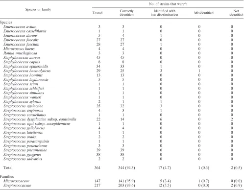
TABLE 1. Performance of the new VITEK 2 GP identification card by species and by family
Species or family
No. of strains that werea:
Tested Correctly
identified
Identified with
low discrimination Misidentified
Not identified
Species
Enterococcus avium 3 3 0 0 0
Enterococcus casseliflavus 1 1 0 0 0
Enterococcus durans 5 4 1 0 0
Enterococcus faecalis 27 27 0 0 0
Enterococcus faecium 28 27 1 0 0
Micrococcus luteus 4 4 0 0 0
Rothia mucilaginosa 3 3 0 0 0
Staphylococcus aureus 45 45 0 0 0
Staphylococcus capitis 8 8 0 0 0
Staphylococcus epidermidis 34 33 1 0 0
Staphylococcus haemolyticus 29 25 3 1 0
Staphylococcus hominis 13 13 0 0 0
Staphylococcus lugdunensis 5 5 0 0 0
Staphylococcus sciuri 1 1 0 0 0
Staphylococcus schleiferi 1 1 0 0 0
Staphylococcus simulans 1 1 0 0 0
Staphylococcus wameri 1 1 0 0 0
Staphylococcus xylosus 2 1 1 0 0
Streptococcus agalactiae 35 32 3 0 0
Streptococcus anginosus 4 3 1 0 0
Streptococcus constellatus 1 1 0 0 0
Streptococcus dysgalactiaesubsp.equisimilis 22 14 6 0 2
Streptococcus equisubsp.zooepidemicus 1 1 0 0 0
Streptococcus gallolyticus 4 4 0 0 0
Streptococcus lutetiensis 1 1 0 0 0
Streptococcus oralis 2 2 0 0 0
Streptococcus parasanguinis 1 1 0 0 0
Streptococcus pasteurianus 3 3 0 0 0
Streptococcus pneumoniae 39 39 0 0 0
Streptococcus pyogenes 38 38 0 0 0
Streptococcus salivarius 2 2 0 0 0
Total 364 344 (94.5) 17 (4.7) 1 (0.3) 2 (0.5)
Families
Micrococcaceae 147 141 (95.9) 5 (3.4) 1 (0.7) 0 (0.0)
Streptococcaceae 217 203 (93.6) 12 (5.5) 0 (0.0) 2 (0.9)
a
Values in parentheses are percentages.
on May 16, 2020 by guest
http://jcm.asm.org/
RESULTS
Overall, we did not encounter any major technical problem using the VITEK 2 instrument and the GP identification card during the 3-month evaluation period. Quality control strains were correctly identified to the species level in every instance, demonstrating the reliability and reproducibility of the tech-nique. The hands-on time remained the same for the GP iden-tification card as for the ID-GPC.
Table 1 lists the performance of the GP identification card for the 31 taxa (18 belonging to the familyStreptococcaceaeand 13 belonging to the familyMicrococcaceae) tested. Of the total of 364 strains (217 belonging to the familyStreptococcaceae
and 147 belonging to the familyMicrococcaceae), 344 strains (94.5%) were correctly identified to the species level, 17 strains (4.7%) were identified with low discrimination, 2 strains (0.5%) remained unidentified, and 1 strain (0.3%) was misi-dentified. Identification results were even better when simple additional tests (see below) were applied to resolve the results for strains with low discrimination. Identification results were slightly better for strains of the familyMicrococcaceae(95.9% correctly identified, 3.4% identified with low discrimination, 0.7% misidentified, and 0.0% not identified) than for strains of the familyStreptococcaceae (93.6% correctly identified, 5.5% identified with low discrimination, 0.0% misidentified, and 0.9% not identified).
The distribution of all taxa tested was not equal but was weighted according to the frequency with which the different species are seen in a routine clinical laboratory. The eight most frequently isolated gram-positive cocci, namely, E. faecalis,
Enterococcus faecium, S. aureus, Staphylococcus epidermidis,
Staphylococcus haemolyticus,Streptococcus agalactiae, Strepto-coccus pneumoniae, and Streptococcus pyogenes represented 75.3% of all strains included in the present study. Of these, 96.7% were correctly identified and 2.9% were identified with low discrimination (only one strain [0.4%] was misidentified). Minor differences in identification results were observed with the different culture media. Of the 175 strains tested on Columbia sheep blood agar from BD, 96.6% were correctly
identified, 2.3% were identified with low discrimination, 0.0% were misidentified, and 1.1% were not identified. Of the strains tested on Columbia sheep blood agar from bioMe´rieux, 88.5% were correctly identified, 10.3% were identified with low dis-crimination, 1.3% were misidentified, and 0.0% were not iden-tified; of the strains tested on Trypticase soy blood agar, 95.5% were correctly identified, 4.5% were identified with low dis-crimination, 0.0% were misidentified, and 0.0% were not iden-tified.
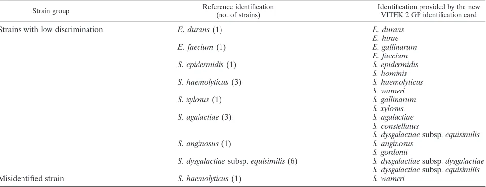
Table 2 lists the strains identified with low discrimination as well as the misidentified strains. The differentiation between
Enterococcus durans (saccharose positive) and Enterococcus hirae(saccharose negative) is readily achieved. Enterococcus gallinarumis motile, butE. faeciumis not.S. epidermidis fer-mentsD-mannose, whereasStaphylococcus hominisis unable to do so.S. haemolyticusis urease negative but expresses pyrro-lidonyl arylamidase activity, whereasStaphylococcus warneriis urease positive and pyrrolidonyl arylamidase negative. Staph-ylococcus xylosusis differentiated fromStaphylococcus gallina-rumby a negative reaction forD-raffinose fermentation. The
-hemolysis reaction of Streptococcus agalactiae is more dis-crete than that ofStreptococcus dysgalactiaesubsp.equisimilis. In addition,S. agalactiae is positive in the Voges-Proskauer reaction, butS. dysgalactiae subsp.equisimilisis not. Strepto-coccus constellatusdoes not hydrolyze hippurate and exhibits the typical diacetyl odor, whereas S. agalactiae is hippurate positive and does not produce the typical “Streptococcus mil-leri” odor.Streptococcus anginosusis Voges-Proskauer positive, butStreptococcus gordoniiis not.S. dysgalactiaesubsp. equisi-milisis beta-hemolytic, whereasS. dysgalactiaesubsp. dysgalac-tiaeis not.
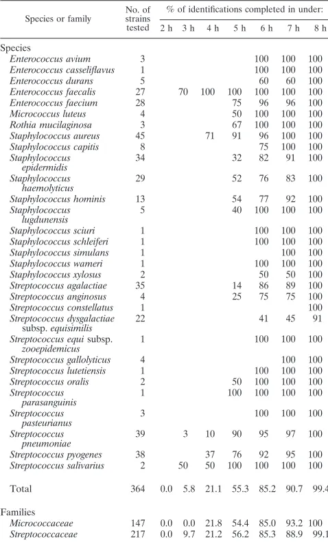
Table 3 gives a detailed report on the exact time required for final identification of the strains tested. Over 90% of all strains were identified within 7 h. No significant difference was ob-served in the times to final identification ofMicroccoccaceae
andStreptoccoccaceae.
[image:3.585.45.542.81.273.2]Table 4 lists the identification results when gram-positive cocci from primary plating media were tested. As with the
TABLE 2. Strains identified with low discrimination or misidentified
Strain group Reference identification
(no. of strains)
Identification provided by the new VITEK 2 GP identification card
Strains with low discrimination E. durans(1) E. durans
E. hirae
E. faecium(1) E. gallinarum
E. faecium
S. epidermidis(1) S. epidermidis
S. hominis
S. haemolyticus(3) S. haemolyticus
S. wameri
S. xylosus(1) S. gallinarum
S. xylosus
S. agalactiae(3) S. agalactiae
S. constellatus
S. dysgalactiaesubsp.equisimilis
S. anginosus(1) S. anginosus
S. gordonii
S. dysgalactiaesubsp.equisimilis(6) S. dysgalactiaesubsp.dysgalactiae S. dysgalactiaesubsp.equisimilis
Misidentified strain S. haemolyticus(1) S. wameri
on May 16, 2020 by guest
http://jcm.asm.org/
overall study, a weighted distribution of isolates was tested. The results for testing of cocci from primary isolation plates were slightly better (97.1% correctly identified) than the re-sults of the overall study.
DISCUSSION
To the best of our knowledge, this is the first study on the performance of the GP identification card in a routine clinical laboratory. Overall, we were impressed by the performance of the system, since more than 94% of the isolates were correctly identified to the species level without application of any further additional tests. This performance is clearly above the de-manded 90% accuracy level that has been discussed by some authorities in the field of commercial clinical microbiology device evaluations. The taxonomy used in the database was up-to-date (e.g., Streptococcus gallolyticus instead of Strepto-coccus bovisbiotype I [12] andStreptococcus pasteurianus in-stead ofS. bovisbiotype II.2 [11]), which is not always the case
for other commercial identification systems designed for GP cocci. In addition, we are not aware of any other commercial identification system for GP cocci claiming to cover so many different taxa.
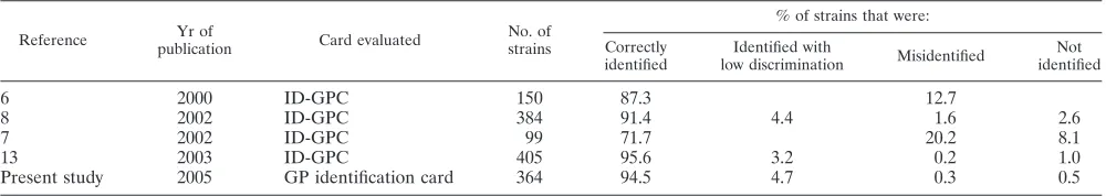
Considering the broad distribution and use of the VITEK 2 instrument in routine clinical microbiology laboratories world-wide, surprisingly few evaluations of the ID-GPC have been published (Table 5). The present study is the third largest on VITEK 2 test cards for identification of GP cocci, and it is the largest with regard to the number of individual taxa that have been included. Despite the extended incubation and reading time, as well as the larger database, the results of our present evaluation were comparable to the results of the two other major studies on VITEK 2 test cards for identification of GP cocci (8, 13). It is important to note that the extension of the database did not lead to poorer identification results. The identification of E. faecium (96.4% of the strains correctly identified in the present study in contrast to only 71.4% in a previous study [8]) as well as the identification ofS. hominis
(100% of the strains correctly identified in the present study in contrast to only 65.6% in a previous study [13]) have been significantly improved. If the kinetic mode is used, the majority of the results obtained with the GP identification card become available about 3 to 4 h later (Table 3) than with the ID-GPC. However, this may be of no consequence for microbiology laboratories not operating on a 24-h schedule. In summary, our data indicate that the GP identification card fulfills the needs of a routine clinical microbiology laboratory serving outpa-tients as well as hospitalized paoutpa-tients. Regarding the cost ef-fectiveness we cannot at present comment on the GP identifi-cation card because the pricing of the GP was not available to us at the time of writing this article.
[image:4.585.43.282.90.486.2]Finally, it is recommended that other evaluations may in-clude a larger number of strains from primary isolation plates in order to study whether the VITEK 2 system in conjunction with the GP identification card performs as acceptably as in the present study when a limited number of first-isolation strains is used. A pure stress test evaluation that includes nearly all taxa present in the database, in particular, the recently delineated,
TABLE 3. Time to final identification by VITEK 2 analysis with the new GP identification card
Species or family
No. of strains tested
% of identifications completed in under: 2 h 3 h 4 h 5 h 6 h 7 h 8 h
Species
Enterococcus avium 3 100 100 100 Enterococcus casseliflavus 1 100 100 100 Enterococcus durans 5 60 60 100 Enterococcus faecalis 27 70 100 100 100 100 100 Enterococcus faecium 28 75 96 96 100 Micrococcus luteus 4 50 100 100 100 Rothia mucilaginosa 3 67 100 100 100 Staphylococcus aureus 45 71 91 96 100 100 Staphylococcus capitis 8 75 100 100 Staphylococcus
epidermidis
34 32 82 91 100
Staphylococcus
haemolyticus 29 52 76 83 100 Staphylococcus hominis 13 54 77 92 100 Staphylococcus
lugdunensis
5 40 100 100 100
Staphylococcus sciuri 1 100 100 100 Staphylococcus schleiferi 1 100 100 100 Staphylococcus simulans 1 100 100 Staphylococcus wameri 1 100 100 100 Staphylococcus xylosus 2 50 50 100 Streptococcus agalactiae 35 14 86 89 100 Streptococcus anginosus 4 25 75 75 100 Streptococcus constellatus 1 100 Streptococcus dysgalactiae
subsp.equisimilis 22 41 45 91
Streptococcus equisubsp. zooepidemicus
1 100 100 100
Streptococcus gallolyticus 4 100 100 Streptococcus lutetiensis 1 100 100 100 Streptococcus oralis 2 50 100 100 100 Streptococcus
parasanguinis 1 100 100 100 100 Streptococcus
pasteurianus
3 100 100 100
Streptococcus
pneumoniae 39 3 10 90 95 97 100 Streptococcus pyogenes 38 37 76 92 95 100 Streptococcus salivarius 2 50 50 100 100 100 100
Total 364 0.0 5.8 21.1 55.3 85.2 90.7 99.4
Families
Micrococcaceae 147 0.0 0.0 21.8 54.4 85.0 93.2 100 Streptococcaceae 217 0.0 9.7 21.2 56.2 85.3 88.9 99.1
TABLE 4. Performance of the new VITEK 2 GP identification card applied to cocci from primary isolation plates
Species
No. of strains that werea:
Tested Correctlyidentified Identifiedwith low discrimination
Mis-identifiedidentifiedNot
Enterococcus avium 1 1 0 0 0 Enterococcus faecalis 26 26 0 0 0 Enterococcus faecium 3 3 0 0 0 Rothia mucilaginosa 1 1 0 0 0 Staphylococcus aureus 26 26 0 0 0 Staphylococcus epidermidis 7 7 0 0 0 Staphylococcus
haemolyticus
2 1 1 0 0
Streptococcus agalactiae 9 9 0 0 0 Streptococcus dysgalactiae
subsp.equisimilis 3 1 1 0 1
Streptococcus pneumoniae 11 11 0 0 0 Streptococcus pyogenes 15 15 0 0 0 Streptococcus salivarius 1 1 0 0 0
Total 105 102 (97.1) 2 (1.9) 0 (0.0) 1 (1.0)
aValues in parentheses are percentages.
on May 16, 2020 by guest
http://jcm.asm.org/
[image:4.585.301.541.539.716.2]infrequently encountered catalase-negative, GP cocci (3, 4) is also recommended, since our evaluation covered only a por-tion of the taxa in the database, though it of course comprised the most frequently found and clinically relevant GP cocci. As with the ID-GPC, it is encouraged and expected that studies by other authors will evaluate the performance of the GP identi-fication card in different countries and under different labora-tory conditions.
ACKNOWLEDGMENTS
bioMe´rieux, La-Balme les Grottes, France, provided the study ma-terials. We thank Sophie Cagne`s, Mireille Desmonceaux, Genevie`ve Bossy, and Daniel Monget for support and helpful critical comments during the evaluation.
REFERENCES
1.Barenfanger, J., C. Drake, and G. Kacich.1999. Clinical and financial ben-efits of rapid bacterial identification and antimicrobial susceptibility testing.
J. Clin. Microbiol.37:1415–1418.
2.Doern, G. V., R. Vautour, M. Gaudet, and B. Levy.1994. Clinical impact of rapid in vitro susceptibility and bacterial identification. J. Clin. Microbiol.
32:1757–1762.
3.Facklam, R.2002. What happened to the streptococci: overview of
taxo-nomic and nomenclature changes. Clin. Microbiol. Rev.15:613–630.
4.Facklam, R., and J. A. Elliott.1995. Identification, classification, and clinical relevance of catalase-negative, gram-positive cocci, excluding the
strepto-cocci and enterostrepto-cocci. Clin. Microbiol. Rev.8:479–495.
5.Funke, G., R. Frodl, and H. Sommer.2004. First comprehensively
docu-mented case ofParacoccus yeeiinfection in a human. J. Clin. Microbiol.
42:3366–3368.
6.Garcia-Garrote, F., E. Cercenado, and E. Bouza.2000. Evaluation of a new system, VITEK 2, for identification and antimicrobial susceptibility testing of
enterococci. J. Clin. Microbiol.38:2108–2111.
7.Gavin, P. J., J. R. Warren, A. A. Obias, S. M. Collins, and L. R. Peterson.
2002. Evaluation of the Vitek 2 system for rapid identification of clinical
isolates of gram-negative bacilli and members of the familyStreptococcaceae.
Eur. J. Clin. Microbiol. Infect. Dis.21:869–874.
8.Ligozzi, M., C. Bernini, M. G. Bonora, M. de Fatima, J. Zuliani, and R. Fontana.2002. Evaluation of the VITEK 2 system for identification and antimicrobial susceptibility testing of medically relevant gram-positive cocci.
J. Clin. Microbiol.40:1681–1686.
9.Miller, J. M.1991. Evaluating biochemical identification systems. J. Clin.
Microbiol.29:1559–1561.
10.Murray, P. R., E. J. Baron, J. H. Jorgensen, M. A. Pfaller, and R. H. Yolken (ed.).2003. Manual of clinical microbiology, 8th ed. American Society for Microbiology, Washington, D.C.
11.Poyart, C., G. Quesne, and P. Trieu-Cuot.2002. Taxonomic dissection of the Streptococcus bovisgroup by analysis of manganese-dependent superoxide
dismutase gene (sodA) sequences: reclassification of ‘Streptococcus
infan-tarius subsp. coli’ asStreptococcus lutetiensissp. nov. and ofStreptococcus bovisbiotype II. 2 asStreptococcus pasteurianussp. nov. Int. J. Syst. Evol.
Microbiol.52:1247–1255.
12.Schlegel, L., F. Grimont, E. Ageron, P. A. D. Grimont, and A. Bouvet.2003.
Reappraisal of the taxonomy of theStreptococcus bovis/Streptococcus equinus
complex and related species: description ofStreptococcus gallolyticus subsp.
gallolyticussubsp. nov.,S. gallolyticussubsp.macedonicussubsp. nov. andS. gallolyticussubsp.pasteurianussubsp. nov. Int. J. Syst. Evol. Microbiol.53:
631–645.
13.Spanu, T., M. Sanguinetti, D. Ciccaglione, T. D’Inzeo, L. Romano, F. Leone, and G. Fadda.2003. Use of the VITEK 2 system for rapid identification of clinical isolates of staphylococci from bloodstream infections. J. Clin.
[image:5.585.43.543.81.170.2]Micro-biol.41:4259–4263.
TABLE 5. Evaluations of identification cards for gram-positive cocci on the VITEK 2 instrument
Reference Yr of
publication Card evaluated
No. of strains
% of strains that were: Correctly
identified
Identified with
low discrimination Misidentified
Not identified
6 2000 ID-GPC 150 87.3 12.7
8 2002 ID-GPC 384 91.4 4.4 1.6 2.6
7 2002 ID-GPC 99 71.7 20.2 8.1
13 2003 ID-GPC 405 95.6 3.2 0.2 1.0
Present study 2005 GP identification card 364 94.5 4.7 0.3 0.5