Copyright © 2004, American Society for Microbiology. All Rights Reserved.
Development of a Single-Tube, Cell Lysis-Based, Genus-Specific PCR
Method for Rapid Identification of Mycobacteria: Optimization of
Cell Lysis, PCR Primers and Conditions, and Restriction
Pattern Analysis
Izhar U. H. Khan and Jagjit S. Yadav*
Molecular Toxicology Division, Department of Environmental Health, University of Cincinnati Medical Center, Cincinnati, Ohio 45267-0056
Received 5 May 2003/Returned for modification 4 June 2003/Accepted 29 September 2003
A single-tube PCR method was developed for efficient identification of nontuberculous mycobacteria (NTM) and their environmental isolates in about 3 h without conventional DNA isolation. The following three steps were optimized or developed: (i) a simple, 6-min direct cell lysis protocol as a PCR prestep for generation of DNA-template, (ii) an improvedMycobacterium-specific PCR amplification protocol with a broader species specificity using newly designed primers targeting a 228-bp region of the 65-kDa heat shock protein (hsp) gene and optimal PCR amplification conditions, and (iii) a genus-specific restriction analysis of the PCR product for conclusive identification of the unknown NTM isolates.
Nontuberculous mycobacteria (NTM) are important causes of nosocomial infections and occupational illnesses. These or-ganisms are commonly associated with natural ecosystems such as water supplies, aerosols, food, and soil (3, 4, 6, 7, 17). NTM that cause nosocomial infections are frequently associated with hospital water supplies and washing equipment. From an oc-cupational health standpoint, NTM are considered causal agents for hypersensitivity pneumonitis, asthma, and bronchitis in machine workers exposed to metalworking fluids (MWF) and their aerosols, which are used in metalworking industries for cooling and lubrication (8, 10, 12, 14, 18). A method for early and reliable detection of mycobacteria from these envi-ronments might help minimize these illnesses. The existing practice of identification of mycobacteria from clinical and environmental sources includes isolation using enrichment and selective agar media, a method which often results in the col-lection of a large number of putative isolates for subsequent screening and confirmation by morphological, culture, and bio-chemical methods (20). Molecular methods such as PCR offer a significant alternative for rapid screening and identification of these bacteria (11). In practical terms, the major limiting steps of the PCR approach for the rapid screening are the extraction of amplifiable-quality genomic DNA and the avail-ability of genus-specific primers with broad specificity for dif-ferent species. Due to the complexity and rigidity of the cell walls of these acid-fast bacteria, several efforts have been re-ported for the rapid isolation of amplifiable genomic DNA by using physical, chemical, or enzymatic strategies or combina-tions thereof (1, 5, 9, 13, 16, 22). However, these cumbersome and/or time-consuming protocols failed to yield effective cell lysis, thereby preventing their use in routine screening and
diagnosis of diverse environmental species ofMycobacterium. The two existing genus-specific PCR-protocols, one based on a 16S rRNA gene (15) and the other based onMycobacterium -specific 65-kDa heat shock protein (hsp), are applied for iden-tification of mycobacteria (16). The latter protocol has re-ceived wider acceptance. However, this protocol fails to reliably amplify the targethspsequence in some environmental species such asMycobacterium immunogenum, which is an im-portant newly identified NTM species widely associated with both clinical infections and pseudo-outbreaks (19) and with MWF implicated in hypersensitivity pneumonitis (18). Consid-ering the above limitations, we designed the present study to develop a simple and rapid cell lysis protocol that would be usable as a prestep in PCR; we also sought to develop an improved genus-specific hspgene-based PCR protocol appli-cable to a broader range of Mycobacterium species coupled with a genus-specific restriction analysis of the amplicon for more reliable detection and identification.
Bacterial strains and isolates.Four reference strains of the genus Mycobacterium, M. chelonae (ATCC 35752),M. smeg-matis(ATCC 19420),M. immunogenum(ATCC 700506), and
M. bovis BCG (ATCC 35741), originating from both clinical and environmental sources, and a total of eight isolates ob-tained from different types of used MWF originating from different industrial plants at diverse geographical locations were used for methods development and comparisons. In
[image:1.603.301.541.644.725.2]ad-* Corresponding author. Mailing address: Molecular Toxicology Di-vision, Department of Environmental Health, University of Cincinnati Medical Center, Kettering Laboratory, 3223 Eden Ave., Room 138, Cincinnati, OH 45267-0056. Phone: (513) 4806. Fax: (513) 558-4397. E-mail: [email protected].
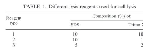
TABLE 1. Different lysis reagents used for cell lysis
Reagent type
Composition (%) of:
SDS Triton X-100
1 10 10
2 10 1
3 5 2
4 2 5
5 2 10
453
on May 15, 2020 by guest
http://jcm.asm.org/
dition, Bacillus sp. (B-22), Escherichia coli (DH 5␣), and
Pseudomonas fluorescens(ATCC 13525) were included as neg-ative controls. For isolation of mycobacteria, samples were plated on Middlebrook 7H10 agar supplemented with oleic acid-albumin-dextrose-catalase enrichment and Lowestein-Jensen agar slants and incubated at 37°C for up to 10 days. Putative Mycobacterium colonies selected based on growth rate, colony morphology, and acid-fast staining reactions were pursued further for confirmation by using the PCR method.
Cell lysis optimization.Five different cell lysis reagents were prepared using various concentrations of sodium dodecyl sul-fate (SDS) and Triton X-100 (Sigma, St. Louis, Mo.) in Tris-EDTA buffer (pH 8.0) as presented in Table 1. Initially, a six-step thermal regime involving different heating-cooling temperatures and incubation times was used in combination with each of the formulated lysis reagents using the GeneAmp PCR system model 9700 (Applied Biosystems, Foster City, Calif.). In order to develop a simpler thermal regime, four other combinations involving shorter heating-cooling regimes were examined, as shown in Table 2. By using a sterile autopi-pette tip, we transferred an isolated mycobacterial colony to an amplification tube (0.2 ml) containing 5l of a selected lysis reagent and resuspended by gentle mixing. The contents were subjected to lysis under the appropriate combinations of chem-ical lysis reagents and thermal regimes listed in Tables 1 and 2 by using the GeneAmp PCR system model 9700. The tube containing the crude DNA-containing lysate was used directly for the subsequent PCR.
Genus-specific PCR.We evaluated two distinct protocols for their applicability to diverse mycobacterial species. The first was an existing genus-specific PCR protocol based on thehsp
gene and involving amplification of a 439-bp region using the recommended primer pair (16); the second was its modified version which uses newly designed broad-spectrum genus-spe-cific PCR primers and optimal amplification conditions (Table 3). Direct cell lysis was used to generate the DNA template.
The PCR amplification reaction with either of the primer pairs was performed by using Ex-Taq DNA polymerase and the compatible PCR reagents (Panvera, Madison, Wis.) in the GeneAmp PCR system model 9700. The reaction mixture (50
l) consisted of 5l of DNA template (cell lysate), 1⫻Ex-Taq
buffer with MgCl2, 200M of each of the four deoxynucleoside
triphosphates, 100 ng of both the forward and reverse primers, and 1.25 U of ex-TaqDNA polymerase. The presence of PCR products was determined by electrophoresing 10 l of the reaction product on a 1% Trevigel gel matrix (Trevigen, Gaith-ersburg, Md.) with 1⫻ Tris-acetate-EDTA buffer containing ethidium bromide (0.5g/ml) and using 5l of a 100-bp DNA size marker (PGC Scientifics [Frederick, Md.] and Invitrogen [Carlsbad, Calif.]). Each PCR product was further quantitated and photographed by using the Kodak EDAS 290 gel docu-mentation system (Kodak, Rochester, N.Y.).
Amplicon identification by restriction analysis and DNA sequencing.The mycobacterial origin of the PCR products was confirmed by restriction analysis. A protocol was optimized based on the uniqueNarI restriction site in these amplicons as determined by aligning the availablehsp gene sequences for this region. A randomly selected PCR amplicon (correspond-ing to the isolate M-JY3) was also sequenced at the University of Cincinnati’s DNA core facility for confirmation. When an-alyzed with a BLAST search against available gene database, the sequence showed homology with hspgene sequences for mycobacteria. The sequence showed closest homology (99%) with theM. chelonae hspsequence.
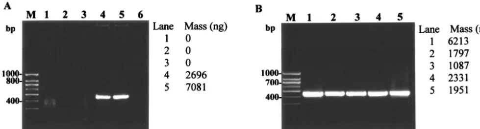
Of the five lysis reagents tested with a six-step heating re-gime (Tables 1 and 2), only reagent 4 (containing 2% SDS and 5% Triton X-100) and reagent 5 (2% SDS and 10% Triton X-100) yielded an effective cell lysis result as a prestep to PCR amplification (Fig. 1A). However, for some mycobacterial iso-lates such as M-JY4, lysis reagent 4 was not as effective as lysis reagent 5 (data not shown). The results showed that the pro-portional ratio of the two chemicals (SDS and Triton X-100) is important for achieving optimal cell lysis without interfering with the subsequent amplification reaction step. Hence, re-agent 5 was selected as the optimum lysis rere-agent and was used for subsequent thermal regime optimizations. The six-step thermal regime was further modified and shortened by exam-ining four other heating-cooling regimes (Fig. 1B). The five regimes tested gave nearly comparable cell lysis results for all reference strains and culture isolates when used with the op-timal lysis reagent 5. Hence, the shortest thermal regime (V) involving heating at 98°C for 5 min followed by cooling at 4°C for 1 min was selected for the optimized protocol. This shows that the heating regime is not as critical as the chemical com-position of the lysis reagent in achieving the desired cell lysis.
TABLE 2. Different thermal regimes used for cell lysis
Step no.
Parameters for thermal regimea:
I II III IV V
H T H T H T H T H T
1 80 1 80 1 80 1 95 1 98 5
2 8 0.5 4 0.5 4 0.5 4 0.5 4 1
3 95 5 95 8 95 5 95 5
4 8 1 4 1 4 1 4 1
5 95 3
6 4 1
[image:2.603.42.549.654.707.2]aH, heating-cooling temperature (°C); T, time (minutes).
TABLE 3. Oligonucleotide primers and conditions used forhsp-based PCRs for mycobacteria
Amplicon length (bp)
Direction and sequence of primer
PCR program Forward (5⬘–3⬘) Reverse (5⬘–3⬘)
439 ACCAACGATGGTGTGTCCAT CTTGTCGAACCGCATACCCT Aa
228 CTGGTCAAGGAAGGTCTGCG GATGACACCCTCGTTGCCAAC Bb
aProgram A consisted of 35 cycles of 94°C for 60 s, 60°C for 60 s, and 72°C for 60 s. bProgram B consisted of 30 cycles of 95°C for 15 s, 58°C for 15 s, and 72°C for 30 s.
on May 15, 2020 by guest
http://jcm.asm.org/
FIG. 1. Optimization of cell lysis protocol for direct PCR-based detection of mycobacteria. (A) Optimization of chemical reagent for cell lysis as a prestep inhsp-based PCR amplification ofM. smegmatis. Lanes 1 to 5, comparison of five different lysis reagents (reagents 1 through 5) containing various concentrations of SDS and Triton X-100 in Tris-EDTA buffer used in combination with thermal regime I (Table 1); lane 6, negative control (no template DNA). (B) Optimization of heating regimes for cell lysis ofM. smegmatisusing lysis reagent 5 (Table 1) in the
hsp-based PCR. Lanes 1 to 5, comparison of thermal regimes 1 through 5 (Table 2); PCR amplification was based on mycobacterium-specifichsp
[image:3.603.48.538.73.205.2]gene with an expected 439-bp amplicon as described in the text. Lane M, 100-bp DNA size marker (PGC Scientifics).
FIG. 2. Evaluation of the optimized protocols for cell lysis andhsp-based PCR for mycobacteria. (A) Evaluation of the optimized cell lysis protocol (lysis reagent 5, thermal regime V) forhsp-based PCR amplification (439 bp) of mycobacterial reference strains and MWF isolates. Panel I: lanes 1 to 4,M. smegmatis,M. chelonae,M. immunogenum, andM. bovis; lane 5, no-template control. Panel II: lanes 1 to 8,M. smegmatis,M. chelonae,M. immunogenum, andMycobacteriumisolates M-JY1, M-JY2, M-JY3, M-JY4 and M-JY5; lane M, 100-bp DNA size marker (PGC Scientifics). PCR primers and conditions were the same as described in the legend of Fig. 1. (B) Evaluation of newhsp-based PCR primers and modified PCR conditions for genus-specific PCR amplification (228 bp) of mycobacteria. Lanes 1 to 3, M. smegmatis,M. chelonae, and M. immunogenum; lanes 4 to 11,Mycobacteriumisolates M-JY1, M-JY2, M-JY3, M-JY4, M-JY5, M-JY6, M-JY7 and M-JY8; lane M, 100-bp DNA size marker (PGC Scientifics). Cells were lysed using the optimized direct cell lysis method as described for panel A, and the lysates were amplified using new PCR primers and conditions described in the text for amplification of a 228-bp PCR product of thehspgene.
on May 15, 2020 by guest
http://jcm.asm.org/
Due to the relatively resistant cell walls of mycobacteria, var-ious combinations of chemicals (such as SDS and Triton X-100), mechanical devices, heat, and solvents are often ap-plied for the conventional extraction and purification of DNA from mycobacteria (1, 2, 15). However, these protocols are cumbersome and often time-consuming, and they fail to yield PCR-quality DNA; they also increase the chances of contam-ination or exposure during analysis. Our developed lysis pro-tocol involves direct single-tube cell lysis in the PCR tube using a one-step heating cycle and has the potential for high-throughput applications as well as offering a safer alternative to conventional protocols. Single-tube cell lysis also makes the protocol ideal for use in quantitative PCR applications. The direct cell lysis procedure was also found to be equally effective against other bacteria, including both negative and gram-positive bacteria (data not shown).
A 439-bp region of thehspgene has been used as a PCR target for the identification and species differentiation in my-cobacteria by restriction enzyme analysis or nucleotide se-quencing (16, 18, 19, 21). In the preceding amplifications using this existinghsp-based PCR method, one of the four reference strains and two of the eight isolates yielded weak amplification signals (Fig. 2A [I and II]). Quantification of the amplified PCR products using the Kodak gel documentation system showed a range of DNA concentrations (Fig. 2A [I and II]). Amplicon signal variability among the reference strains (Fig. 1 and 2A) was traced to nucleotide sequence variation in the primer binding regions based on multiple alignment of their
hspsequences using MegAlign 5.0 software (DNASTAR, Inc., Madison, Wis.). Hence, in order to achieve unambiguous PCR amplification for all NTM species, including the three strains showing weak amplification, a new genus-specific primer pair based on the sequence alignment of the available hsp gene sequences was designed. We selected modified PCR amplifi-cation conditions compatible with the melting point values for the new primers as determined by using Gene Runner soft-ware. When the optimized lysis protocol was used, the modi-fied protocol yielded a 228-bp PCR product of comparable
intensity and concentration for all tested isolates and strains (Fig. 2B). All control strains of various nonmycobacterial gen-era were negative, thus confirming the genus-specific nature of the developed protocol. Moreover, the modified PCR condi-tions optimized for the new primers resulted in a relatively rapid amplification compared to that of the existing method (Table 3).
TheNarI digestion of the amplicons, performed using rec-ommended digestion conditions (New England Biolabs, Bev-erly, Mass.), yielded two fragments (192 and 36 bp) for the reference strains and the eight isolates of mycobacteria (Fig. 3). The developed genus-specific protocol for restriction pat-tern analysis of the amplicons enabled rapid confirmation of the mycobacterial identity of the environmental isolates.
In conclusion, the developed direct cell lysis-based PCR protocol requires less than 2 h for identification of a putative mycobacterial isolate, involving a 6-min lysis step followed by a 50-min amplification and a 30- to 60-min gel analysis. The single-tube protocol is potentially adaptable as a diagnostic tool in routine analytical and clinical laboratories for rapid or high-throughput screening with minimized risks of contamina-tion or exposure. Confirmacontamina-tion of the mycobacterial origin of the PCR product may require an additional hour for restriction analysis. The detection time (2.5 to 3.0 h) could be further shortened in situations in which the real-time format for PCR is available, as the use of such a format would allow early detection of the amplicon as well as confirmation of the my-cobacterial origin of the amplicon based on melting-curve analysis.
Nucleotide sequence accession number.The sequence de-scribed herein has been submitted to GenBank (accession number AY322157).
The study was supported by a Centers for Disease Control and Prevention National Institute of Occupational Safety and Health grant (number 1R01OH007364-01A1 to J.S.Y.).
We thank Milacron management for providing the used MWF sam-ples for isolation of mycobacteria.
FIG. 3. Confirmation of the mycobacterial origin of PCR amplicons (228 bp) based onNarI restriction patterns (192 and 36 bp). Shown are gel patterns forM. chelonaereference strain (lane 1 and 2) and for NTM field isolates M-JY1 (lanes 3 and 4), M-JY2 (lanes 5 and 6), M-JY3 (lanes 7 and 8), M-JY4 (lanes 9 and 10), M-JY5 (lanes 11 and 12), M-JY6 (lanes 13 and 14), M-JY7 (lanes 15 and 16), and M-JY8 (lanes 17 and 18); lane M, 100-bp DNA size marker (Invitrogen). For each isolate, the two lanes represent the original amplicon (8l) and itsNarI restriction digestion product, respectively.
on May 15, 2020 by guest
http://jcm.asm.org/
[image:4.603.139.446.68.222.2]REFERENCES
1. Armand, M. O., M. Mestdagh, and F. Porteals.2001. DNA isolation from chloroform/methanol-treated mycobacterial cells without lysozyme and pro-teinase K. BioTechniques30:272–274.
2. Bollet, C., M. J. Gevaudan, X. de Lamballerie, C. Zandotti, and P. D. Micco.
1991. A simple method for the isolation of chromosomal DNA from gram positive or acid-fast bacteria. Nucleic Acids Res.19:1955.
3. Covert, T. C., M. R. Rodgers, A. L. Reyes, and G. N. Stelma, Jr.1999. Occurrence of nontuberculous mycobacteria in environmental samples. Appl. Environ. Microbiol.65:2492–2496.
4. Dailloux, M., C. Laurain, M. Weber, and P. Hartemann.1999. Water and nontuberculosis mycobacteria. Water Res.33:2219–2228.
5. De Baere, T., R. de Mendonca, G. Claeys, G. Verschraegen, W. Mijs, R. Verhelst, S. Rottiers, L. Van Simaey, C. De Ganck, and M. Vaneechoutte.
2002. Evaluation of amplified rDNA restriction analysis (ARDRA) for the identification of cultured mycobacteria in a diagnostic laboratory. BMC Microbiol.2:4–15.
6. Falkinham, J. O., III.2002. Nontuberculous mycobacteria in the environ-ment. Clin. Chest Med.23:529–551.
7. Fraser, V., and R. J. Wallace, Jr.1996. Nontuberculous mycobacteria, p. 1224–1338.InC. Glen. Mayhall (ed.), Hospital epidemiology and infection controls. Williams & Wilkins, Baltimore, Md.
8. Freeman, A., J. Lockey, P. Hawley, P. Biddinger, and D. Trout.1998. Hy-persensitivity pneumonitis in a machinist. Am. J. Ind. Med.34:387–392. 9. Kirschner, P., B. Springer, U. Vogel, A. Meier, A. Wrede, M. Kiekenbeck,
F. C. Bange, and E. C. Bottger.1993. Genotypic identification of mycobac-teria by nucleic acid sequence determination: report of a 2-year experience in a clinical laboratory. J. Clin. Microbiol.31:2882–2889.
10. Lummus, Z. L., J. E. Lockey, and I. L. Bernstein.1998. Microbial flora of metalworking fluids associated with occupational respiratory disorders. J. Allergy Clin. Immunol.1:166.
11. Miyazaki, Y., H. Koga, S. Kohno, and M. Kaku.1993. Predictive value of PCR applied to clinical samples forMycobacterium tuberculosisdetection. J. Clin. Microbiol.31:2228–2232.
12. Muilenberg, M. L., H. A. Berge, and T. Sweet.1993. Hypersensitivity pneu-monitis and exposure to acid-fast bacilli in coolant aerosols. J. Allergy Clin. Immunol.91:311.
13. Reischl, U., M. Pulz, W. Ehret, and H. Wolf.1994. PCR-based detection of
Mycobacteriumin sputum samples using a simple and reliable DNA extrac-tion protocol. BioTechniques17:844–845.
14. Shelton, G. B., W. D. Flanders, and K. G. Morris.1999.Mycobacteriumsp. as a possible cause of hypersensitivity pneumonitis in machine workers. Emerg. Infect. Dis.5:270–273.
15. Talaat, A. M., R. Reimschuessel, and M. Trucksis.1997. Identification of mycobacteria infecting fish to the species level using polymerase chain re-action and restriction enzyme analysis. Vet. Microbiol.58:229–237. 16. Telenti, A., F. Marchesi, M. Balz, F. Bally, E. C. Bottger, and T. Bodmer.
1993. Rapid identification of mycobacteria to the species level by polymerase chain reaction and restriction enzyme analysis. J. Clin. Microbiol.31:175– 178.
17. Wallace, R. J., Jr., V. A. Silcox, M. Tsukamura, B. A. Brown, J. O. Kilburn, W. R. Butler, and G. Onyi.1993. Clinical significance, biochemical features, and susceptibility patterns of sporadic isolates of theMycobacterium chelo-nae-like organism. J. Clin. Microbiol.31:3231–3239.
18. Wallace, R. J., Jr., Y. Zhang, R. W. Wilson, L. Mann, and H. Rossmoore.
2002. Presence of a single genotype of the newly described species Myco-bacterium immunogenumin industrial metalworking fluids associated with hypersensitivity pneumonitis. Appl. Environ. Microbiol.68:5580–5584. 19. Wilson, R. W., V. A. Steingrube, E. C. Bottger, B. Springer, B. A.
Brown-Elliott, V. Vincent, K. C. Jost, Jr., Y. Zhang, M. J. Garcia, S. H. Chiu, G. O. Onyi, H. Rossmoore, D. R. Nash, and R. J. Wallace, Jr.2001.Mycobacterium immunogenumsp. nov., a novel species related toMycobacterium abscessus
and associated with clinical disease, pseudo-outbreaks and contaminated metalworking fluids: an international cooperative study on mycobacterial taxonomy. Int. J. Syst. Evol. Microbiol.51:1751–1764.
20. Yadav, J. S., I. U. H. Khan, F. Fakhari, and M. B. Soellner.2003. DNA-based methodologies for rapid detection, quantification, and species- or strain-level identification of respiratory pathogens (Mycobacteria and Pseudomonads) in metalworking fluids. Appl. Occup. Environ. Hyg.18:966– 975.
21. Yakrus, M. A., S. M. Hernandez, M. M. Floyd, D. Sikes, W. R. Butler, and B. Metchock.2001. Comparison of methods for identification of Mycobac-terium abscessusandM. chelonaeisolates. J. Clin. Microbiol.39:4103–4110. 22. Yoder, S., C. Argueta, A. Holtzman, T. Aronson, O. G. Berlin, P. Tomasek, N. Glover, S. Froman, and G. Stelma, Jr.1999. PCR comparison of Myco-bacterium aviumisolates obtained from patients and foods. Appl. Environ. Microbiol.65:2650–2653.

![N (2 Hydroxyethyl) 2 [3 (p tolyl)triazen 1 yl]benzamide](data:image/gif;base64,R0lGODlhAQABAIAAAP///wAAACH5BAEAAAAALAAAAAABAAEAAAICRAEAOw==)