ScholarWorks @ Georgia State University
ScholarWorks @ Georgia State University
Biology Dissertations Department of Biology
4-26-2010
Anatomy and Physiology of the Nucleus Paragigantocellularis:
Anatomy and Physiology of the Nucleus Paragigantocellularis:
Neural Regulation of Genital Reflexes in Male and Female Rats
Neural Regulation of Genital Reflexes in Male and Female Rats
Joseph Jeremy Normandin
Georgia State University
Follow this and additional works at: https://scholarworks.gsu.edu/biology_diss
Part of the Biology Commons
Recommended Citation Recommended Citation
Normandin, Joseph Jeremy, "Anatomy and Physiology of the Nucleus Paragigantocellularis: Neural Regulation of Genital Reflexes in Male and Female Rats." Dissertation, Georgia State University, 2010. https://scholarworks.gsu.edu/biology_diss/73
ANATOMY AND PHYSIOLOGY OF THE NUCLEUS PARAGIGANTOCELLULARIS:
NEURAL REGULATION OF GENITAL REFLEXES IN MALE AND FEMALE RATS
By
JOSEPH J. NORMANDIN
Under the Direction of Dr. Anne Z. Murphy
ABSTRACT
The supraspinal control of descending inhibition of genital reflexes (such as ejaculation) is poorly
understood but is important in our global comprehension of how neural signals are integrated to
pro-duce sexual behavior, and in our understanding of sexual dysfunction. Sexual dysfunctions, such as
premature ejaculation/delayed ejaculation in men, and involuntary vaginal spasms, dyspareunia, and
anorgasmia in women, are common. An underlying dysregulation of genital reflexes may produce these
dysfunctions, especially in those individuals being treated for depression and anxiety with serotonergic
drugs. The nucleus paragigantocellularis (nPGi) of the rat medulla has been described as a descending
inhibitory system for genital reflexes in rats, and a homologue is known in humans. Through retrograde
tracing of nPGi afferents with the tracer Fluorogold in rats, we found that a number of brain regions
implicated in sexual behavior, such as the medial preoptic area, paraventricular nucleus of the
hypotha-lamus, and periaqueductal gray (PAG) provide sexually dimorphic projections to the nPGi, and that many
found that excitotoxic lesions of the nPGi with N-methyl-D-aspartate facilitate male sexual behavior by
reducing the number of intromissions required for ejaculation, and decreasing ejaculation latency. In
females, such lesions attenuated sexual behavior by reducing the amount of time the female spent
mat-ing and reducmat-ing the reinforcement value of vaginocervical stimulation. Lastly, we found that by
remov-ing the source of serotonin to the nPGi (from the ventrolateral PAG) with the serotonergic neurotoxin
5,7-DHT in male rats, we were able to mimic the effects of nPGi lesions and facilitated male sexual
be-havior indicating that serotonin neurotransmission at the level of the nPGi is critical for genital reflex
control. Taken together our results indicate that the nPGi is an important site of integration of internal
signals for the regulation of sexual behavior that is sexually dimorphic and under serotonergic control.
Our understanding of normal and dysfunction genital reflex control, and possible treatment options in
people, is complemented by these results.
ANATOMY AND PHYSIOLOGY OF THE NUCLEUS PARAGIGANTOCELLULARIS:
NEURAL REGULATION OF GENITAL REFLEXES IN MALE AND FEMALE RATS
by
JOSEPH J. NORMANDIN
A Dissertation Submitted in Partial Fulfillment of the Requirements for the Degree of
Doctor of Philosophy
in the College of Arts and Sciences
Georgia State University
Copyright by Joseph Jeremy Normandin
ANATOMY AND PHYSIOLOGY OF THE NUCLEUS PARAGIGANTOCELLULARIS:
NEURAL REGULATION OF GENITAL REFLEXES IN MALE AND FEMALE RATS
by
JOSEPH J. NORMANDIN
Committee Chair: Anne Murphy
Committee: Andrew Clancy
Charles Derby
Aras Petrulis
James Pfaus
Electronic Version Approved:
Office of Graduate Studies
College of Arts and Sciences
Georgia State University
DEDICATION
“Ocian in view O! The Joy!"
-Lewis and Clark Expedition, November, 1805
An academic life is an endless journey. Approaching the end of this phase of my career, I was
struck by this quote from the Lewis & Clark expedition and how it spoke to me of finding the end of
one’s journey (in their case, the Pacific Ocean), only to be confronted with a great expanse of possibility.
This work is dedicated to the mentors that have inspired and supported me (and in some cases, taken a
chance on me) throughout my academic life, and who have given me the opportunity to forever be
look-ing over the horizon.
Dr. Richard C. Pillard
Dr. Mary S. Erskine
ACKNOWLEDGEMENTS
Science is not a one-man job, and there have been many who have helped with this work both
directly and indirectly. Thank you to Dr. Anne Murphy for her love, guidance, and fervent support.
Thank you to my dissertation committee for all your help and encouragement: Dr. Andrew Clancy, Dr.
Charles Derby, Dr. Aras Petrulis, and Dr James Pfaus. I am indebted to Dr. Laura Carruth, Dr. Matthew
Grober, Dr. Tim Bartness, and Dr. Elliott Albers for their advice, assistance, teachings, and support. I also
wish to thank my army of minions (Murphy lab undergraduates) for their technical contributions to my
work: Tayisha Vilceus, Hau P. Bui, Alex Iskhakov, Erica Famojure, Brittany Grandy, Amal Abdulahi, Hila
Eichenbaum, and Vincent Laufer.
I enjoyed the excellent administrative support of Latesha Morrison-Warren of the Department
of Biology, and Dixie Elmore and Fatima Adams of the Neuroscience Institute. Thank you to all of the
members of the Animal Resource Program especially Dr. Michael Hart, Dean Blake, and Matthew Davis.
Moral support provided by Lori Eidson, Nicole Victoria, Elizabeth Jeffress, Rick Hanberry, Pam
Maras, Laura Been, Marc Badura, Amanda Arnold, Luis Martinez, Kyle Gobrogge, Dr. Kelli Duncan, Dr.
Dayna Loyd, and Dr. Jamie LaPrairie. Unconditional love provided by my mother Patricia Normandin, my
brother and brother-in-law Michael Normandin and Rob Villegas, and mon coeur Patrick Colot.
This work would not have been possible without the financial support of the National Institutes
of Health (Grant DA16272 to Anne Z. Murphy) the National Science Foundation (Grant IBN9876754 to
the Center for Behavioral Neuroscience) and the Georgia State University William M. Suttles Fellowship
to Joseph J. Normandin.
TABLE OF CONTENTS
ACKNOWLEDGEMENTS ... v
LIST OF TABLES ... ix
LIST OF FIGURES ... x
1 GENERAL INTRODUCTION ... 1
1.1 The Study of Genital Reflexes ... 1
1.2 Neural control of Genital Reflexes ... 2
1.2.1 Anatomy & Physiology ... 2
1.2.2 The Nucleus Paragigantocellularis ... 3
1.2.3 The Influence of Serotonin on Sexual Behavior ... 4
1.2.4 Serotonin and the Nucleus Paragigantocellularis ... 4
1.3 Implications for Human Health ... 5
1.4 Experimental Rationale ... 6
1.4.1 Experiment 1: Nucleus paragigantocellularis afferents in male and female rats: organization, gonadal steroid receptor expression, and activation during sexual behavior ... 6
1.4.2 Experiment 2: Excitotoxic lesions of the nucleus paragigantocellularis facilitate male sexual behavior but attenuate female sexual behavior in rats ... 6
1.4.3 Experiment 3: Serotonergic lesions of the midbrain source of serotonin to the nucleus paragigantocellularis facilitate sexual behavior in male rats ... 7
2 NUCLEUS PARAGIGANTOCELLULARIS AFFERENTS IN MALE AND FEMALE RATS: ORGANIZATION, GONADAL STEROID RECEPTOR EXPRESSION, AND ACTIVATION DURING SEXUAL BEHAVIOR ... 8
2.1 Introduction ... 8
2.2 Materials and Methods ... 10
2.2.1 Subjects ... 10
2.2.2 Fluorogold injections ... 10
2.2.3 Sexual behavior ... 11
2.2.4 Perfusion fixation ... 11
2.2.5 Immunocytochemistry ... 11
2.2.6 Data analysis and presentation ... 13
2.3 Results.. ... 15
2.3.1 Injection sites ... 15
2.3.3 Organization and gonadal steroid receptor expression of nPGi afferents ... 15
2.3.4 Activation of nPGi afferents during sexual behavior ... 19
2.4 Discussion ... 21
2.4.1 Preoptic area ... 21
2.4.2 Amygdala and bed nucleus of the stria terminalis ... 23
2.4.3 Hypothalamus ... 24
2.4.4 Periaqueductal gray ... 25
2.4.5 Superior and inferior colliculus ... 26
2.4.6 The nPGi as a central integrator for arousal ... 27
2.5 Conclusions... 29
3 EXCITOTOXIC LESIONS OF THE NUCLEUS PARAGIGANTOCELLULARIS FACILITATE MALE SEXUAL BEHAVIOR BUT ATTENUATE FEMALE SEXUAL BEHAVIOR IN RATS ... 45
3.1 Introduction ... 45
3.2 Materials and methods ... 47
3.2.1 Subjects ... 47
3.2.2 Ovariectomy and gonadal-steroid replacement ... 47
3.2.3 nPGi lesions ... 47
3.2.4 Sexual behavior ... 48
3.2.5 Conditioned place preference for aVCS ... 48
3.2.6 Perfusion / fixation / tissue preparation ... 50
3.2.7 Immunohistochemistry ... 50
3.2.8 Lesion analysis ... 51
3.2.9 Sexual behavior- and aVCS-induced Fos expression analysis ... 51
3.2.10 Statistical analysis ... 51
3.3 Results.. ... 52
3.3.1 Lesion verification ... 52
3.3.2 Male sexual behavior ... 52
3.3.3 Female sexual behavior ... 52
3.3.4 Female CPP for aVCS ... 53
3.3.5 Male sexual behavior-induced Fos expression ... 54
3.3.6 Female sexual behavior-induced Fos expression ... 54
3.4 Discussion ... 54
3.4.1 Males ... 54
3.4.2 Females ... 56
3.5 Conclusions... 58
4 SEROTONERGIC LESIONS OF THE MIDBRAIN SOURCE OF SEROTONIN TO THE NUCLEUS PARAGIGANTOCELLULARIS FACILITATE SEXUAL BEHAVIOR IN MALE RATS ... 71
4.1 Introduction ... 71
4.2 Methods and materials ... 73
4.2.1 Subjects ... 73
4.2.2 Ovariectomy and gonadal-steroid replacement in stimulus females ... 73
4.2.3 vlPAG 5-HT lesions ... 73
4.2.4 Sexual behavior tests ... 74
4.2.5 Perfusion / fixation / tissue preparation ... 74
4.2.6 Immunohistochemistry ... 75
4.2.7 Lesion analysis ... 75
4.2.8 Statistical analysis ... 76
4.3 Results.. ... 76
4.3.1 Lesion Verification ... 76
4.3.2 Sexual Behavior ... 76
4.4 Discussion ... 77
4.5 Conclusions... 78
5 GENERAL CONCLUSIONS ... 84
REFERENCES ... 88
APPENDICES ... 105
Appendix A – Preliminary data: Manipulation of serotonin at the nucleus paragigantocellularis of male rats produces paradoxical effects on sexual behavior ... 105
Appendix B – Preliminary data: Trans-synaptic tracing of functional genitosensory pathways in male and female rats ... 109
Appendix C – Preliminary data: The paraventricular nucleus of the hypothalamus contains oxytocinergic cells integrated into ascending and descending genital circuitry ... 116
LIST OF TABLES
Table 2.1 Distribution of FG+ cells, and FG+ cells that also contained ERα, AR, or sex behavior-induced Fos following injection of FG into the nPGi ... 30 Table 2.2 Sexual Behavior Measures (mean) in Tracer-injected Male and Female Rats ... 31 Table 3.1 Sexual behavior measures (mean +/- standard error of the mean (SEM)) in control group and
lesion group males ... 59 Table 3.2 Sexual behavior measures (mean +/- SEM) in control group and lesion group females ... 59 Table 3.3 Conditioned place preference measures (mean +/- SEM) in control group and lesion group
females ... 59 Table 4.1 Sexual behavior measures (mean +/- standard error of the mean) in sham group and lesion
group males ... 80 Table B.1 Abundance of H129 labeling in supraspinal sites of male and female rats as a result of genital
LIST OF FIGURES
Figure 2.1 Photomicrograph of Fluorogold injection sites ... 32
Figure 2.2 Distribution of FG+ cells and co-localization with gonadal steroid receptors ... 33
Figure 2.3 Graphs of FG+ cell counts and co-localization with gonadal steroid receptors in the MPO ... 35
Figure 2.4 Graphs of FG+ cell counts and co-localization with gonadal steroid receptors throughout the brain ... 36
Figure 2.5 Photomicrograph of FG+ cells and co-localization with gonadal steroid receptors in the PVN 37 Figure 2.6 Graphs of FG+ cell counts and co-localization with gonadal steroid receptors in the PAG ... 38
Figure 2.7 Distribution of FG+ cells and co-localization with sexual behavior-induced Fos ... 39
Figure 2.8 Graph of FG+ cell counts and co-localization with sexual behavior-induced Fos in the MPO .. 41
Figure 2.9 Graph of FG+ cell counts and co-localization with sexual behavior-induced Fos throughout the brain ... 41
Figure 2.10 Photomicrograph of FG+ cells and co-localization with sexual behavior-induced Fos in the PeF ... 42
Figure 2.11 Photomicrograph of FG+ cells and co-localization with sexual behavior-induced Fos in the PVN ... 42
Figure 2.12 Graph of FG+ cell counts and co-localization with sexual behavior-induced Fos in the PAG .. 43
Figure 2.13 Diagram of regions providing dense input to the nPGi ... 44
Figure 3.1 Photomicrograph of sham lesion and lesion sites ... 60
Figure 3.2 Diagram of lesion sites ... 61
Figure 3.3 Measures of male sexual behaviors related to genital reflex function ... 62
Figure 3.4 Measures of male sexual behavior unrelated to genital reflex function ... 63
Figure 3.5 Measures of stimulus males’ sexual behavior during mating with experimental females ... 64
Figure 3.6 Measures of female receptive and proceptive sexual behavior ... 65
Figure 3.7 Measures of female paced-mating behaviors during sexual behavior ... 66
Figure 3.8 Measures of condition placed preference for aVCS in females ... 67
Figure 3.9 Sexual behavior- and aVCS-induced Fos expression in male and female rats ... 68
Figure 4.1 Photomicrograph of sham lesion and lesion sites ... 81
Figure A.1 Sexual behaviors of male rats treated with mCPP, SB-242,084, or vehicle ... 108
Figure B.1 Photomicrographs of H129 labeling in the brainstem of a male and a female rat ... 114
Figure B.2 Photomicrographs of H129 labeling in the midbrain of a male and a female rat ... 114
Figure B.3 Photomicrographs of H129 labeling in the thalamus, hypothalamus, and amygdala of a male and a female rat ... 115
Figure B.4 Photomicrographs of H129 labeling in the cerebral cortex of a male rat ... 115
Figure C.1 Photomicrograph of PVN cells labeled with FG or H129 ... 121
Figure C.2 Photomicrograph of PVN cells labeled for FG and oxytocin ... 122
Figure C.3 Photomicrograph of PVN cells labeled for H129 and oxytocin ... 122
Figure C.4 Photomicrograph of PVN cells labeled for FG and oxytocin ... 123
1 GENERAL INTRODUCTION
1.1 The Study of Genital Reflexes
The neural circuits underlying genital response to internal and external cues have not been fully
elucidated. In both men and women, internally or externally derived stimulation results in increased
blood flow and engorgement of genital tissue, and rhythmic contractions of genital muscles often
asso-ciated with orgasm (Argiolas and Melis, 2003). All of these processes are reflexive and under the control
of sympathetic, parasympathetic, and somatic spinal efferents (Giuliano and Clement, 2005; McKenna,
2002; Temel et al., 2005); these efferents are subsequently under both excitatory and inhibitory control
from numerous supraspinal sites including the paraventricular hypothalamic nucleus (PVN), nucleus
paragigantocellularis (nPGi), and lumbar spinal cord spinothalamic cells (Liu et al., 1997a; Marson and
McKenna, 1990; Truitt and Coolen, 2002). Coordination of the various circuits controlling genital
reflex-es has been studied extensively in men, with recent interreflex-est in defining homologous circuits in women.
Non-human animal models, in particular the rat, have been particularly informative in elucidating the
basic anatomy and physiology of these circuits (Pfaus et al., 2003). From this work, a general
under-standing of the discrete processes underlying genital reflex control has been established. First, the
au-tonomic nervous system plays the key role in providing the necessary signals for increased blood flow to
the genitalia for both men (erection) and women (genital engorgement) as well as providing signals for
the production of secretive fluids used for lubrication and seminal transfer (McKenna, 2002; Temel et
al., 2005; Yang and Jiang, 2009). Second, the somatic nervous system provides the signals for rhythmic
contractions of the genital musculature in both men (ejaculation: expulsion phase) and women
(vagino-cervical contractions), which are often associated with orgasm (Giuliano and Clement, 2005; Levin, 1998;
Yang and Jiang, 2009). This dissertation is concerned with the latter of these processes in which there
has been considerable interest in understanding how supraspinal sites impose inhibitory and excitatory
1.2 Neural control of Genital Reflexes
1.2.1 Anatomy & Physiology
The bulbospongiosus (also referred to as the bulbocavernosus) and ischiocavernosus muscles
are the primary genital muscles in mammals providing the rhythmic contractions needed for somatic
genital reflexes (Gerstenberg et al., 1990; Meston et al., 2004; Sachs, 1982). They are innervated by the
pelvic and pudendal nerves (Pacheco et al., 1989; Pastelin et al., 2008) from the lower lumbar and upper
sacral divisions (L5-S1) of the spinal cord (de Araujo et al., 1982; Katagiri et al., 1986; Roppolo et al.,
1985). In rats, the spinal motoneuron pools associated with these nerves are referred to as the
dorso-medial nucleus (DM; also referred to as the spinal nucleus of the bulbocavernosus) and dorsolateral
nucleus (DL) of L5-S1 ventral horn (Collins et al., 1991; Katagiri et al., 1986; McKenna and Nadelhaft,
1986; Peshori et al., 1995; Schroder, 1980). The DM and DL are considered homologues of Onuf’s
nuc-leus in humans and rhesus monkeys (Breedlove and Arnold, 1980; Roppolo et al., 1985; Schroder, 1981).
Interneurons within the spinal cord appear to connect primary afferent somatosensory information with
the DM and DL (Collins et al., 1991; Peshori et al., 1995; Wiedey et al., 2008). Descending projections
from other parts of the spinal cord, as well as from supraspinal sites, provide both excitatory and
inhibi-tory drive to these motoneuron pools (Allard et al., 2005; Coolen, 2005).
Supraspinal inhibitory and excitatory modulatory inputs to the DM and DL have been described
in rats (Liu et al., 1997a; Marson and McKenna, 1990; Truitt and Coolen, 2002), cats (Kausz, 1990), and
birds (Berk and Finkelstein, 1983). These excitatory and inhibitory drives compete at the level of the
spinal cord motoneuronal pools to regulate genital reflexes within specific behavioral contexts. A
hy-pothesized ejaculation generator, constituting a central pattern generator for the muscles of ejaculation,
has been described in the lumbar spinal cord of rats, which provides input to the DM and DL (Truitt and
Coolen, 2002; Truitt et al., 2003). Lesions of these lumbar spinothalamic cells abolish the ability of male
crit-ical for ejaculation (Truitt and Coolen, 2002). In addition, the PVN has direct connections to the DM and
DL (Wagner and Clemens, 1991). The PVN has been previously implicated in the control of genital
ref-lexes, and is most likely a source of descending excitatory input to the genital musculature (Chen et al.,
1997; Eaton et al., 1991). With respect to descending inhibition, the nPGi of the medulla provides dense
input to the DM and DL (Tang et al., 1999) and is the hypothesized source of descending inhibition to
genital reflexes (Marson and McKenna, 1990).
1.2.2 The Nucleus Paragigantocellularis
Numerous lines of evidence suggest that the nPGi is the primary source of descending inhibition
of genital reflexes in male rats. The nPGi sends direct descending projections to the L5-S1 motoneuronal
pools that innervate the bulbospongiosus and ischiocavernosus muscles in both male and female rats
(Hermann et al., 2003; Marson and Carson 3rd, 1999; Marson and McKenna, 1996; Tang et al., 1999). In
humans, the homologous structure is referred to as the nucleus paragigantocellularis lateralis (Zec and
Kinney, 2001) and is also believed to be associated with descending inhibition of genital reflexes
(Johnson, 2006).
Lesions of the nPGi in rats consistently result in the facilitation of sexual behavior, as indicated
by a decrease in mount and intromission frequency, ejaculation latency, and an increase the number of
ejaculations to satiety (Yells et al., 1992; Yells et al., 1994). nPGi lesions also decrease the latency to-
and increase the number of ex copula erections (Marson et al., 1992; Marson and McKenna, 1990).
Similarly, electrical stimulation of nPGi neurons produces increased firing latency and decreased
ampli-tude of firing in the DM (Johnson and Hubscher, 1998), consistent with the role of the nPGi as a source
of tonic descending inhibitions of genital reflexes.
To date, the role of the nPGi in female sexual behavior has not been directly tested. However,
anatomical evidence suggests that the nPGi is important for sexual behavior in females. Retrograde
and cervix (Lee and Erskine, 2000) produce labeling in the nPGi of females. In addition, a number of
brain regions associated with sexual behavior project to the nPGi female rats (Marson and Foley, 2004;
Marson and Murphy, 2006; Murphy and Hoffman, 2001; Normandin and Murphy, 2008), suggesting a
role for the nPGi in female sexual behavior.
1.2.3 The Influence of Serotonin on Sexual Behavior
Many behavioral systems in mammals are modulated by serotonin (5-HT), including mood
(Dayan and Huys, 2009) and sexual behavior (Pfaus, 2009). Increased 5-HT levels through systemic
ad-ministration of selective serotonin reuptake inhibitors (SSRIs) produces delayed ejaculation in male rats
(Ahlenius et al., 1980; de Jong et al., 2005a; Vega Matuszcyk et al., 1998) and humans (Hsu and Shen,
1995). Importantly, the effect of 5-HT on sexual behavior is brain region and receptor dependent,
pro-ducing both facilitation and attenuation of sexual behavior. For example, administration of the 5-HT1A
agonist 8-hydroxy-2-(di-n-propylamino) tetralin facilitates male sexual behavior when injected into the
medial preoptic area of the hypothalamus or nucleus accumbens (de Castilhos et al., 2006;
Fernandez-Guasti et al., 1992), while administration of trifluoromethyl-phenyl-piperazine, a 5-HT1B/C agonist to the
same regions inhibits male sexual behavior (Fernandez-Guasti et al., 1992; Hillegaart et al., 1991). To
date, site-specific manipulations of 5-HT have not been used extensively, but are critical for delineating
inhibitory and facilitatory circuits modulating sexual behavior.
1.2.4 Serotonin and the Nucleus Paragigantocellularis
Serotonergic cells within the nPGi send direct descending projections to the DM and DL (Azmitia
and Gannon, 1986; Marson and McKenna, 1992). The human homologue, the nucleus
paragigantocellu-laris lateralis, also contains serotonergic cells (Azmitia and Gannon, 1986). Application of 5-HT to the
spinal targets of the nPGi blocks the urethrogenital reflex (an artificial measure of genital reflexes), an
effect reversed by application of a 5-HT antagonist (Marson and McKenna, 1992). In addition, the SSRI
et al., 2007). These studies make it clear that 5-HT acts at the level of the spinal cord to inhibit genital
reflexes, and that 5-HT from the nPGi is a necessary antecedent for normal inhibition of genital reflexes.
It is unclear, however, whether 5-HT may be acting at the level of the nPGi itself.
The nPGi contains receptors for 5-HT including the 5-HT1A (Thor et al., 1990; 1992), 5-HT1c
(Hoffman and Mezey, 1989), 5-HT2A (Fay and Kubin, 2000; Fonseca et al., 2001), 5-HT2C (Fonseca et al.,
2001), and 5-HT3 (Fonseca et al., 2001) subtypes, although only the 5-HT1A and 5-HT2C subtypes are
found in abundance (Fonseca et al., 2001; Thor et al., 1990). The ventrolateral periaqueductal gray
(vlPAG) is the primary source of 5-HT to the nPGi (Li et al., 2001; Lu et al., 2010; Underwood et al., 1999;
Zeng et al., 1991). To date, the effect of 5-HT neurotransmission at the level of the nPGi has not been
studied.
1.3 Implications for Human Health
Sexual dysfunction is a common problem in both men and women. Up to 31% of men and 43%
of women will experience sexual dysfunction in their lifetime (Laumann et al., 1999). Such dysfunctions
can include loss of sex drive (libido), premature ejaculation, delayed ejaculation, anorgasmia,
involunta-ry vaginal spasms, and dyspareunia (vaginal pain during intercourse; (Breiner, 2004). These dysfunctions
have a large impact on fertility and quality of life experiences (Cameron and Tomlin, 2007). More
alar-mingly, SSRIs used to treat depression and anxiety in people have well-documented sexual dysfunction
side effects that include decreased libido, delayed ejaculation, and anorgasmia (Kennedy and Rizvi,
2009; Schweitzer et al., 2009). These sexual dysfunctions all have an underlying dysregulation of genital
reflexes in common, with 5-HT a key mediator. By identifying the regions of the brain that regulate nPGi
function in males and females, and the role of 5-HT within the nPGi, we can provide new insights and
1.4 Experimental Rationale
Our experiments were designed to address three general gaps in knowledge of how the nPGi
provides descending inhibition of genital reflexes. First, anatomical tract tracing was used to provide a
complete description of brain regions that project to the nPGi in males and females (Experiment 1).
Second, excitotoxic lesions were used to delineate the role of the nPGi in male and female sexual
beha-vior (Experiment 2). The final series of experiments examined the role of 5-HT drive from the vlPAG to
the nPGi in male sexual behavior using 5-HT specific lesions of the vlPAG (Experiment 3). Together these
experiments will provide detailed analysis of nPGi afferents, provide information as to how the nPGi
affects sexual behavior in males and females, and determine whether serotonergic drive to the nPGi is
important in sexual behavior.
1.4.1 Experiment 1: Nucleus paragigantocellularis afferents in male and female rats:
organi-zation, gonadal steroid receptor expression, and activation during sexual behavior
Key to understanding how genital reflexes are regulated, and how this might be disrupted in
sexual dysfunction, is knowledge of what regions of the brain can influence nPGi activity. In Experiment
1, we used anatomical tract tracing to delineate the afferents projections to the nPGi in male and female
rats. As gonadal steroids are key modulators of sexual behavior (van Dis and van de Poll, 1974; Wrobel
and Karasek, 2008), we also examined whether nPGi afferents contained receptors for estrogens and
androgens. We also examined whether sites providing afferent drive to the nPGi were active during
sexual behavior, using the protein Fos (as a marker of neural activity).
1.4.2 Experiment 2: Excitotoxic lesions of the nucleus paragigantocellularis facilitate male
sexual behavior but attenuate female sexual behavior in rats
Previous studies using lesions to examine the role of the nPGi in male sexual behavior used
elec-trolytic (fiber destroying) lesions. Therefore, it is not clear if nPGi neurons and not fibers-of-passage
studies have pointed to the likelihood of its importance to female sexual behavior through anatomical
tract tracing (Lee and Erskine, 2000; Marson and Murphy, 2006). In Experiment 2, we used excitotoxic
lesions of the nPGi in male and female rats to examine the role of nPGi neurons in male and female
sex-ual behavior.
1.4.3 Experiment 3: Serotonergic lesions of the midbrain source of serotonin to the nucleus
paragigantocellularis facilitate sexual behavior in male rats
The nPGi contains receptors for 5-HT (Fonseca et al., 2001; Thor et al., 1990), and serotonergic
input to the nPGi is provided by the vlPAG (Li et al., 2001; Lu et al., 2010; Underwood et al., 1999; Zeng
et al., 1991). However, the role of 5-HT drive to the nPGi in male sexual behavior is unknown. To this
end, in Experiment 3, we produced 5-HT-specific lesions of the vlPAG to the nPGi in male rats and
2 NUCLEUS PARAGIGANTOCELLULARIS AFFERENTS IN MALE AND FEMALE RATS:
ORGANIZA-TION, GONADAL STEROID RECEPTOR EXPRESSION, AND ACTIVATION DURING SEXUAL
BEHA-VIOR
2.1 Introduction
Genital reflexes in males and females subserve critical functions in reproductive biology.
De-spite this obvious importance, little is known about the supraspinal control of these reflexes.
Dysfunc-tions within these systems contribute to, or are the basis of, sexual dysfuncDysfunc-tions that produce profound
disruptions not only in fertility, but also in quality of life experiences (Cameron and Tomlin, 2007;
Laumann et al., 1999). Across the lifespan, approximately 31% percent of men will experience sexual
dysfunction, including an inability to achieve an erection and/or premature or delayed ejaculation
(Laumann et al., 1999). Similarly, 43% of women will experience some form of female sexual
dysfunc-tion, including involuntary vaginal spasms, painful sensations during penetradysfunc-tion, and/or early or
de-layed orgasm (Laumann et al., 1999). Our understanding of the supraspinal control of genital reflexes is
an important contribution to understanding and treating sexual disorders.
Studies in male rats have identified the nucleus paragigantocellularis (nPGi) of the brainstem as
the primary source of tonic descending inhibition of erectile and ejaculatory reflexes. Projections from
the nPGi terminate onto the spinal motor neurons that innervate the bulbospongiosus and
ischiocaver-nosus muscles (Hermann et al., 2003; Murphy and Marson, 2000; Tang et al., 1999); these muscles are
critical to the control of erection and ejaculation (Holmes et al., 1991; McKenna and Nadelhaft, 1986).
Bilateral lesions of the nPGi in male rats decrease mount and intromission frequency, ejaculation
laten-cy, and increase the number of ejaculations to satiety (Yells et al., 1992; Yells et al., 1994). Lesions of
the nPGi also decrease the latency, and increase the number of ex copula erections (Marson et al.,
amplitude of firing in motor neurons associated with genital reflexes (Johnson and Hubscher, 1998).
Interestingly, nPGi lesions do not alter the number of non-contact erections in males exposed to females
behind a wire mesh screen (Liu and Sachs, 1999), suggesting that nPGi modulation of penile reflexes is
context dependent. How this context is signaled to the nPGi remains to be elucidated.
The central regulation of the nPGi is poorly understood. In males rats, both the medial preoptic
area (Murphy et al., 1999a) and the midbrain periaqueductal gray (Lovick, 1986; Murphy and Hoffman,
2001) send direct projections to the nPGi. Interestingly, MPO projections to the PAG terminate in close
apposition to PAG projections to the nPGi, forming an indirect MPO-PAG-nPGi pathway in addition to
the direct MPO-nPGi pathway (Murphy and Hoffman, 2001). In females, remarkably little is known
re-garding the anatomy and physiology of the nPGi. Spinally-projecting nPGi efferents terminate among
the motor neurons involved in the urethrogenital reflex (Marson et al., 2003), and trans-synaptic
retro-grade tracer injection into rat clitoris (Marson, 1995), cervix (Lee and Erskine, 2000), and vagina/clitoris
(Marson and Murphy, 2006) results in dense retrograde labeling in the nPGi. Similar to males, direct
MPO projections to the nPGi of female rats have been reported (Marson and Foley, 2004) and nPGi cells
retrogradely labeled from rat vagina/clitoris with a trans-neuronal tracer were associated with terminals
from the MPO and PAG (Marson and Murphy, 2006). Contributions of other central sites of input to the
nPGi in females remain to be described.
The nPGi can be conceptualized as the final common output from supraspinal sites involved in
sexual behavior to the spinal cord motor neurons controlling genital reflexes. An integration of
priate external and internal signals related to sexual behavior must occur to determine when it is
appro-priate for genital reflexes to be produced. The nPGi may be one such area where an integration of
sig-nals occurs. This study was conducted to comprehensively characterize nPGi afferents as to their
fe-male rats. Possible sex-differences in the anatomical and/or physiological organization of this circuit
were also examined.
2.2 Materials and Methods
2.2.1 Subjects
Adult male (n=56) and female (n=88) Sprague-Dawley rats (Zivic Laboratories Inc., Pittsburgh,
PA) were used in these experiments. Animals were weight-matched (250-300 g) and housed in same-sex
pairs in separate rooms on a 12:12 hour light:dark cycle (lights on at 7:00AM). Animals used in the
sex-ual behavior studies were housed in reverse light cycle (lights off at 7:00AM). Access to food and water
was ad libitum throughout the experiment, except during surgery. These studies were performed in
strict compliance with the Institutional Animal Care and Use Committee at Georgia State University. All
efforts were made to minimize any possible suffering by the animal, and to reduce the number of
ani-mals used.
2.2.2 Fluorogold injections
Rats were anesthetized with ketamine/xylazine/acepromazine (50 mg/kg, 3.3 mg/kg, 3.3 mg/kg;
i.p.; Henry Schein, Melville, NY) and placed in a stereotaxic frame upon achieving a deep surgical plane
of anesthesia. The skull was adjusted so that Bregma and lambda were at the same dorsoventral plane.
Glass micropipettes (10-20 μm) were filled with the retrograde tracer Fluorogold (FG; 2% soln. w/v in
saline; Fluorochrome LLC, Denver, CO) and lowered into the nPGi at the following coordinates (in mm):
AP: -2.0 Lambda; ML: -1.0; DV: -8.5. FG was iontophoresed (50% duty cycle, 7.5 μA current) for 20 min.
Pipettes remained in place for five minutes after injection to prevent backflow of tracer along the
pi-pette tract and to facilitate neuronal uptake. Special care was taken to ensure that all injection protocols
were comparable for males and females. Following surgery, the animals were allowed to recover under
a heat lamp in clean cages, and returned to their original housing facilities upon recovery from the
2.2.3 Sexual behavior
A subset of our FG injected intact males (n=44) and females (n=36) rats engaged in three one
hour mating bouts with stimulus animals before injection of FG. All females were ovariectomized and
hormone-primed (10 µg estradiol in sesame oil 48 hours before mating, 500 µg progesterone in sesame
oil) 4 hours before mating. Mating took place in a 60 cm x 30 cm transparent acrylic arena.
Experimen-tal males had free access to stimulus females, whereas experimenExperimen-tal females engaged in paced-mating
with a separator in the arena (with two 4 cm diameter holes through which only females could fit).
Fol-lowing retrograde tracer injections, the rats engaged in three additional one hour mating bouts, and
were sacrificed on the last mating bout one hour after producing or receiving (male and females
respec-tively) an ejaculation.
2.2.4 Perfusion fixation
Animals were given a euthanizing dose of sodium pentobarbital (160 mg/kg, i.p.; Sigma-Aldrich,
St. Louis, MO) and perfused transcardially with 100-200 ml of 0.9% sodium chloride containing 2%
so-dium nitrite as a vasodilator, followed immediately by 250 ml of 4% paraformaldehyde in 0.1M
phos-phate buffer containing 2.5% acrolein (Polysciences Inc., Warrington, PA). Following fixation, a final rinse
(100-200 ml) with the sodium chloride/sodium nitrate solution was used to remove any residual acrolein
from the animal. Immediately following perfusion, the brains were removed and stored at 4°C in 30%
sucrose solution until sectioned. The brains were cut into 25 µm coronal sections with a Leica 2000R
freezing microtome and stored free-floating in cryoprotectant-antifreeze solution (Watson et al., 1986)
at –20°C until immunocytochemical processing was initiated.
2.2.5 Immunocytochemistry
A 1:6 series through the rostrocaudal axis of each brain was processed for FG and estrogen
re-ceptor alpha (ERα), FG and androgen receptor (AR), or FG and the immediate-early gene product Fos as
re-moved from the cryoprotectant-antifreeze solution, rinsed extensively in potassium phosphate-buffered
saline (pH 7.4), and then reacted for 20 minutes in 1% sodium borohydride to remove excess aldehydes.
Sections were then incubated in primary antibody solution directed against either ERα, AR, or Fos in
po-tassium phosphate buffered saline (KPBS) containing 0.1% Triton-X for 1 hour at room temperature
fol-lowed by 48 hours at 4°C. Cells containing ERα were identified using a polyclonal rabbit anti-ERα antibody
(Santa Cruz Biotech Inc., sc-542, Lot: B1505, Santa Cruz, CA) at a concentration of 1:20,000. This rabbit
antiserum was prepared against a peptide mapping at the C-terminus of ERα of mouse origin
(HSLQTYYIPPEAEGFPNTI) corresponding to amino acids 580-599 (manufacturer’s technical information),
and specificity of this antibody has been confirmed by preabsorbtion with epitope peptide (Quesada et
al., 2007). Cells containing AR were identified using the polyclonal rabbit anti-AR antibody (Santa Cruz
Biotech Inc., sc-816, Lot: B0204, Santa Cruz, CA) at a concentration of 1:10,000. This rabbit antiserum
was prepared against a peptide mapping at the N-terminus of AR of human origin
(MEVQLGLGRVYPRPPSKTYRG) corresponding to amino acids 2-21 (manufacturer’s technical
informa-tion), and specificity of this antibody has been confirmed by preabsorbtion with epitope peptide (Creutz
and Kritzer, 2004). Cells containing Fos were identified using the polyclonal rabbit anti-Fos antibody
(Calbiochem, PC38, Lot: 4191, San Diego, CA) at a concentration of 1:700,000. This rabbit antiserum was
prepared against a synthetic peptide (SGFNADYEASSSRC) corresponding to amino acids 4-17 of human
c-Fos, and specificity of this antibody has been confirmed by preabsorbtion with epitope peptide (Sun et
al., 2007). In Western blots, this antibody recognizes the ~55 kDa c-Fos and ~62 kDa c-Fos proteins, and
does not cross-react with the ~39 kDa Jun protein (manufacturer’s technical information).
After primary antibody incubation, the tissue was rinsed in KPBS, incubated for 1 hour in
biotiny-lated goat anti-rabbit IgG (Jackson Immunoresearch Labs Inc., West Grove, PA) at a concentration of
1:600, rinsed in KPBS, followed by a 1 hour incubation in avidin-biotin peroxidase complex (Vector Labs,
ace-tate (0.175 M; pH 6.5), ERα, AR, and Fos were visualized as a black reaction product using nickel sulfate
intensified 3,3’-diaminobenzidine solution containing 0.08% hydrogen peroxide in sodium acetate
buf-fer. The reaction product was terminated after approximately 30 minutes by rinsing in sodium acetate
buffer. Tissue was then processed for FG immunoreactivity as above. Cells containing FG were
identi-fied using the polyclonal rabbit anti-Fluorogold antibody (Chemicon, AB153, Lot: 25060005, Temecula,
CA) at a concentration of 1:30,000. This antibody was raised against the chemical compound
hydroxys-tildamidine (Fluorogold; manufacturer’s technical information). No cytoplasmic FG+ staining was
present in animals in which the tracer failed to be ejected from the electrode. FG was visualized as a
brown reaction product using 3,3’-diaminobenzidine containing 0.08% hydrogen peroxide in Tris buffer
(pH 7.2). The reaction product was terminated after approximately 30 minutes by rinsing in Tris buffer.
Sections were mounted out of saline onto gelatin-subbed slides, air dried overnight, dehydrated in a
series of graded alcohols, cleared in Histoclear, and cover-slipped using Histomount.
2.2.6 Data analysis and presentation
A single individual conducted all analyses. Animals were coded to blind the counter to sex and
animal number. Injection sites were subjectively graded with respect to their location and spread. Only
those animals with injections that corresponded to a focal point within the nPGi were selected for cell
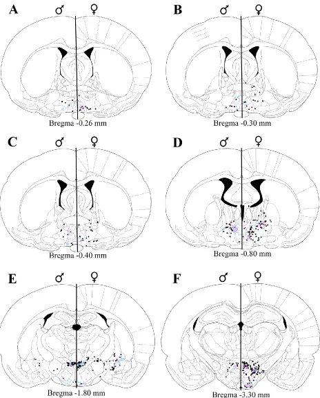
counting (see Figure 1). Thirteen males and 15 females had injections that met our strict and
conserva-tive criteria. Of these animals, all were used for our retrograde tracing analysis, 6 males and 7 females
were used in our retrograde tracer/gonadal-steroid receptor expression analysis, and 5 males and 6
females were used in our retrograde tracer/sexual behavior-induced Fos expression analysis.
Missed injections dorsal or lateral to the nPGi (i.e. into the pyramidal tract) were examined for
specificity of retrograde tracing in target injections. All regions containing FG labeling were examined.
The MPO and PAG were additionally subdivided into four (Bregma -0.26, -0.30, -0.40, -0.80) and six
ERα/FG+, AR/FG+, or Fos/FG+ cells were determined on the side ipsilateral to the injection site, and FG+
labeling on the side contralateral to the injection site were noted, but not counted. FG+ cells were
easi-ly identifiable based on the brown reaction product in the cytoplasm. FG+ cells that contained ERα, AR,
or Fos were readily identifiable based on the black reaction product restricted to the nucleus. The
min-imum distance between any two sections analyzed was 40 µm (Bregma -0.26 and -0.30). The majority of
retrograde labeling in this area was observed in the MPO, where the maximum diameter of the largest
cells can be expected to be approximately 12 µm (calculated from (Madeira et al., 1999); therefore cell
counts between these sections (and any two sections) are independent and do not reflect duplicate
counting of cells.
Single and dual-labeled cells were plotted and quantified across the rostrocaudal axis of the rat
brain based on the atlas of (Paxinos and Watson, 1997). The mean and standard error of the mean
(SEM) of FG+, ERα/FG+, AR/FG+, and Fos/FG+ cells for each sex, section, and region of interest were
cal-culated. We use the term “density” within this manuscript as shorthand for the number or proportion
of cells observed within a given brain region. The relative density of FG+ labeled cells was defined as
sparse when <25 labeled cells were observed and dense when ≥25 cells were observed. The relative
density of the percent co-localization of FG+ cells with ERα, AR, or Fos was defined as low when <10%,
moderate when between 10% and 24%, and high when >25%.
Behavioral measures for our sexual behavior-induced Fos experiments include the mean
num-ber of mounts (M), intromissions (I), and ejaculations (E) in males, and the mean numnum-ber of M/I/E
re-ceived, lordosis quotient (LQ; lordosis/M+I+E received), and lordosis rating (LR; magnitude of
dorsoflex-ion; 0 = none, 1 = slight, 2 = full) as defined in (Pfaus, 1996).
Statistical comparisons of FG+, ERα/FG+, AR/FG+, and Fos/FG+ cell numbers were made between
males and females for regions providing dense input to the nPGi. T-tests were used for mean
MPO and PAG. The family-wise alpha level (≤0.05) was adjusted to ≤0.0031 using the Bonferroni method
for the sixteen unplanned comparisons.
For data presentation, a representative animal from each experimental group was selected and
the distribution of FG+, ERα/FG+, AR/FG+, and Fos/FG+ cells were plotted using a Nikon Drawing Tube
attached to a Nikon Optiphot microscope. Plots were scanned, imported to a computer, and finalized
using Adobe Illustrator 10. Photomicrographs were generated using a Synsys digital camera attached to
a Nikon Eclipse E800 microscope. Images were captured with QCapture and finalized using Adobe
Pho-toshop 7.0. Alterations to the images were strictly limited to enhancement of brightness/contrast.
2.3 Results
2.3.1 Injection sites
Injections were centered within the rostral portion of the nPGi with moderate spread to the
py-ramidal tract. Subjective blind ratings of injection quality did not differ between the sexes. Figure 1
shows a representative FG injection site within the nPGi of a male and female rat.
2.3.2 General patterns in labeling
Table 1lists all regions that contained FG+ cells in male and female rats, weighted by density of
FG+ cells and co-localization with ERα, AR, or Fos. Regions ipsilateral to the injection site showed a
quali-tatively higher degree of retrograde labeling than contralateral regions. There were a few notable
ex-ceptions: the rostral parvocellular red nucleus (RPC) and rostral portion of the ventral nucleus of the
lateral lemniscus in males, and the caudal portion of the RPC and the paralemniscal nucleus (PL) in
fe-males had a qualitatively higher degree of retrograde labeling on the side contralateral to the injection
site.
2.3.3 Organization and gonadal steroid receptor expression of nPGi afferents
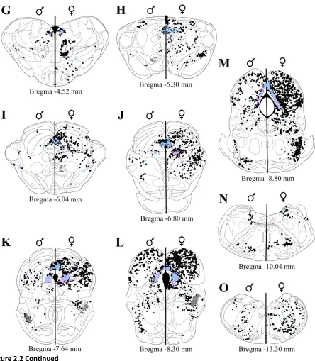
Diencephalon. Representative plots of FG+, ERα/FG+, and AR/FG+ cells in the diencephalic
MPO (Figure 2A-B). However, within the caudal half of MPO (Figure 2C-D), the number of FG+ cells
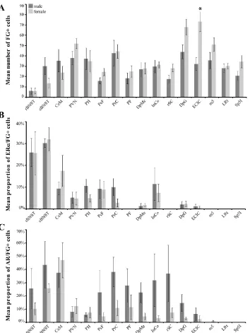
in-creased significantly in males [p=0.014; Figure 3A]. FG labeling in the caudal MPO in males formed a
discrete cell group that was not consistently observed in females (Figure 2D). The proportion of FG+
cells that contained ERα varied for males and females along the rostrocaudal axis of MPO (Figure 3B). In
the rostral pole of MPO, 33% of FG+ cells in females also contained ERα, whereas only 9% of FG+ cells
contained ERα in males (Figure 3B). For the androgen receptor, at most rostrocaudal levels, males
gen-erally had a higher percent co-localization than females (mean 37% AR/FG+ cells versus 18% in females),
and males differed from females significantly at two rostrocaudal levels [Bregma -0.26, p=0.024 and
Bregma -0.40, p=0.037; Figure 3C]. The lateral preoptic area was sparsely labeled for FG in both sexes.
In the bed nucleus of the stria terminalis (BNST; Figure 2A-D), retrograde labeling was primarily
restricted to the caudal regions (Figure 2C-D) in both males and females (Figure 4A). ERα and AR
co-localization with FG+ caudal BNST cells was comparable in both sexes (approximately 30%; Figure 4B).
With respect to the androgen receptor, 43% of FG+ caudal BNST cells contained AR in males, and 26%
co-localized in females (Figure 4C).
Within the amygdala (Figure 2E-F), the medial amygdala exhibited sparse FG+ labeling in both
sexes. In contrast, dense FG+ labeling was present in the central amygdala (CeM) in males, with less
labeling observed in females (Figure 4A). ERα co-localization with FG+ CeM cells was low in males (9%),
and moderate in females (17%; Figure 4B). For AR, higher levels of co-localization in the CeM was
ob-served in both sexes (between 40-50%; Figure 4C).
Dense FG+ labeling in the hypothalamus (Figure 2E-F) was observed in the paraventricular
nuc-leus (PVN) and posterior hypothalamus (PH) of both sexes, and the perifornical nucnuc-leus (PeF) in females
(Figure 4A). Figure 5 shows an example of FG labeled cells in the PVN for males and females. These
re-gions exhibited low levels of ERα (Figure 4B) or AR (Figure 4C and 5) co-localization, with the exception of
the anterior hypothalamus, lateral hypothalamus, retrochiasmatic area, dorsomedial hypothalamus,
ventromedial hypothalamus, arcuate nucleus, tuber cinereum area, and the magnocellular nucleus of
the lateral hypothalamus in both sexes.
In the thalamus (Figure 2E-G) dense FG+ labeling was observed in the precommissural nucleus
(PrC) in both sexes and the parafascicular thalamic nucleus (PF) in females (Figure 4A). ERα
co-localization with FG+ PrC cells was moderate in males (10%) and low in females (3%; Figure 4B). By
con-trast, AR co-localization with FG+ PrC cells was higher (males, 38%; females, 11%; Figure 4C). In the PF,
while ERα was present, there were no ERα/FG+ cells observed. However, AR/FG+ cells were observed in
males (28%) and females (12%; Figure 4C) in this same region. Sparse FG+ labeling was observed in the
rostral interstitial nucleus of the medial longitudinal fasciculus, prerubral field, parvocellular
subparafas-cicular nucleus, subincertal nucleus, and zona incerta in both sexes.
Mesencephalon. Representative plots of FG+, ERα/FG+, and AR/FG+ labeling in the
mesence-phalic regions are shown in Figure 2. In the midbrain (Figure 2G-M), dense FG+ labeling was observed in
the deep mesencephalic nucleus (DpMe) and intercollicular nucleus (InCo) in both sexes (Figure 4A),
with low to moderate levels of ERα co-localization (Figure 4B). A greater percentage of AR/FG+ cells
were observed in males within the DpMe (23% versus 5% in females (Figure 4C). Sparse FG+ labeling
was observed in the oculomotor nucleus, Edinger-Wetsphal nucleus, nucleus of the posterior
commis-sure, magnocellular nucleus of the posterior commiscommis-sure, interstitial nucleus of the medial longitudinal
fasciculus, RPC, retrorubral field, posterior intralaminar thalamic nucleus, peripeduncular nucleus,
later-al substantia nigra, reticular substantia nigra, parabrachilater-al pigmented nucleus, dorslater-al substantia nigra
compacta, nucleus of the optic tract, posterior limitans thalamic nucleus, subbrachial nucleus, and the
ventral tegmental area (VTA).
In both sexes, dense FG+ labeling was observed throughout the rostrocaudal axis of the PAG
cells at all rostrocaudal levels [Bregma -5.30, p=0.009, Bregma -6.80, p=0.009, Bregma -7.64, p=0.001
and Bregma -8.30, p=0.029; Figure 6A]. Similar sex differences (females > males) were observed in the
lateral [lPAG; Bregma -8.30, p=0.022] and ventrolateral regions of the PAG (Figure 6A). For the dmPAG
and lPAG, the percentage of FG+ cells that also contained ERα increased moving caudally through the
PAG in both males and females. Interestingly, while females had a greater number of FG+ cells
through-out the rostrocaudal axis of PAG, co-localization with ERα or AR was generally higher in males (Figure 6B
and 6C). Sparse FG+ labeling was observed in the dorsolateral PAG.
In the superior colliculus (Figure 2H-K) dense FG+ labeling was observed in the deep gray layer
(DpG) in both sexes, and the rostral superior colliculus (rSC) in females (Figure 4A). Females had a
greater number of FG+ cells in both the DpG and rSC. Little ERα expression is present within the superior
colliculus, and consequently steroid receptor co-localization with superior collicular FG+ cells was low or
absent (Figure 4B). Despite the low amount of AR present in this region, AR/FG+ co-localization was
higher in males than females in the DpG and rSC (Figure 4C). Sparse FG+ labeling was observed in the
optic nerve layer of the superior colliculus and intermediate gray layer of the superior colliculus.
Dense FG+ labeling was present in the inferior colliculus (Figure 2H-M) of both sexes. In the
ex-ternal cortex of the inferior colliculus (ECIC) the number of FG+ cells was significantly greater in females
[p=0.002; Figure 4A]. Little ERα and AR expression is present within the inferior colliculus in either sex,
and consequently low levels ERα or AR co-localization with FG+ cells in the ECIC were observed in both
sexes (Figures 4B and 4C). Sparse labeling was observed in the brachium of the inferior colliculus in both
sexes.
Metencephalon. Representative plots of FG+, ERα/FG+, and AR/FG+ labeling in the
metence-phalic regions are shown in Figure 2. Dense pontine (Figure 2K-M) FG+ labeling was observed in the
region of the motor root of the trigeminal nerve, possibly encompassing a caudal portion of the PL, in
stero-id receptor co-localization with pontine FG+ cells was low or absent (Figures 4B and 4C). Sparse FG+
labeling was observed in the retrorubral nucleus, pedunculopontine tegmental nucleus, cuneiform
nuc-leus, dorsal tegmental bundle, parabrachial nucnuc-leus, laterodorsal tegmental nucnuc-leus, oral pontine
reticu-lar nucleus, motor trigeminal nucleus, ventrolateral tegmental area, and the olivary nuclei.
Myelencephalon. Representative plots of FG+, ERα/FG+, and AR/FG+ labeling in the
myelence-phalic regions are shown in Figure 2. Dense medullary (Figure 2N-O) FG+ labeling was observed in the
lateral reticular nucleus in both sexes and interpolar spinal trigeminal nucleus in females (Figure 4A).
Little ERα and AR expression is present within the myelencephalon and consequently steroid receptor
co-localization with medullary FG+ cells was absent (Figure 4B and 4C). Sparse FG+ labeling was
ob-served in the locus coeruleus, nucleus of the solitary tract, cuneate nucleus, intermediate reticular
nuc-leus, and the parvocellular reticular nucleus.
2.3.4 Activation of nPGi afferents during sexual behavior
Several sexual behavior measures were quantified for the last mating bout (Table 2). These
val-ues are consistent with previous studies examining male and female sexual behavior (Pfaus, 1996).
Diencephalon. Representative plots of Fos/FG+ labeling in the diencephalic regions are shown in
Figure 7. In the preoptic area (Figure 7A-D), the proportion of sex-induced Fos present in FG+ cells in
males and females differed along the rostrocaudal axis. In the rostral MPO, females had a higher
per-centage of co-localization than males (24% versus 13%, respectively; Fig 8). By contrast, in the caudal
MPO males had a higher percentage of Fos/FG+ cells than females (Figure 8). At mid levels of the MPO,
43% of FG+ cells contained Fos in males, in comparison to 20% in females [p=0.035]. Significant
differ-ences were also noted in the caudal MPO (32% Fos/FG+ cells in males, versus 15% in females [p=0.042]).
In males, FG+ cells that co-localized with Fos formed a discrete group of cells that was rarely observed in
In the BNST (Figure 7A-D), while high levels of Fos was observed in both males and females, very
few were present in FG+ cells (Figure 9). Similarly, in the CeM (Figure 7E-F), while moderate amounts of
Fos were present in males and females, low levels of co-localization were observed in both sexes (Figure
9).
In the hypothalamus (Figure 7E-F), males and females differed in the proportion of sex-induced
Fos in FG+ cells. For example, in the PeF, females had a higher level of co-localization than males (44%
versus 18% in males, Figures 9 and 10). Moderate amounts of Fos/FG+ cells were observed in both
males and females in the PVN (Figures 9 and 11) and PH (Figure 9).
In the thalamus (Figure 7E-G) moderate levels of Fos/FG+ co-localization were present in the PrC
for males and females. In the PF, females had a greater percentage of Fos/FG+ co-localization than
males (16% versus 2%; Figure 9).
Mesencephalon. Representative plots of Fos/FG+ labeling in the mesencephalic regions are
shown in Figure 7. In the midbrain (Figure 7G-M), moderate levels of Fos/FG co-localization were
ob-served in the DpMe and InCo in both sexes (Figure 9). Interestingly, while the PAG (Figure 7H-M)
con-tained high levels of both Fos and FG, low levels of co-localization (on average 13%) were observed in
both males and females (Figure 12). This was true even though females had significantly higher levels of
retrogradely labeled cells (Figure 6A).
High levels of Fos were only present in the outermost layers of the superior and inferior
collicu-lus of both sexes, where retrograde labeling was not observed. Consequently, low to moderate levels of
Fos/FG co-localization were observed in the superior and inferior colliculus (Figure 7H-M).
Metencephalon and Myelencephalon. Representative plots of Fos/FG+ labeling in the
metence-phalic regions are shown in Figure 7K-M and for the myelencephalon in Figure 7N-O. Overall,
2.4 Discussion
In this study, we characterized nPGi afferents as to their location, gonadal steroid receptor
ex-pression, and activation during sexual behavior. The general anatomical organization of nPGi afferents
was similar between the sexes in that there were no regions projecting to the nPGi in only one sex.
However, qualitative and quantitative sex differences were observed within specific subregions of the
brain (see Figure 13). ERα expression in nPGi afferents was highly variable between the sexes with no
consistent pattern overall. Conversely, AR expression in nPGi afferents was almost exclusively higher in
males than females. Activation of nPGi afferents during sex was observed throughout the brain in
al-most all regions that provide dense input to the nPGi, and subregion-specific sex differences in nPGi
afferent activation during sex were observed. Sex differences in FG co-localization with ERα, AR, or
sinduced Fos may be reflective of sex differences in basal expression of these proteins in the regions
ex-amined. Nevertheless, any sex differences noted in cells projecting to the nPGi have implications for
how males and females may sex-specifically regulate genital reflexes. Those regions known to be
impor-tant to sexual behavior, or that exhibited large sex differences in organization or activity, are discussed
below.
2.4.1 Preoptic area
Our analysis revealed that male rats have significantly more nPGi afferents from the caudal MPO
than females, and that these projections contain significantly more AR and sex-induced Fos than
fe-males. We also observed that females had more nPGi afferents from the rostral MPO than males, and
these projections contained more ERα and sex-induced Fos.
The rostrocaudal difference between the sexes in the relative number of nPGi afferents from
the MPO, their gonadal steroid receptor expression, and their activation during sex may represent a
fundamental principle in the control of sexual behavior, at least with regards to genital reflexes. Other
behavior. In male rats, lesions restricted to the caudal MPO disrupt sexual behavior (Van De Poll and
Van Dis, 1979), while lesions restricted to the rostral MPO do not effect non-contact erections (a genital
reflex) although other aspects of sexual behavior (including ejaculation) are impaired (Liu et al., 1997b).
In female rats, Fos immunoreactivity by mounts or intromissions alone is induced in ERα expressing
ro-stral, but not caudal preoptic cells (Greco et al., 2003). Our findings are in agreement with this work
where nPGi afferents from the rostral MPO have a female-bias in number, ERα expression, and
sex-induced Fos. By contrast, nPGi afferents from the caudal MPO have a male-bias in number, AR
expres-sion, and sex-induced Fos.
A subregion of the caudal MPO in male rats, in the dorsolateral portion of the MPO, near the
striohypothalamic nucleus may be critical to the control of genital reflexes in males, but not females, as
retrograde labeling in this group of cells is consistently observed in males but not females. Sex-induced
Fos was co-localized to a high degree in this region in males. The striohypothalamic nucleus is known to
receive projections from the amygdala (Perez-Clausell et al., 1989), and the retrograde labeling we
ob-served may be part of this complex. It is conceivable that projections from the striohypothalamic
nuc-leus inhibit nPGi function upon activation of relevant pheromonal signals processed by the amygdala.
Support for this idea comes from work in hamsters where the magnocellular preoptic nucleus, which
may be a homologue to the region described here, has been described as part of a BNST-amygdala
net-work that transduces pheromonal signals to sexual behavior motor output (Swann et al., 2003; Wood
and Newman, 1995). Furthermore, in male rats, MPO lesions that also encompassed this region
pro-duce deficits in the percent of males that mounted, intrommitted, and ejaculated, during a sexual
beha-vior test (Liu et al., 1997b). Work in quail also supports the importance of this subregion of the MPO in
male sexual behavior. Although a clear homologue to the quail medial preoptic nucleus (POM) has not
been established, the behavioral effects and anatomical connectivity (Balthazart and Ball, 2007; Carere
ap-pears to be critical for the consummatory aspects of male copulatory behavior in quail (Balthazart et al.,
1998). Taken together, the evidence is mounting that this subregion of the MPO is the critical
compo-nent to male consummatory sexual behavior across species.
In males, the MPO can also affect the nPGi through a relay in the PAG (Murphy et al., 1999a). It
is not clear how the projections from the MPO to the nPGi and the MPO to PAG to nPGi work together.
We found that MPO efferents to the nPGi were active during sexual behavior, but that PAG afferents to
the nPGi were not, despite the large contribution of this region in input to the nPGi. It is possible that
the MPO has a pro-sexual effect by inhibiting the nPGi directly, and also by inhibiting PAG cells that
normally enhance nPGi activity.
2.4.2 Amygdala and bed nucleus of the stria terminalis
We observed a greater number of nPGi afferents from the caudal BNST in males than females.
These afferents expressed AR to a higher degree in males than females, but expressed ERα similarly in
both sexes, and neither sex expressed sex-induced Fos to a large degree. The BNST of rodents is part of
a highly interconnected network with the amygdala and MPO where the contextual relevancy of
phe-romones is determined (Fiber et al., 1993; Kollack-Walker and Newman, 1997; Wood and Newman,
1995). BNST efferents to the nPGi may signal this relevancy, modulating nPGi activity and thereby
pro-viding the nPGi external contextual information (i.e. sexual odor vs. food odor). Although we have
shown that the caudal portion of the BNST provides dense input to the nPGi, the majority of these
affe-rents did not express Fos following sexual behavior. These regions may preferentially signal appetitive
aspects of sexual behavior to the nPGi, which our sexual behavior model would not have necessarily
been able to detect, or alternatively, were inhibited by sexual behavior and therefore would not be
ex-pected to show Fos immunoreactivity.
The number of cells projecting from the CeM to the nPGi, their steroid receptor expression, and
be active in other behavioral contexts. For example, the CeM is known to be involved in the processing
of stressful stimuli (McEwen, 2007), and CeM-nPGi projections have previously been hypothesized to
regulate behavioral defense responses in cats (Hopkins and Holstege, 1978). It is possible that this
re-gion, when active during stressful situations, inhibits genital reflexes by enhancing nPGi activity.
Interes-tingly, the amygdala in men is inhibited during ejaculation, as measured by functional magnetic
reson-ance imaging (Holstege et al., 2003), suggesting that its activity may be part of an inhibitory network.
The CeM also appears to be part of a genitosensory network. Recent work in our lab using an
grade trans-neuronal tracer injected into the genitals of male and female rats has found dense
antero-grade labeling in the central amygdala (Normandin and Murphy, 2007), providing a functional pathway
for the modulation of nPGi activity by the CeM through the integration of genitosensory information.
2.4.3 Hypothalamus
The PVN provides dense projections to the nPGi, which show little expression of ERα or AR, and
are highly active following sexual activity in both sexes. The robust input to the nPGi from the PVN in,
and its associated activity during sexual behavior in, indicates that this region may be critical in both
sexes in the control of genital reflexes in both sexes. The PVN has been previously implicated in sexual
behavior. Oxytocin fibers from the PVN are found in the spinal cord regions mediating genital reflexes in
male rats (Tang et al., 1998), and there is evidence that the PVN can directly affect motoneuron pools
associated with sexual behavior (Perez et al., 2005; Wagner and Clemens, 1991). PVN efferents to the
nPGi may inhibit nPGi activity to reduce the overall inhibitory tone to genital reflexes, while at the same
time producing direct excitation of spinal motor neurons. Indeed, PVN activity has been associated
with both erectile and ejaculatory behavior in male rats (Chen et al., 1997), and both males and females
show an increase in Fos immunoreactivity in the PVN following mating (Flanagan et al., 1993; Rowe and
The PeF projections to the nPGi were highly active in females but not males, indicating that this
region may be important for sex-specific the control of genital reflexes. This neuropeptide rich region,
containing orexin, dynorphin, and neuropeptide W (Nambu et al., 1999; Niimi and Murao, 2005;
Zardetto-Smith et al., 1988) has previously been associated with feeding behavior (Sweet et al., 1999),
as well as arousal (Suntsova et al., 2007; Uschakov et al., 2006) and suggests that this nucleus may be
part of a general motivational circuit. Interestingly, the PeF has also been implicated as part of the
do-paminergic system of rats, as it projects heavily to dodo-paminergic neurons of the VTA (Fadel and Deutch,
2002), suggesting a role of the PeF in reward/reinforcement systems. In fact, at least in males, systemic
orexin antagonsists impair copulation (Muschamp et al., 2007). Furthermore, orexin-A administration
increases the firing rate of VTA cells, and tyrosine hydroxylase-positive VTA cells also express
mating-induced Fos and are in close apposition to orexinergic fibers (Muschamp et al., 2007). Presumably, PeF
activity would also inhibit nPGi function, enabling pro-sexual behavior, and our data suggests that PeF
signaling to the nPGi may be more important in females.
2.4.4 Periaqueductal gray
Throughout the rostrocaudal extent of the PAG, there were significantly more retrogradely
la-beled cells in females in comparison to males. Co-localization with ERα was comparable between the
sexes while AR expression in these cells was greater in males than females at all levels examined. Sex
differences in PAG output to the brainstem have been previously reported (Loyd and Murphy, 2006),
and suggest an overarching principle of PAG-brainstem organization whereby females in general have a
larger number of output neurons utilized in a specific circuit than males. Given the large population of
gonadal steroid receptors localized within the PAG (Murphy et al., 1999b), these observed sex
differenc-es could be due to circulating gonadal steroids during a critical period or alternatively, reprdifferenc-esent
plastici-ty within the female PAG due to changes in gonadal steroid levels across the estrous cycle (Griffiths and