ScholarWorks @ Georgia State University
ScholarWorks @ Georgia State University
Biology Dissertations Department of Biology
12-18-2014
Maintenance of Neuron Activity by Homeostatic Alterations in
Maintenance of Neuron Activity by Homeostatic Alterations in
Receptors and Ion Channels in a Rett Syndrome Mouse Model
Receptors and Ion Channels in a Rett Syndrome Mouse Model
Max OginskyFollow this and additional works at: https://scholarworks.gsu.edu/biology_diss
Recommended Citation Recommended Citation
Oginsky, Max, "Maintenance of Neuron Activity by Homeostatic Alterations in Receptors and Ion Channels in a Rett Syndrome Mouse Model." Dissertation, Georgia State University, 2014.
https://scholarworks.gsu.edu/biology_diss/152
RECEPTORS AND ION CHANNELS IN A RETT SYNDROME MOUSE MODEL
by
MAX F. OGINSKY
Under the Direction of Chun Jiang, PhD
ABSTRACT
Rett Syndrome (RTT) is a developmental disorder that affects numerous
neuronal systems that underlie problems with breathing, movement, cognition and
sleep. RTT is caused by mutations in the methyl-CpG-binding protein 2 (Mecp2)
gene. MeCP2 is a ubiquitous protein that is found in all mature neurons and binds to
methylated DNA to repress transcription; thus regulating protein expression levels in
neurons. The mutations in Mecp2 affect a large number of proteins that are crucial
for regulating neuronal activity. Despite the abnormal expression of many of these
proteins, mice with a total loss of MeCP2 can live to adulthood and some people
with RTT can live to a very late age as well. It is possible that mutations in the
hypothesis we performed these studies in which we focused on how synaptic and
membrane currents were altered to maintain normal neuronal activity in Mecp2-null
mice. We show two examples from different neurons where neuroadaptations of ion
channel expression allowed the neuron to remain viable. First, the properties of the
nicotinic acetylcholine receptor (nAChR) current were altered in LC neurons in
Mecp2-null mice. This was caused by changes in the nicotinic receptor subunit
expression. Despite the changes in the nAChR current, the cholinergic modulation of
LC neuron activity in WT and Mecp2-null mice were similar. Secondly, we show that
the fast Na+ voltage-gated and the hyperpolarization-activated currents were altered
in mesencephalic trigeminal V (Me5) propriosensory neurons. The changes in the
hyperpolarization-activated current caused a smaller sag and post-inhibitory
rebound. Opposite to what we expected, these cells were hyperexcitable. The
hyperexcitability was due to changes in the fast Na+ voltage-gated current causing a
decreased action potential threshold. Alterations in the ionic currents in Me5 neurons
seem to be due to changes in subunit expression patterns. These results indicate
that despite the complications caused by defects in the Mecp2 gene, neurons
respond by rearranging receptor / ion channel expression. This reorganization allows
neurons to remain viable despite the MeCP2 deficiency.
INDEX WORDS: Rett syndrome, Homeostasis, Nicotinic receptor,
RECEPTORS AND ION CHANNELS IN A RETT SYNDROME MOUSE MODEL
by
Max F. Oginsky
A Dissertation Submitted in Partial Fulfillment of the Requirements for the Degree of
Doctor of Philosophy
in the College of Arts and Sciences
Georgia State University
Copyright by Max F. Oginsky
RECEPTORS AND ION CHANNELS IN A RETT SYNDROME MOUSE MODEL
by
Max F. Oginsky
Committee Chair: Chun Jiang
Committee: William Walthall
Vincent Rehder
Electronic Version Approved:
Office of Graduate Studies
College of Arts and Sciences
Georgia State University
DEDICATION
To my Wife Dana and my Daughter Amelia.
Both of who I am eternally blessed and forever grateful to have in my life.
To my Mother Patricia
A lovely, lovely woman who has always been there for me and showed me how to persevere through all of life’s trials, a significant key to my success.
To my Father Robert and step-Mother Rosanne
A couple who has instilled a hard work ethic and love of music in me, both of which have been crucial to my well-being
To my Brother Robert
A wonderful source of inspiration at a critical time in my life
To my Brother Daniel My Rock of Gibraltar
To my in-laws Russell and Eileen
ACKNOWLEDGEMENTS
I will do my best to mention everyone that has helped me and guided me through
this entire process of earning my PhD. Please forgive me if I forget to acknowledge
someone. There are so many people at GSU that have provided either words of
wisdom, helped me with a problem or pushed me to excel. For that, I am forever
grateful.
First and foremost, I would like to express my appreciation for my Ph.D. advisor,
Chun Jiang. He has been a constant source of guidance and motivation throughout my
time in his lab. Each day he pushed me to become a better scientist. Without his
support and mentorship, I would not be where I am today.
Secondly, I would like to thank my committee members, Dr. William Walthall and
Dr. Vincent Rehder. Dr. Walthall was the first person I met here at GSU when I started
as a Master’s student. He has always been supportive and gone above and beyond with
helping me during my time here at GSU. Dr. Rehder is wonderful person who has
offered me sage advice and helpful comments during my dissertation experience.
I would also like to thank other faculty members at GSU. Thanks to Dr. Deborah
Baro for giving me a start in the GSU PhD program. I am not sure I would be having the
career I am having without her help and for that I am very grateful. Thanks to Dr. Yuan
Liu for her guidance when I first came to GSU as a Master’s student and during my
qualification exam. Thanks to Dr. Gennady Cymbalyuk for taking a lot of time out of his
schedule to teach me computational neuroscience skills. I am confident this will very
useful in my future. Thank you to Dr. Julia Hilliard for being very helpful when it was
a postdoc position. Dr. Robert Simmons for his help with my microscopy experiments
and good conversations. Dr. Blaustein and Nancy Russell for providing a wonderful
student teaching experience.
I would also like to thank the staff at GSU. LaTesha Warren has always been
there to provide professional assistance and helpful guidance. Barry Grant for his help
with some maintenance repairs. Elizabeth Weaver for her help with all Brains and
Behavior events and issues. Debby Walthall, Sonja Young and Ping Jiang have been
very helpful throughout the years.
Thank you to the many friends and colleagues I have made during my time at
GSU. From the early days, Dr. Wulf Krenz, Dr. Edmund Rodgers, Dr. Hongmei Zhang,
Amarallys Citron, Kimi Kent, Anna Huff and Jingjing Fu. Members of the Jiang lab, Dr.
Ningren Cui, Dr. Xin Jin, Dr. Yang Yang, Dr. Xiaotao Jin, Dr. Shanshan Li, Christopher
M. Johnson, Shuang Zhang, Vivian Zhong, Dawn Wu, Casey Trower. Also, Johnny
Garretson for all his help with problems, difficulties, issues, complications, crises and
conundrums, Dr. Lei Zhong, Stephen Estes, Dr. Liana Artinian, William Barnett, Hasti
Ghabel, Jill Weathington, Devaleena Pradham, Lori Eidson, Dr. Tim Balmer, Katherine
TABLE OF CONTENTS
ACKNOWLEDGEMENTS ... v
LIST OF TABLES ... xii
LIST OF FIGURES ... xiii
1SPECIFIC AIMS OF THE DISSERTATION ... 1
2INTRODUCTION ... 5
Definition of key concepts ... 5
Signaling in the CNS is governed by synaptic transmission and neuron activity ... 6
Overview of chemical synaptic transmission in the mammalian CNS ... 7
Presynaptic mechanisms of neurotransmitter release... 8
Postsynaptic mechanisms mediating the chemical message ... 9
Intrinsic Membrane ionic currents affect neuronal activity ... 11
Overview of K+ currents role in neuronal activity ... 13
Role of IH in neuron firing properties ... 16
Voltage-gated Na+ channels are crucial for action potential generation ... 17
Membrane ionic current regulation of mesencephalic trigeminal V neuron activity ... 18
LC-ACh-GABA brainstem circuit ... 19
Neuronal activity during autonomic behavior ... 21
Expression of nAChRs in LC neurons ... 22
Rett Syndrome ... 24
History ... 24
Progression and symptoms of RTT ... 24
Mutations in the MECP2 gene underlies RTT ... 25
NE deficiencies contribute to RTT symptoms ... 26
Motor system deficiencies underlie RTT symptoms ... 27
Mecp2–/Y mice as a model system ... 28
Benefits and drawbacks of the system ... 29
3SIGNIFICANCE ... 30
4METHODS ... 32
Animals ... 32
Brain Slice Preparation ... 32
Identification of LC neurons ... 33
Identification of Me5 neurons... 33
Electrophysiology in LC neurons ... 33
Electrophysiology in Me5 neurons ... 35
Single-cell PCR ... 37
Quantitative PCR ... 38
Data Analysis ... 38
5ALTERATIONS IN THE CHOLINERGIC SYSTEM OF BRAINSTEM NEURONS IN A MOUSE MODEL OF RETT SYNDROME ... 39
Acknowledgements ... 39
Abstract ... 39
Introduction ... 40
Results ... 44
The whole-cell nAChR currents were altered in Mecp2−/Y mice .. 44
5 The increase in decay time was attributable to alterations in nAChR subunit expression ... 45
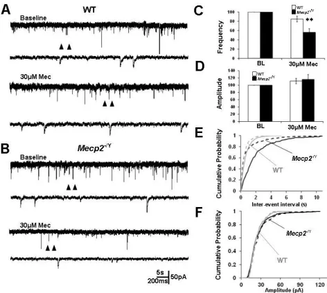
GABA-ergic input but not glutamatergic input was augmented by nicotinic presynaptic modulation in Mecp2−/Y mice ... 49
Cholinergic modulation of LC neuronal activity was sustained in Mecp2−/Y mice despite a large decrease in nAChR currents ... 50
Discussion ... 51
Changes in nAChR subunit expression may be responsible for altered current ... 51
Adaptations in LC neurons of Mecp2−/Y mice ... 53
6HOMEOSTATIC REORGANIZATION OF HCN AND VOLTAGE-GATED NA+
CHANNELS IN MESENCEPHALIC TRIGEMINAL PROPRIOSENSORY
NEURONS OF A RETT SYNDROME MOUSE MODEL AND ITS IMPACT ON
MEMBRANE EXCITABILITY ... 72
Acknowledgements ... 72
Abstract ... 72
Introduction ... 73
Results ... 75
Post-inhibitory rebound and sag in WT mice ... 75
Decreases of PIR and sag in Mecp2−/Y mice ... 76
Reduction in IH in Mecp2−/Y mice ... 77
Impact on firing and repetitive firing activity ... 78
Alteration of voltage-gated Na+ currents in Mecp2−/Y mice ... 79
Evidence for altered expression of HCN and Na+ channels in Mecp2−/Y mice ... 80
Discussion ... 82
6 IH alterations in Mecp2−/Y mice ... 82
INa alterations in Mecp2−/Y mice ... 83
Homeostatic reorganization of HCN and voltage-gated Na+ channels in Mecp2−/Y mice ... 84
Figures ... 87
7GENERAL DISCUSSION ... 103
Homeostatic compensation in animal models ... 103
Gene knockout mouse models ... 103
Parkinson’s disease mouse models ... 104
Rett Syndrome mouse models ... 105
Evolutionary considerations ... 107
Degeneracy of ionic currents ... 108
Ionic currents change depending on the presence of modulatory input ... 109
Protein expression from cell to cell and animal to animal ... 109
Similarities between WT animals and animal models of disease ... 110
Conclusion ... 111
REFERENCES ... 111
LIST OF TABLES
Table 5.1 Nicotinic Receptor PCR Primers ... 71
LIST OF FIGURES
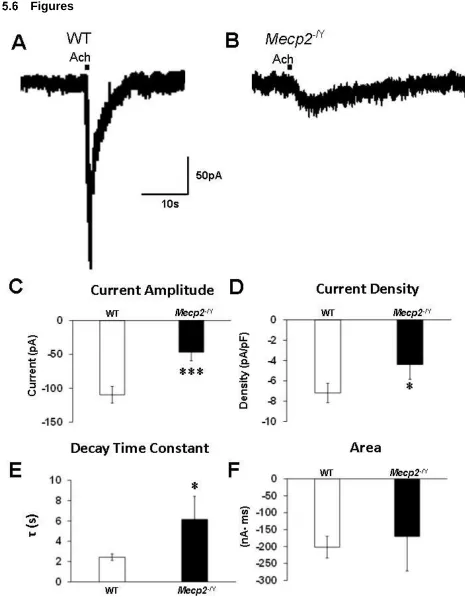
Figure 5.1 The nAChR currents were altered in LC neurons from Mecp2−/Y
mice. ... 58
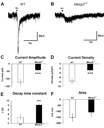
Figure 5.2 Nicotine elicited a similar current to ACh in WT and Mecp2−/Y
mice. ... 60
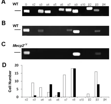
Figure 5.3 Receptor subunit expression in identified LC neurons. ... 62
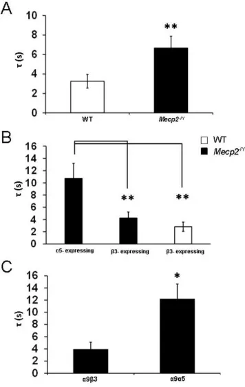
Figure 5.4 LC neurons from Mecp2−/Y mice expressing the α5 subunit had
longer decay times than β3-expressing neurons. ... 63
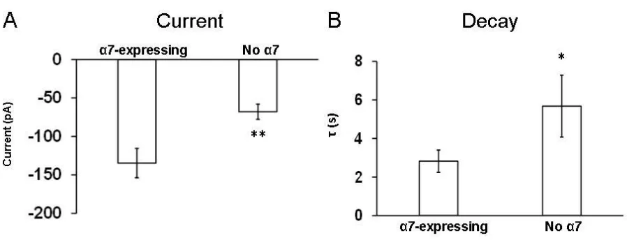
Figure 5.5 Presence of the α7 subunit affects the current amplitude and
decay time constant in LC neurons from WT mice. ... 64
Figure 5.6 Cholinergic modulation of GABA-ergic input to LC neurons with
nAChR agonist in Mecp2−/Y mice is enhanced compared to WT mice. ... 65
Figure 5.7 Cholinergic modulation of GABA inputs to the LC neurons with a
nAChR antagonist is enhanced in Mecp2−/Y mice. ... 67
Figure 5.8 There is a small but insignificant difference in nicotinic
modulation of LC neurons from WT and Mecp2−/Y mice. ... 69
Figure 6.1 ZD7288 blocks the strong sag and PIR in Me5 trigeminal
neurons. ... 87
Figure 6.2 PIR and sag are less in Me5 trigeminal neurons from Mecp2−/Y
mice. ... 89
Figure 6.3 The hyperpolarization-activated current in Me5 trigeminal
Figure 6.4 The hyperpolarization-activated current was altered in Me5
trigeminal neurons from Mecp2−/Y mice. ... 93
Figure 6.5 Excitability was increased in Me5 neurons of Mecp2−/Y mice. .... 95
Figure 6.6 The steady state half-activation of voltage-gated sodium current
was shifted in the hyperpolarized direction in Mecp2−/Y mice. ... 97
Figure 6.7 Alterations in HCN subunit expression in Me5 neurons. ... 98
Figure 6.8 Alterations in NaV and SCN subunit expression in Me5
1 SPECIFIC AIMS OF THE DISSERTATION
The loss of proper Methyl-CpG-binding protein 2 (MeCP2) function is the
molecular basis of Rett syndrome (RTT). RTT is characterized by abnormalities in
breathing, motor function, cognition and sleep, in addition to other autistic features.
These problems have largely been linked to dysfunction in signaling between neurons
as well as neuronal intrinsic membrane properties. The decrease in GABA and
norepinephrine (NE) signaling has been shown to be associated with breathing
disorders, characteristics of RTT that underlie the high (26%) sudden and unexpected
death rates (Abdala et al. 2010; Viemari et al. 2005). This may be partially due to the
decrease in the expression of the NE -synthesizing enzymes, tyrosine hydroxylase and
dopamine-β- hydroxylase in locus coeruleus (LC) neurons, and the GABA-synthesizing
enzyme glutamic acid decarboxylase in GABAergic neurons (Chao et al. 2010; Zhang et
al. 2010b). Abnormalities in LC neuronal intrinsic membrane properties also contribute
to the defects in breathing (Zhang et al. 2010a; Zhang et al. 2011). The coexistence of
the defects in NE and GABA systems suggest that brainstem neuronal networks are
affected by the Mecp2 disruption, which may involve other neurotransmitter systems
such as serotonin and acetylcholine (ACh) as well. ACh regulates the expression of
enzymes for NE and GABA biosynthesis and may be involved in the problems
associated with the NE and GABAergic systems (Gueorguiev et al. 2000; Maloku et al.
2011). Indeed, ACh signaling has been shown to be defective in RTT (Nag and
Berger-Sweeney 2007; Weng et al. 2011; Wenk and Hauss-Wegrzyniak 1999; Wenk and
including arousal state and sleep/wake cycles. Experimental evidence suggests that the
modulation of the NE system by GABA is deficient and contributes to neuronal
hyperexcitability. A similar defect may occur in the cholinergic system, a hypothesis that
we have proposed experiments to test.
The involvement of multiple neurotransmitter systems in the development of
RTT-like disorders in Mecp2-null mice suggests that an appropriate regulation of these
neurotransmitter systems is necessary to maintain them in a homeostatic state.
Homeostasis of neurotransmitter systems will not only allow a better control of
neurotransmission, but also enable potential compensatory neuroadaptations to the
defects in receptors and ion channels. The latter may be in action in RTT mouse models
to limit the impact of the loss of MeCP2 function, a novel hypothesis that we have
proposed in these studies.
The homeostatic adaptive mechanism is known to occur in response to genetic
perturbation. In animals with a specific ion channel gene knocked out, neurons alter the
expression of other ion channels to maintain normal function. (Bonin et al. 2013; Kim
and Hoffman 2012; Ortinski et al. 2006). There is evidence for neuroadaptations in
Parkinson’s disease animal models as well (Golden et al. 2013; Lloyd 1977; McCallum
et al. 2006). Therefore, it is possible that similar types of alterations in channel
expression may be occurring in RTT mouse models to maintain normal function.
Despite the belief of its existence, it was unclear how the homeostatic compensatory
mechanism would work. Since RTT affects signaling between neurons, we investigated
the mechanisms for neuronal activity and communication, in which receptors and ionic
To validate the homeostatic neuroadaptation hypothesis, we investigated
receptors and ion channels in two different neuron types, i.e., cholinergic synaptic
currents in LC neurons and membrane ionic currents in mesencephalic trigeminal V
(Me5) neurons. The LC is the center for NE production in the CNS, whereas the Me5
neurons are propriosensory cells that are crucial for coordinated movement. They
function in autonomic regulation and motor behaviors, respectively. These two systems
show the most severe defects in RTT. Therefore, we have proposed studies to address
two specific aims:
Specific Aim 1 (tested in Chapter 5): How does ACh modulate LC neurons
through presynaptic and postsynaptic mechanisms in WT and Mecp2-/Y mice?
NE is deficient in humans with RTT and mouse models (Panayotis et al. 2011;
Zoghbi et al. 1989). This seems to be due to inherent defects and abnormal synaptic
inputs in LC neurons (Jin et al. 2013a; Zhang et al. 2010a; Zhang et al. 2011). ACh has
several functional overlaps with the NE system. ACh is a classical neurotransmitter
involved with many functions in the CNS and peripheral nervous system. It plays a role
in CNS function by regulating attention, memory function, arousal, transitions in sleep
cycles and mood. These behaviors are dysregulated in RTT (Carotenuto et al. 2013;
Maloku et al. 2011; Schaevitz et al. 2012). The loss of MeCP2 function on cholinergic
signaling is unclear. Few have focused on the dysfunction of the cholinergic system
caused by the knockout of the Mecp2 gene. However, it is clear that NE and ACh
regulate similar behaviors. In one example, LC neurons and ACh neurons regulate each
however, whether the modulation of LC neurons by ACh is altered in Mecp2-/Y mice.
Therefore, in this study we investigated the cholinergic modulation of LC neurons in
these mice. These experiments may provide insight into how the knockout of the Mecp2
gene alters the expression of nicotinic acetylcholine receptors (nAChR) in LC neurons,
and whether neurons can alter the expression of synaptic receptors to maintain normal
modulation.
Specific Aim 2 (tested in Chapter 6): How does the loss of MeCP2 affect ionic
currents and firing activity in mesencephalic trigeminal V propriosensory
neurons?
Motor dysfunction is a characteristic of RTT, which may result from motor,
extrapyramidal and propriosensory neuron dysfunction. Propriosensory neurons are
important for relaying information to the brain about the position of the limb in space.
This is critically important for coordinated and directed movement such as locomotion
and mastication. Girls with RTT have problems chewing and swallowing which leads to
malnutrition and poor health (Motil et al. 2012). Whereas most work has been focused
on motor neurons to determine the underlying causes of movement problems, little is
known about how the loss of MeCP2 impairs propriosensory function. In these
experiments we took advantage of the presence of Me5 neurons in the brainstem to test
the homeostatic neuroadaptation hypothesis by focusing on ion channels and intrinsic
member properties. The experiments herein test the hypothesis that the Me5 neurons
insight into the membrane ionic currents that affect the intrinsic membrane properties of
Me5 neurons in Mecp2-/Y mice.
Taken together, this dissertation addresses the effects of the Mecp2 knockout on
synaptic and membrane ionic currents. It addresses two important questions: 1) how
does the disruption of Mecp2 impair ion channels and receptors? 2) Do neurons
respond to these defects by rearranging other ion channels and receptors to minimize
and compensate for the defects? The NE-ergic system and the Me5 propriosensory
system were chosen as they represent major defective functions in RTT. Ion channels
and receptors were studied in these neurons as they underlie the intrinsic membrane
properties of neurons as well as neuronal communication, both of which are known to
be defective in RTT. Therefore, this dissertation provides evidence that the loss of
proper MeCP2 function affects neuronal communication by changing synaptic
transmission and by altering membrane ionic currents. It is possible that these types of
neuroadaptations may exist in other diseases with a basis in genetic defects.
2 INTRODUCTION
Definition of key concepts
There is growing evidence suggesting the reason that some diseases progress
through different stages, instead of the victims dying quickly, is because neurons are
able to compensate for the problems caused by the disease. Compensation can be
described as a neuroadaptive mechanism by which one attribute is altered to limit the
problems created by a deficiency in another attribute. For example, in Parkinson’s
nigra pars compacta neurons. The remaining dopaminergic neurons compensate by
increasing their production of dopamine. This is done by increasing expression of
tyrosine hydroxylase, the rate-limiting enzyme for dopamine synthesis (Zigmond et al.
1984). Here, we provide evidence of compensation done by homeostatic reorganization
of receptors and ion channels. The reorganization refers to the increase in one subunit
of the receptor/ion channel family while another is decreased, or vice versa. These
changes in expression seem to be a homeostatic mechanism because it provides a way
for the neuron to remain viable. These concepts provide a basis for understanding how
neurons adapt to perturbation caused by a disease.
Signaling in the CNS is governed by synaptic transmission and neuron
activity
There are two major components that regulate how a neuron communicates to
other neurons. First, the release of neurotransmitters from a presynaptic neuron and the
subsequent binding of the transmitter to its receptor on the postsynaptic neuron has
been widely studied. Secondly, the intrinsic membrane properties of the neuron governs
how it fires action potentials and how the neuron responds to input from presynaptic
neurons. While not a focus of the thesis, electrical synapses are found in a small
number of neurons. These synapses are formed by gap junctions and allow neurons to
communicate by directly passing electrical current from one cell to another. Because
there is no chemical intermediary, the communication is very fast and usually involved
in simple behaviors such as the escape reflex in lobsters and crayfish. Understanding
how these processes underlie normal communication is essential for understanding
Overview of chemical synaptic transmission in the mammalian CNS
The synapse is the basic structure of chemical neurotransmission in the CNS.
The synapse consists of a presynaptic neuron that releases transmitters and a
postsynaptic neuron that has ligand-gated receptors that convert the chemical message
into an electrical signal. Depending on whether the electrical signal is made up of
cations or anions, the neuron becomes excited or inhibited.
The synaptic communication between two neurons is modified by modulatory
inputs. These inputs can change the amount of neurotransmitter released at the
presynaptic terminal (Garcia-Ramirez et al. 2014). The modulatory inputs can influence
the movement of receptors in and out of the synapse in the postsynaptic neuron (Gao et
al. 2006; Zou et al. 2005). These mechanisms are involved with increasing or
decreasing the synaptic strength and ultimately are thought to underlie compensatory
neuroadaptations such as long term potentiation (LTP) and long term depression (LTD)
(Bassani et al. 2013; Castillo 2012) .
It is unclear how certain genetic diseases that affect signaling between neurons
affect these normal neuromodulatory processes. Experimental evidence from transgenic
animals indicate that the defect in a given gene produces a phenotype that lacks
function (Drago et al. 2003). Whereas in other animals, molecular and systemic
compensation occurs by activating other gene(s) that have similar functions, reducing
and in some cases even masking the defect (Bonin et al. 2013). Evolutionarily, it is
reasonable to believe that compensatory neuroadaptations are necessary for survival or
The transcription repressor protein, methyl-CpG-binding protein 2 (Mecp2) plays
a role in the expression of enzymes crucial for the synthesis of neurotransmitters
(Maloku et al. 2011). As a result, its defect can affect a large number of downstream
genes including several neurotransmitter systems (Chao et al. 2010; Zhang et al.
2010b). In this thesis we elucidate some of the effects that the loss of functioning
MeCP2 has on neuronal activity.
Presynaptic mechanisms of neurotransmitter release
The release of neurotransmitters from a nerve terminal occurs through action
potential dependent and action potential independent mechanisms. The action potential
induced release of neurotransmitters from a presynaptic terminal involves many events
to occur in a choreographed manner. The invasion of the action potential into the
presynaptic terminal causes local depolarization and opening of voltage-gated Ca2+
channels. The influx of Ca2+ into the cell is critical for neurotransmitter release and
blocking of these channels or reducing extracellular Ca2+ prevents the release of the
transmitters. Further, even when the action potential is blocked with tetrodotoxin,
neurotransmitter release at the presynaptic terminal can occur. Depolarization of the
membrane potential at the presynaptic terminal, without the influence from an action
potential, can activate Ca2+ channels (Nakamura and Jang 2010; Yang et al. 2011).
Therefore, action potential independent mechanisms are in place to regulate the release
of neurotransmitters.
Ca2+ binds to molecular targets in the terminal causing the release of
neurotransmitters into the synapse. The vesicles that store the neurotransmitter at the
to fuse to the membrane and release the neurotransmitters into the synapse (Sudhof
2012). However, since some neurons can respond to stimuli by firing at > 200Hz, it is
important to know how this is possible. Vesicles are tethered and stored in
compartments that are distant from the active zone. Ca2+ binds to synapsin, a protein
that tethers the vesicles, and initiates the vesicle movement to the active zone (Kile et
al. 2010).
Neurotransmitters are released as discrete units called quanta. Each quantum
results in a fixed postsynaptic potential. A single quantum has been linked to the
release of neurotransmitters in a single vesicle. After the action potential invades the
terminal, many quanta are released at once and can affect the membrane potential of
the postsynaptic neuron. However, the release of neurotransmitters independent of an
action potential can be modulated by other inputs at the presynaptic terminal.
Acetylcholine alters the amount of GABA and glutamate released at terminals by
depolarizing the membrane potential. This causes an increase in the frequency of
quanta release. Therefore, increasing the quanta release enhances the effect the
presynaptic neuron has on the postsynaptic neuron.
Postsynaptic mechanisms mediating the chemical message
The ligand-gated ionotropic receptors found in the postsynaptic membrane are
the most common mode of converting the chemical signal from the presynaptic neuron
to an electrical signal in the postsynaptic neuron. In the mammalian CNS, glutamate
mediates the excitatory signal by binding to NMDA and AMPA receptors in the
postsynaptic membrane. Opening of these receptors allows Na+ and Ca2+ to enter and
causing Cl- to pass into the cell. Also, GABA may bind to GABA
B receptors and initiate a
second messenger cascade resulting in activation of K+ currents. In both instances,
GABA can hyperpolarize the cell. Another class of signaling molecule is acetylcholine
(ACh), which is most commonly associated with signaling between motoneurons and
muscles in the peripheral nervous system. In the CNS, ACh acts through the ionotropic
nicotinic acetylcholine receptors (nAChRs) and muscarinic acetylcholine receptors and
is involved with diverse behaviors such as attention, cognition, sleep as well as others
(Romanelli et al. 2007).
Glutamate, GABA and ACh elicit postsynaptic currents that affect the firing
activity of the postsynaptic neuron. Since NMDA, AMPA and nicotinic acetylcholine
receptors allow Na+ and Ca2+ into the cell, the cell depolarizes increasing the likelihood
of an action potential and increasing the firing frequency. GABA- elicited inhibition
through GABAA receptors is caused by Cl- moving into the cell causing the membrane
potential to become hyperpolarized. Hyperpolarization of the membrane potential
increases the likelihood of the cell to be silent or at least reduce the firing frequency.
The activation of these receptors will affect the firing activity of neurons by directly
altering the membrane potential
Long-term potentiation (LTP) Long-term Depression (LTD) are the major
mechanisms of changing the synaptic strength in the CNS. Most often these
mechanisms are associated with excitatory synapses but have been found at inhibitory
synapses as well (Arendt et al. 2013; Cohen et al. 1999; Nugent and Kauer 2008). Pre-
and postsynaptic mechanisms have been described. In brief, the presynaptic
LTD. Postsynaptically, the ligand-gated receptors move into the synapse during LTP
and are removed during LTD. Many methods have been used to stimulate LTP and
LTD, whether its high/low frequency stimulation of the presynaptic neuron (Martin et al.
2013; Mizuno et al. 2001) or coincidental firing of an action potential between the pre
and postsynaptic neurons (Banerjee et al. 2014; Kodangattil et al. 2013). LTP and LTD
are important for normal cognitive function the brain and disruption of these
mechanisms because of disease leads to improper communication between neurons
(Han et al. 2014; Liu et al. 2008; Mori et al. 2014). This suggests people with diseases
that have altered signaling processes in neurons have impaired learning and memory.
Intrinsic membrane ionic currents affect neuronal activity
Besides synaptic transmission, the intrinsic membrane properties of neurons
governs their firing properties. These intrinsic membrane properties have been
extensively studied and can be generally attributed to specific ionic membrane currents.
Here, we will introduce specific firing properties in the neurons studied and how these
properties affect signaling and function.
The relationship between the injected current and the voltage response of the
neuron is very important for understanding the input resistance of the neuron. The input
resistance is a good measure of the number of channels that are either open or closed
in the membrane of the neuron (John and Manchanda 2011). More specifically, it is a
valuable tool in determining how a drug may affect the membrane ion channels thus
altering the activity of the neuron (Podda et al. 2010). Consequently, the opening and
closing of ion channels alters the excitability of neurons. According to Ohm’s law, the
large, a current injection is going to change the membrane potential more than if the
input resistance is small. Therefore, membrane ion channels alter the response of the
postsynaptic neuron to presynaptic currents by changing the input resistance of the
neuron.
The delayed excitation found in many neurons affects the interspike interval. The
time it takes from the end of one action potential to the start of the next is regulated by
subthreshold currents. A major contributor to the interspike interval is the fast-transient
K+ current (IA) (McDermott and Schrader 2011). This current is mediated by Kv(4.1-4.3)
channels in mammals and is inactivated during an action potential. Kv4 channels
become deinactivated at subthreshold potentials and activate during the interspike
interval allowing K+ to flow out of the cell. The movement of K+ out of the cell causes the
depolarization of the cell toward threshold to take longer than it would if IA was not
present. Therefore, this particular K+ current regulates the interspike interval and allows
cells to spontaneously fire action potentials at low frequencies.
The post-inhibitory rebound (PIR) is very important to how neurons respond to
the release from inhibition. The phenomenon is very important in half-center oscillators
found in central pattern generators and in reciprocal inhibition found in locomotion. In
both cases, the firing of one neuron causes a second neuron to be inhibited from firing
(Harris-Warrick et al. 1995). When the first neuron stops firing action potentials, the
second neuron rebounds from silence and fires action potentials. Without the PIR, the
neuron would just return to its resting membrane potential without firing an action
potential. Therefore, the PIR depolarizes the neuron past its resting membrane potential
The underlying mechanism for the PIR has been attributed to the
hyperpolarization- activated current (IH) (Ascoli et al. 2010). IH is a nonspecific cation
current that depolarizes the cell after it has been inhibited. The removal of the inhibition
happens much faster than the deactivation of the HCN channels. This allows cations to
keep moving into the cell driving the voltage past the resting membrane potential to
threshold (Dean et al. 1989).
The spike frequency adaptation (SFA) is a phenomenon that describes how
neurons respond to a sustained depolarizing pulse. When a neuron is subjected to a
depolarizing pulse, a train of action potentials will be elicited. If the SFA is present, then
the frequency of action potentials will decrease during the time of depolarizing pulse
(Chen et al. 2014). This is found in both sensory and motor neurons and has been
related to different mechanisms. The most prominent is the action potential-dependent
SFA. In this case, Ca2+ is brought into the cell during the action potential and Ca2+
-dependent K+ currents (I
KCa) are activated. The activation of these currents increases
the afterhyperpolarization (AHP). The larger AHP slows the depolarization of the
membrane potential. With each successive action potential, the net concentration of
Ca2+ in the cell grows, thus activating more and more K
Ca channels (Vandael et al.
2012). Therefore, the interspike interval becomes longer during the depolarizing pulse.
This is how the neuron is able to accommodate to the depolarization and slow the
action potential frequency.
Overview of K+ currents role in neuronal activity
Two of the most important functions performed by K+ currents in neurons is the
the interspike interval. The resting membrane potential is mainly set by K+ leak currents
mediated by TWIK-related acid sensitive K+ (TASK) and K
ir channels (Butt and Kalsi
2006). These channels are not voltage or ligand-gated. The conductance of K+ obeys
Ohm’s law. Consequently, the driving force created by the concentration gradient of K+
and the membrane potential of the neuron causes K+ to move out of the neuron. For
most cells, the resting membrane potential is very near the equilibrium potential of the
K+ leak current. When the cell is hyperpolarized below the K+ equilibrium potential the
polyamine block is removed and the channels conduct K+ into the cell. Usually if IKir
plays a large role in setting the resting membrane potential, the potential is usually near
the K+ reversal potential.
The delayed rectifier K+ current (I
K) repolarizes neurons during the action
potential. IK activates slowly so that it only influences the membrane potential after the
voltage-gated Na+ current (I
Na) is inactivated. With no inactivation gate, once the
membrane potential becomes hyperpolarized, the driving force for K+ movement is lost
and thus stops conducting K+. As mentioned before, IKCa regulates spike frequency
adaptation. This is accomplished by its regulation of the AHP of the action potential.
With the exception of the IKCa mediated by SK channels, IKCa is voltage-activated and
modulated by Ca2+. Just as increasing the voltage increases the K+ conductance, so
does increasing the Ca2+. There are three main channel types, BK, IK and SK that
possess different conductance values and Ca2+ dependences (Vergara et al. 1998).
Whereas the BK channel has a voltage dependence independent of Ca2+, the IK and
Therefore, these channels have a different effect on the afterhyperpolarization and
consequently different SFA characteristics.
IA is a subthreshold K+ current that opposes the depolarization of the membrane
potential thus affecting a neuron’s response to presynaptic signaling. This current is
interesting because a hyperpolarized membrane potential deinactivates the channel.
Then when the membrane potential is quickly depolarized, the activation gate opens
allowing K+ to flow out of the cell. The current ceases when the inactivation gate closes
which happens quickly giving the current its transient characteristic. These channels are
located in the somatodendritic compartment of neurons and their intrinsic gating
mechanisms make them suitable to regulate signaling between the dendrites and the
soma (Johnston et al. 2000). For many cells, IA acts as a shunting mechanism to limit
the propagation of postsynaptic potentials to the soma. Therefore, it regulates how
much presynaptic neurons affect the firing of the postsynaptic neuron. Another way that
the expression of these channels affects neuronal communication is in coincidence
detection. The back-propagation of action potentials in the postsynaptic neuron is
necessary for spike timing-dependent LTP. Since IA acts as a shunt, it can limit the
back-propagation of action potentials (Harnett et al. 2013) thus limiting the coincidence
detection that occurs to induce LTP (Ramakers and Storm 2002).
In sum, K+ currents are vital to normal neuronal activity. Not only do they affect
the intrinsic membrane properties during and in between action potentials, but also
signaling between neurons as well. Therefore, the expression levels of the K+ channels
Role of IH in neuron firing properties
As mentioned before, IH isresponsible for the PIR found in many neurons. IH is a
nonselective cation currentthat conducts mostly Na+, but also some K+, when it is
activated by hyperpolarization. This results in the depolarization of the membrane
potential back towards the threshold. Not only is this property of the channel critical in
half-center oscillators and locomotion, but also in pacemaking of neuronal activity,
setting the resting membrane potential and integration of excitatory postsynaptic
currents (Altomare et al. 2003; He et al. 2014).
IH has been well-studied in heart myocytes and contributes to the pacemaking of
the cells (Baruscotti et al. 2005). Just as IA opposed the depolarization of the membrane
potential in between action potentials to make the interspike interval longer, IH enhances
the depolarization of the membrane potential to decrease the interspike interval. IH plays
a large role in setting the interval and it is because of this it is considered a pacemaker
current (Deng et al. 2014). When IA and IH are both present, both contribute to the
interspike interval and can coregulate the interval.
IH is mediated by hyperpolarization and cyclic nucleotide-activated (HCN)
channels. Depending on which HCN channels are expressed, the half-activation may be
anywhere from -70mV to -110mV. Therefore, they do contribute to the setting of the
resting membrane potential. Further, when IH is blocked by its specific blocker, ZD7288,
the resting membrane potential becomes hyperpolarized.
Similar to IA, IH has the ability to affect the propagation of EPSPs from the
dendrites to the soma. Since HCN channels are partially activated at the resting
is much larger in the distal dendrites and much smaller in the proximal dendrites.
Depending on how the HCN channels are expressed in the dendrites they affect how
postsynaptic potentials propagate to the soma (Rusznak et al. 2013).
Voltage-gated Na+ channels are crucial for action potential generation
The voltage-gated Na+ current (INa) is required for the fast depolarization phase
of the action potential. INa is a transient current that depolarizes the cell toward the Na+
equilibrium potential. Functional Na+ channels contain one α pore-forming subunit and
two β auxiliary subunits. The α subunit consists of SCN(1-9)A and the β consists of
SCN(1-4)B gene transcripts (Aman et al. 2009). The threshold of the action potential is,
at least partially, regulated by subunits expressed in the neuron. Regulation of the
threshold ultimately affects the excitability of the neuron. With a lower threshold, less
contribution of depolarizing subthreshold currents is required to fire an action potential.
The Na+ channels that mediate INa are mainly expressed in the axonal
compartment. Therefore, when the action potential is initiated in the axon hillock, the
signal can be propagated down the axon to the terminal, thus initiating the release of
neurotransmitters. The propagation of the action potential is a series of electrical
self-stimulation. The movement of Na+ into the cell depolarizes the local area of the
membrane. This depolarization of the membrane causes the Na+ channels to open next
to it and the process repeats itself allowing the movement of the signal down the axon
Membrane ionic current regulation of mesencephalic trigeminal V neuron
activity
Mesencephalic trigeminal V (Me5) neurons are propriosensory neurons that
monitor the stretch and tension of jaw masseter muscles thus sending critical
information to the motor neurons needed for coordinated movement. These neurons are
the only propriosensory neurons that are located in the CNS. Most propriosensory
neurons are located in the dorsal root ganglia. Me5 neurons receive signals from the
muscle spindles and relay the information of the limb placement to the motor neurons
(Hidaka et al. 1999; Luo et al. 2001). This controls movement of the limb by
communicating with the inhibitory neurons innervating the muscles that control the
antagonizing muscle.
The membrane currents found in the Me5 neurons are important for their
response to stimuli from the jaw and synaptic inputs from cortical areas. The
voltage-gated K+ currents that are sensitive to tetraethylammonium (TEA) and 4-aminopyridine
(4-AP) play a role in how the neuron responds to stimuli. Depending on the resting
membrane potential at the time of depolarization, these neurons respond with either
single action potentials or multiple action potentials (Del Negro and Chandler 1997).
Blocking the current with 4-AP, caused the cells to respond to depolarization with only
multiple action potentials. Therefore, the K+ currents do play a role in Me5 neuron
excitability.
A clear response of the Me5 neurons to a hyperpolarizing current injection is a
strong sag and PIR. The sag is a depolarization of the membrane potential despite a
the sag is IH. The deactivation of IH after the hyperpolarizing current injection is
responsible for the PIR. Also, the presence of HCN channels provides a basis for
dendritic integration and postsynaptic potential propagation to the soma. Since Me5
neurons receive input from muscle spindle fibers and synaptic inputs, IH may play a
large role with how Me5 neurons respond to stimuli.
There are several types of Na+ currents in the Me5 neurons. Besides I
Na that
controls the action potential initiation and propagation, there are the persistent Na+
current (INaP) and the resurgent current (INaR) (Enomoto et al. 2007). Unlike, INa that
inactivates very quickly, INaP does not inactivate. INaP induces bursting in these neurons
and regulates resonance properties (Enomoto et al. 2006). INaR is much less studied
than INa and INaP. INaR is a subthreshold current that is activated during repolarization but
still is quickly inactivated. Studies indicate that INaR plays a role in spontaneous firing in
neurons and multipeaked action potentials (Raman and Bean 1997).
Brainstem circuitry underlying autonomic behaviors
LC-ACh-GABA brainstem circuit
Within the dorsal pons of the brainstem near the lateral edge of the 4th ventricle is
the locus coeruleus (LC). These neurons produce about 95% of the entire
norepinephrine (NE) in the mammalian brain. NE has been shown to be involved with
learning and memory, proper sleep, pain analgesia, breathing and attention (Brightwell
and Taylor 2009; Doi and Ramirez 2008; O'Donnell et al. 2012). Therefore, the proper
activity of LC neurons is very important to the maintenance of NE in the brain, thus
The relationship between LC neurons and ACh neurons in the brainstem is very
important for the transitions between rapid eye movement (REM) sleep and non-REM
sleep. During REM sleep cholinergic neurons are very active. ACh is able to suppress
the firing of LC neurons. Eventually, cholinergic neuron activity is decreased and LC
neurons are able to start firing more causing the transition to a non-REM state during
sleep.
GABAergic neurons are the main inhibitory neurons in the mammalian CNS.
These neurons regulate neuronal excitability through synaptic transmission and through
tonic release of GABA. During the transition from non-REM to REM sleep, cholinergic
neurons excite GABAergic neurons which in turn silence the LC neurons through
GABAA receptor- mediated synaptic transmission. GABAA receptors also mediate the
tonic inhibition in the LC (Kawahara et al. 1999) which has been shown to be mediated
by the Δ (Ye et al. 2013), α5 (Groen et al. 2014) and Θ subunits in other brain regions.
Throughout the brain, GABAergic neurons play many roles by regulating neuronal
activity. Within the brainstem, there are areas that regulate the neurons that are
involved with breathing, pain analgesia, cranial motoneuron activity and heart rate.
Besides these behaviors, GABAergic neurons from the forebrain send projections to the
LC nucleus. This is important because NE plays a role in all of these behaviors.
GABAergic neurons projecting from the central amygdala and the posterior lateral
hypothalamic area seem to synapse on LC neurons at the dendrites (i.e., in the peri-LC
region) (Dimitrov et al. 2013). Much less enter into the LC proper. Lastly, there is a local
region with many in the dendritic field (Aston-Jones et al. 2004). These neurons may be
important for integrating information from afferents.
Neuronal activity during autonomic behavior
The autonomic system is involved in regulating involuntary behaviors such as
breathing, body temperature and heart rate. NE, ACh and GABA have all shown to play
a role in breathing. These neurotransmitters modulate the activity of pre-Bötzinger
complex (PBC) neurons in the medulla oblongata (Doi and Ramirez 2008). PBC
neurons are considered the center for breathing rhythmogenesis because the bursting
of these neurons consistently corresponds to extracellular recordings of activity from the
phrenic, vagus and hypoglossal nerves. All of which are involved in breathing but not
rhythmogenesis.
NE, ACh and GABA affect the activity of PBC neurons. LC neurons fire tonically
and the interspike interval seems to be related to the breathing rate (Zhang et al. 2011).
Indeed, disruption in LC activity alters breathing. NE alters the activity of PBC neurons
through two different mechanisms. First, the activation of α2 noradrenergic receptors is
required for cadmium-insensitive bursting PBC neurons (Viemari et al. 2011). Second
the activation of β-noradrenergic receptors increases the frequency of sighs during
breathing. Further, NE modulation through β-noradrenergic receptors increases the
frequency in PBC neurons by modulating INaP (Viemari et al. 2013). GABAergic neurons
play a vital role in breathing as well. Blocking GABAA receptors increases the frequency
and amplitude of the activity recorded from phrenic and hypoglossal nerves (Bongianni
et al. 2010; Shao and Feldman 1997). This suggests that there is a baseline inhibitory
neurotransmitter that affects PBC activity and breathing. Activation of muscarinic
receptors in the PBC leads to an increase in breathing frequency (Muere et al. 2013). It
also decreases the normal PBC activity and increases the amount of sigh-causing
bursts (Tryba et al. 2008).
It is clear that NE plays a large role in modulating breathing. Since about 95% of
NE is produce in LC neurons, the modulation of LC activity by GABA and ACh is
important. The majority of the GABA input to the LC is mediated by GABAA receptors.
Indeed, GABAA agonists reduce the firing frequency, decrease input resistance and
hyperpolarize the membrane potential in LC neurons (Jin et al. 2013a). Cholinergic
modulation of LC activity has been shown to be mediated by nicotinic acetylcholine
receptors (nAChR) (Cucchiaro et al. 2006; Ganesh et al. 2008). However, the activation
of muscarinic receptors increases LC activity as well (Ennis and Shipley 1992; Yang et
al. 2000). Our preliminary experiments showed that most of the cholinergic modulation
of the increase in LC firing frequency was mediated by the nAChRs. Therefore, we
studied how ACh modulates LC activity both through nAChRs postsynaptically and
presynaptically at GABAergic neurons.
Expression of nAChRs in LC neurons
nAChRs are ligand-gated ion channels that are ionotropic receptors. In
mammals, the nAChRs are found in the peripheral nervous system as well as the CNS.
Each receptor is made up of five subunits with each subunit having four transmembrane
domains. In the CNS of mammalian systems, two types of subunits (α and β) are
expressed: α2-α7, α9-α10 and β2-β4 (Gotti et al. 2005). These eleven subunits can
subunits come together to form heteromeric receptors with the most prevalent
consisting of α4β2 and α6β4 in the mammalian brain or they can be homomeric with
most abundant channel being the α7 (Alkondon et al. 1997; Garduno et al. 2012; Yang
et al. 2011). However, it is possible for the α9 receptor to form homomeric channels, but
much of the time it is expressed with the α10 subunit (Boffi et al. 2013). The nAChR
channel opens when two (in heteromers) or five molecules (in homomers) of
acetylcholine (ACh) binds to the receptors allowing Na+ to flow into the cell. In some
cases, certain receptors may be more permeable to Ca2+. The major effect of the
nAChR activation on the cell is the depolarization of the membrane by the movement of
cations into the cell. However, the Ca2+-permeable receptors such as the α7, may also
induce second messenger systems that affect various cellular processes (Gueorguiev et
al. 2000; Nordman et al. 2014).
The LC has been shown to have many types of nAChR subunits expressed.
Using immunohistochemistry (IHC), the α3, α4, α7 and β3 subunits were found
throughout the LC nucleus suggesting these receptors play a strong role in cholinergic
modulation of LC activity (Vincler and Eisenach 2003). To a much lesser extent, the α5,
β2 and β4 were expressed as well but mostly in the dorsal portion of the LC. Also, it has
been shown that the expression of nAChRs in the LC may be based on different cells
within the nucleus (Lena et al. 1999). In rats, larger LC cells (capacitance > 65pF) had a
larger response to cytisine, a nAChR agonist, than smaller LC cells (capacitance
<45pF) suggesting differential expression of nAChRs. Indeed, single cell PCR showed
that the larger cells showed a preference for expressing α6 and β3 whereas the smaller
expressed other receptors but without the consistency of the ones mentioned. There is
a wide diversity of nAChRs expressed in the LC. Based on IHC and electrophysiological
data, LC activity should be significantly modulated by nAChRs.
Rett Syndrome
History
Rett Syndrome (RTT) was first described by Dr. Andreas Rett in 1966 and later
by Dr. Bengt Hagberg (Hagberg et al. 1983). Hagberg was unaware of Rett’s findings
because they were published in German. Nonetheless, from the early findings of Rett to
the later evidence provided by Hagberg, RTT was recognized as a major
neurodevelopmental disorder. Until the 1983 publication by Hagberg, many physicians
in the United States were unaware of the disease. As interest grew in determining the
underlying causes of the disease, the Baylor Rett Syndrome clinic was established.
The Zogbhi lab at Baylor determined that mutations in the MECP2 gene underlies the
problems found in RTT (Amir et al. 1999). Since this discovery, mouse models have
been developed to study the consequences of the loss of proper MeCP2 function in an
attempt to find a cure for the disease.
Progression and symptoms of RTT
RTT is neurodevelopmental disorder meaning it occurs through a progression of
stages that can be clearly delineated by physicians during clinical exams. Normal
development occurs for the first 6 months of life. In stage I, from 6 months to 1.5 years,
the onset begins characterized by a stagnation of development in language and
between 1 and 4 years of age. The loss of communication skills and symptoms of
mental retardation are evident as well. Many times during stage III, which can last up to
many years, there is a period of the reacquiring of communication skills. By the onset of
stage IV, patients lose the ability to walk and usually need wheelchairs for mobility. This
last stage can last up to decades. In some of the less severe cases, women can live up
to fifty years of age.
Mutations in the MECP2 gene underlies RTT
As mentioned above, mutations in the MECP2 gene underlies RTT. There have
been about 600 mutations detected in the MECP2 gene. Most of these result in
missense and nonsense mutants. Also, variations in the 5’ UTR and 3’ UTR, intron
variations, insertions and deletions have been found. However, most of the mutations
are found among exons 3 and 4. Further, most of the well-known mutations found in
RTT are found in the methyl-CpG- binding domain and the transcriptional repression
domain.
Random X-inactivation is a process that silences one of the 2 chromosomes
found in females to avoid having twice the amount of proteins created than are found in
males which only have one X chromosome. The designation of which X chromosome
will be inactivated is random in mammals resulting in not only being inactive for the
entirety of the cell’s life but also to all its descendants as well. The inactivation is
accomplished by condensing the X chromosome into heterochromatin that has structure
in such a way that it is unable to be transcribed. Since most of the girls with RTT are
heterozygous for the mutations in the Mecp2 gene, the random X-inactivation plays a
chromosome is inactivated and in which cell type, the inactivation has a large effect on
the severity and variety of symptoms.
NE deficiencies contribute to RTT symptoms
The reduction in NE is a hallmark of RTT. NE metabolites were diminished in
cerebrospinal fluid in RTT victims (Zoghbi et al. 1989; Zoghbi et al. 1985). In Mecp2-/Y
mouse models, NE reductions were seen at 14 days and 1 month after birth (Ide et al.
2005; Viemari et al. 2005). These reductions have been associated with breathing
problems. The use of desipramine, a NE reuptake blocker, has been used to reduce
breathing problems in Mecp2-/Y mice (Roux et al. 2007; Zanella et al. 2008; Zhang et al.
2011) . These data indicate that NE signaling is important for proper breathing.
The locus coeruleus (LC) provides about 95% of the norepinephrine to the brain.
Therefore, disruption in the normal function of neurons from this nucleus can have
adverse effects throughout the brain. We have shown in the Jiang lab that LC neurons
from Mecp2-/Y mice are hyperexcitable, smaller in size than WT, have a lower action
potential threshold and increased input resistance (Zhang et al. 2010a). The
norepinephrine synthesizing enzymes tyrosine hydroxylase (TH) and
dopamine-β-hydroxylase (DBH) are expressed much less in Mecp2-/Y mice (Zhang et al. 2010b). This
indicates that the activity of LC neurons and subsequent release of NE are deficient in
Mecp2-/Y mice. LC neurons are chemosensory neurons. This is thought to occur
primarily by the sensing of pH by Kir channels. The ability of LC neurons to sense
changes in pH is defective in Mecp2-/Y mice. This has been attributed to alterations in
from the LC may be defective and likely due to insufficient NE release in the
pre-Bötzinger complex, neurons thought to initiate breathing.
The GABA system is defective in RTT. Low GABA has been shown in
cerebrospinal fluid of RTT patients (Perry et al. 1988). Also, there is an increase in
GABA receptor expression in the basal ganglia of RTT victims (Blue et al. 1999). In
Mecp2-/Y mice, GABA signaling is reduced in these mice and has been attributed to low
glutamic acid decarboxylase (GAD) expression (Medrihan et al. 2008). The GABA
modulation of LC neurons in Mecp2-/Y mice is defective as well. The GABAB
receptor-mediated reduction in the Kir current in Mecp2-/Y mice is defective and the mIPSC
frequency and amplitude are reduced (Jin et al. 2013a). These defects seem to account
for the decreased inhibition of LC firing activity in Mecp2-/Y mice. Further, the
allopregnanolone modulation of GABAA receptor currents is defective which contribute
to the hyperexcitability of LC neurons in Mecp2-/Y mice.
Motor system deficiencies underlie RTT symptoms
The most common feature of RTT investigated has been defects in breathing.
However, the motor problems in RTT have received much less attention. These girls
with RTT develop normally but the earliest symptoms involve the loss of muscle tone.
Eventually, these girls have difficulties with coordinated and purposeful hand
movements including a stereotypical hand-wringing movement or they place their hands
in their mouths. As these girls become older, their posture is compromised and walking
becomes almost impossible. Many times, these women become wheel chair bound.
Coordinated movement is not only controlled by motor neurons. The requirement
propriosensory neuron systems. The pyramidal neurons control the contraction of
muscles. The extrapyramidal system provides information to the motor neurons that
help control muscle tone, joint angle and posture. Lastly, the propriosensory neurons
provide feedback to the other systems about the place of the limb in space, the muscle
force, and joint angles. Whereas motor neurons have been studied in RTT,
extrapyramidal and propriosensory neurons have not.
It has been shown that the movement of limbs is deficient in RTT patients. These
girls also have problems chewing and swallowing (Motil et al. 2012) and the difficulty in
eating causes malnutrition. Therefore, the understanding of the complications caused
by MECP2 mutations in the motor system is important to developing therapies to help
RTT patients. Propriosensory neurons of the mesencephalic trigeminal V (Me5) neurons
are crucial for the coordinated movement required during mastication. Thus, we
investigated the dysfunction of these neurons in RTT and provide evidence that Me5
neurons in Mecp2-/Y mice have altered membrane currents which seem to lead to
hyperexcitability.
Mecp2–/Y mice as a model system
In order to study the ramifications of the various mutations in exons 3 and 4,
many mouse model systems have been created. Some have been created that have
specific mutations. The Mecp2tm1Vnar or Mecp2*a140v consists of a missense mutation that
results in abnormal cellular morphologies in brain regions but no behavioral phenotypes
in males. However, phenotypes in females are arbitrary because of random
X-chromosome activation. A nonsense point mutation in Mecp2R168X mice results in a
many of the behavioral phenotypes found in other strains that have been widely used.
Also, when exons 3 and 4 were knocked out of GABAergic neurons only in Viaat-Mecp2
mice, the behavioral phenotypes found in these mice were similar to other mouse
models (Chao et al. 2010). It is worth noting that not only do mice with limited MeCP2
expression, but the overexpression of MeCP2 has proven to be detrimental to animals
as well. The overexpression of MeCP2 in mice causes abnormal nervous system
phenotypes, cognitive behaviors and seizures (Na et al. 2012; Taylor and Doshi 2012) .
The model system chosen for this study is commonly referred to as the “bird
model” named after the lab that originally created the strain. The Mecp2tm1.1Bird strain
created with a deletion of exons 3 and 4 (Guy et al. 2001). The deletion of both exons in
this model recapitulates all of the classical symptoms of RTT. The purpose of our
studies was to study the general impact of the Mecp2 gene mutations in neurons.
Therefore, we used a common mouse model where exons 3 and 4 have been removed
from the gene.
Benefits and drawbacks of the system
Every animal used in science as a model system has its positives and negatives.
These mice have been created by removing exons 3 and 4. We know from human
studies that over 600 known mutations have been discovered in girls suffering from RTT
(Weng et al. 2011). Each mutation will have its own, varying effect on the system.
We use male animals to study a disease that affects females primarily. Human
males still suffer from the disease, but they usually die at a very young age. That is the
reason for us to think of it as a female disease. We study males because the abnormal
decrease in the severity of the female symptoms is presumably due to the random
X-inactivation described above. Conducting experiments on female Mecp2-/Y mice may
lead to confounding results. For example, it is very difficult to know if the cell that one is
patch clamping expresses the normal Mecp2 gene or the knockout in female mice.
Behavioral studies in female mice have seemed to be less conclusive as well.
Therefore, male mice may not be a perfect model for the disease, but it is appropriate
for studies that are not sex-related.
3 SIGNIFICANCE
RTT is a devastating disease with a high prevalence among females (Kerr et al.
1997). Not only do mutations in the MECP2 gene cause autism-like features in RTT, it is
also the origin of the breathing irregularities and motor dysfunction that underlie
long-term disability and sudden death. Therefore, developing strategies to limit the impact of
MECP2 mutations on respiratory and movement centers in the CNS is necessary.
A cure for RTT has not been found. Many of the treatments are
symptom-specific. NE and GABA reuptake blockers improve breathing in mouse models (Abdala
et al. 2010; Roux et al. 2007; Zanella et al. 2008). Each of these drugs only improves a
certain symptom, suggesting that multiple systems are affected. Indeed, recent studies
suggest serotonergic agonists seem helpful for breathing in humans with RTT (Andaku
et al. 2005; Gokben et al. 2012). Another important neurotransmitter system in the
brainstem is the cholinergic system. Increasing cholinergic signaling improves
movement problems (Nag and Berger-Sweeney 2007; Schaevitz et al. 2012). The