ORIGINAL RESEARCH ARTICLE
EBOLA AND ZIKA VIRUS
*Rupali Yevale, Priyanka Kalamkar, Kirtibala Pawar and Nilofar Khan
K.G. Rahul Dharkar College of Pharmacy and Research Institute, Karjat, (M. S.) India
ARTICLE INFO ABSTRACT
Ebola haemorrhagic fever (EHF) is a zoonosis affecting both human and non-human primates (NHP).The virus causing the outbreak has been characterized as Zaire Ebolavirus (EBOV). EBOV belongs to the genus Ebolavirus which together with the genus Marburgvirus forms the family of Filoviridae. Managing Ebola patients in the African setting was a major challenge since there was no effective antiviral drug and no specific vaccine available. Only supportive care could be administered, to sustain cardiac and renal functions with prudent use of perfusion. Zika virus is usually spread to people through the bite of an infected mosquito. The virus can also be spread from a man to his sexual partner during unprotected sexual contact and from a pregnant woman to her baby during pregnancy or around the time of birth. The symptoms of Zika are similar to that of dengue and chikungunya, which are diseases caused by other viruses spread by the same type of mosquitoes. No specific treatment available for Zika. Symptoms are treated by getting rest, drinking fluids to prevent dehydration and taking medicines such as acetaminophen or paracetamol to relieve fever and pain.
*Corresponding author
Copyright ©2017,Rupali Yevale et al. This is an open access article distributed under the Creative Commons Attribution License, which permits unrestricted use, distribution, and reproduction in any medium, provided the original work is properly cited.
INTRODUCTION
Ebola virus
The Ebola virus causing the devastating outbreak in West Africa 38 years ago when it first surfaced and caused a mysterious illness among villagers in Zaire, now the Democratic Republic of Congo. The international team of scientists who were done with investigating that 1976 Ebola outbreak were shocked at the sight of the virus and the disease it caused. The scientists had looked at blood samples sent from Africa under the microscope and the virus looked like a worm or a long string. Once the team got on the ground in Zaire, they saw how rapidly the virus spread and how quickly it killed its victims.On 8 August 2014 the World Health Organisation (WHO) declared the Ebola virus disease (EVD) outbreak in West Africa a Public Health Emergency of International Concern (PHEIC). (Briand et al., 2014) Ebola haemorrhagic fever (EHF) is caused by five genetically distinct members of the Filoviridae family: Zaire ebolavirus (ZEBOV), Sudan ebolavirus (SEBOV), Côte d’Ivoire ebolavirus (CEBOV), Bundibugyoebolavirus (BEBOV) and Reston Ebolavirus
(REBOV). The virus causing the Zaire Ebolavirus (EBOV), belongs to the genus Ebolaviruswhich together with the genus
Marburgvirusforms the family of the Filoviridae.
Disease agent characteristics
• Family: Filoviridae; Genus: Ebolavirus
• Virion morphology and size: Enveloped, helical, cross-striated nucleocapsid, filamentous or pleomorphic virionsthat are flexible with extensive branching, 80 nm in diameter and 970-1200 nm in length
• Nucleic acid: Linear, negative-sense, single-stranded RNA, ~18,900 kb in length
• Physicochemical properties: Stable at room temperature and can resist desiccation; inactivated at 60°C for 30 minutes; infectivity greatly reduced or destroyed by UV light and gamma irradiation, lipid solvents, b-propiolactone, formaldehyde, sodium hypochlorite, and phenolic disinfectants.
For the West Africa outbreak the total number of cases is subject to change due to ongoing reclassification, retrospective
ISSN: 2230-9926
International Journal of Development Research
Vol. 07, Issue, 10, pp.15735-15740, October, 2017
Article History:
Received 16th July, 2017
Received in revised form
20th August, 2017
Accepted 27th September, 2017
Published online 10th October, 2017
Citation: Rupali Yevale, Priyanka Kalamkar, Kirtibala Pawar and Nilofar Khan, 2017. “Ebola and Zika virus”, International Journal of Development
Research, 7, (10), 15735-15740.
ORIGINAL RESEARCH ARTICLE OPEN ACCESS
Keywords:
investigation and the availability of laboratory results. A second, non-related, EVD o utbreak has been reported in the DemocraticRepublic of Congo with currently a total of 62 confirmed and suspected cases. (http://apps.who.int/iris/ bitstream/10665/133833/1/roadmapsitrep4_eng.pdf?ua=1; Nunes-Alves, 2014)
Virology
The virus causing the outbreak has been characterized as Zaire Ebolavirus (EBOV). EBOV belongs to the genus
Ebolaviruswhich together with the genus Marburgvirus forms the family of Filoviridae. This family belongs to the order of the Mononegaviraleswhich further contains members of
Bornaviridae, Paramyxoviridae and Rhabdoviridae.
Ebolavirusesa re l inear, n egativestranded, RNA viruses with a genome of approximately 19 kilobases. Morphologically, when studied under an electron microscope, the viral particles look like long stretched filaments with some particles tending to curve into an appearance looking like the number 6. At this moment the genus Ebolavirusconsists of five species: EBOV, Sudan ebolavirus (SUDV), Tai forest ebolavirus (TAFV), Bundibugyoebolavirus (BDBV) and Reston ebolavirus (RESTV). RESTV is considered to be non-pathogenic to humans. (Feldmann and Geisbert, 2011) The Filoviridaefamily in the order Mononegavirales is separated from other Mononegavirales on the basis of morphological, physiochemical, and biological features (Feldmann et al., 2003; Kiley et al., 1982) and more latterly genomic analyses (Carroll et al., 2013). Filoviruses are non-segmented, negative-strand RNA viruses. The viruses are filamentous (Filo- derived from the Latin filumor thread) enveloped particles of variable length. The filovirus genomes are typically approximately 19 kb in length (Feldmann et al., 2003; Sanchez et al., 2007). The proteins expressed by the filoviruses are: nucleoprotein (NP), glycoprotein (GP), RNA-dependent RNA polymerase (L), and four structural proteins: VP24, VP30, VP35, and VP40 (Sanchez et al., 2007; Feldmann and Kiley, 1999).
Ebolavirusis able to express a truncated soluble glycoprotein (sGP) through RNA editing. The ribonucleoprotein is derived from the RNA genome, NP, VP30, VP35, and L protein, though Marburgvirusis reported to be able replicate in the absence of VP30. The VP35 protein is known to block interferon induction in both Marburg and Ebola viruses (Brauburger et al., 2012), and the discovery of the open reading frame for this protein integrated into bat genomes is an area for future research exploration to better understand host-virus interactions and immunity (Taylor et al., 2011).
Filovirus Outbreaks in Humans—Brief History Including Known Links to Bat Exposure
Lake Victoria marburgvirus was the first filovirus discovered in 1967, when laboratory workers in Marburg, Germany and Belgrade, Yugoslavia (now Republic of Serbia) were contact with infected, imported green monkeys (Chlorocebus spp.) Subsequently, a number of small human outbreaks of Marburgvirus (both Marburg virus and Ravn virus) occurred sporadically between1975–1997, some of which had some link to bat caves (Taylor et al., 2011; Brauburger et al., 2012). The two largest outbreaks of Marburg virus happened in the Democratic Republic of Congo (DRC) 1998–2000 where 128/154 infected people died in Angola in 2004–2005 where 227/252 patients succumbed to the virus (Brauburger et al., 2012). The DRC outbreak was linked to gold mining in
Goroumbwa cave (Bausch et al., 2003), and origins of the Angola outbreak are not certain. Both Kitaka and Python cave are known to harbor large bat populations, and have been sites for follow up studies on Marburg ecology (Towner et al., 2009; Amman et al., 2012). The history of Ebolavirus outbreaks in Africa including an excellent summary of outbreaks up until 2005 (Pourrut et al., 2005).
Transmission
Natural reservoir of Ebola virus has not yet been identified, it is unknown how the virus first appears in a human at the start of an outbreak. However, researchers believe that the first Person becomes infected through contact with an infected animal, such as a fruit bat or nonhuman primate. Ebola is transmitted through direct contact (through broken skin or unprotected mucous membranes in, for example, the eyes, nose, or mouth) with
1. Blood or body fluids (including but not limited to feces, saliva, sweat, urine, vomit, breast milk, and semen) of a person.
2. Objects (like needles and syringes) that have been contaminated with the virus,
3. Infected fruit bats or primates (apes and monkeys), and 4. Possibly from contact with semen from a man who has recovered from ebola (for example, by having oral, vaginal, or anal sex)
Ebola is not spread through the air, water or in general, by food. In Africa, Ebola may be spread as a result of handling bush meat (wild animals hunted for food) and contact with infected bats. There is no evidence that mosquitos or other insects can transmit Ebola virus. Only a few species of mammals (for example, humans, monkeys, and apes) have shown the ability to become infected with and spread Ebola virus.
Signs and symptoms
A person infected with Ebola virus is not contagious until symptoms appear. Signs and symptoms of Ebola include:
1. Fever
2. Severe headache 3. Fatigue
4. Muscle pain 5. Weakness 6. Diarrhea 7. Vomiting 8. Stomach pain
9. Unexplained bleeding or bruising
Symptoms may appear anywhere from 2 to 21 days after exposure to the virus, but the average is 8 to 10 days.
The public can prevent the spread of ebola virus disease within the community by
1. Taking all persons with any of these signs and symptoms of Ebola Virus Disease to the health facility immediately for medical attention.
3. Wear protective clothing such as gowns, gloves, face mask and goggles each time you visit an Ebola virus disease patient in the health facility to protect you from getting infected with Ebola virus disease
4. Reporting the death of suspected Ebola Virus Disease patients to the nearest health facility.
5. Avoiding traditional burial practices such as embalming and washing of the dead body of suspected Ebola virus disease patient.
6. Informing family members, neighbours and friends about the signs, symptoms and simple preventive measures against Ebola Virus disease such as:
i. Keeping the house and environment clean always; ii. Maintaining good personal hygiene practices such
as washing the hands with soap and water always iii. Avoiding eating improperly cooked “bush meat” iv. Avoiding contact with the blood, saliva, faeces and
urine of animals such as fruit bats, chimpanzees, gorillas, monkeys, forest antelope, etc (dead or alive)
v. Avoiding contact with the blood, saliva and urine of an infected person (dead or alive)
7. Ensuring that everyone in your community is educated on the signs, symptoms and how to prevent Ebola Virus Disease through the mosques, churches, schools, market places, Associations, Town hall meetings, etc
Clinical anagement and treatement
Besides activity against influenza virus infection, this drug also has documented activity against RNA viruses including
Ebolaviruses. (Smither et al., 2014; Furuta et al., 2013) Favipiravir prevented death in mice infected with EBOV when treatment was started six dayspost infection. (Oestereich et al., 2014) BCX-4430 is also a nucleoside analogue with broad spectrum activity against RNA viruses and has proven to be effective against the Marburg virus in a non-human primate model and Ebola virus in a mouse model. (Warren et al., 2014)
Zika virus
Zika is a viral infection transmitted by the bite of an infected mosquito. It can sometimes be spread by having sex with an infected man. Anyone who gets bitten by an infected mosquito, or who has unprotected sex with an infected man can become infected with Zika. On 1 February 2016, the World Health organization (WHO) declared that the recent cluster of microcephaly cases and other neurological disorders reported in the America’s, where an outbreak with Zika virus (ZIKV) is ongoing, constitutes a Public Health Emergency of International Concern (PHEIC) (WHO, 2016). Zika virus (ZIKV) is a mosquito-borne virus (genus Flavivirus, family
Flaviviridae) related to yellow fever, dengue, ZIKV was first isolated in 1947 from Rhesus macaques living in the eponymous forest in Uganda (Dick et al., 1952). Up to 2006, only sporadic cases of ZIKV human infections were reported in literature (Hayes, 2009). Accordingly, ZIKV was long considered a low-impact human pathogen, which might explain the limited, compared to other mosquito-borne viruses such as dengue virus (9187 references), West Nile virus (5949 references) or chikungunya virus (2183 references) (Martinez-Pulgarin et al., 2015) Virions of ZIKV are 40–60 nm in diameter, spherical in shape and contain a lipid envelope. Its genome consists of a positive sense RNA of approximately 11 kb. The virions consist of a single capsid (C) and two membrane-associated envelope proteins (M, E). The nonstructural proteins (NS1-NS5) contain sequence motifs
[image:3.595.81.512.408.525.2]characteristic of a serine protease, RNA helicase and RdRp (NS5). The genomic RNA contains a single long ORF flanked by 5’- and 3’-terminal non-coding regions (NCRs) that form specific secondary structures required for genome replication and translation. Translation-initiation of genomic RNA is cap-dependent. Viral proteins are synthesized as part of a polyprotein that is co- and post-translationally cleaved by viral and cellular proteases. RNA synthesis occurs in the cytoplasm in association with modified cellular membranes via synthesis of full-length negative-strand intermediates. Virion assembly, including acquisition of the glycoprotein-containing lipid Table 1. Diagnosis of diease: This is done by using ELISA, PCR
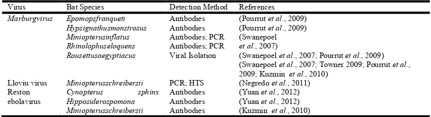
Virus Bat Species Detection Method References
Marburgvirus Epomopsfranqueti Hypsignathusmonstrosus Miniopterusinflatus Rhinolophuseloquens Rousettusaegyptiacus
Antibodies Antibodies Antibodies; PCR Antibodies; PCR Viral Isolation
(Pourrut et al., 2009) (Pourrut et al., 2009) (Swanepoel
et al., 2007)
(Swanepoel et al., 2007; Pourrut et al., 2009)
(Swanepoel et al., 2007; Towner 2009; Pourrut et al.,
2009; Kuzmin et al., 2010)
Lloviu virus Miniopterusschreibersii PCR; HTS (Negredo et al., 2011)
Reston ebolavirus
Cynopterus sphinx Hipposiderospomona Miniopterusschreibersii
Antibodies Antibodies Antibodies
(Yuan et al., 2012) (Yuan et al., 2012)
(Kuzmin et al., 2010)
Table 2. Drug and Mode of Action
S.No Drug Drug Type Mode of Action
1. Favipiravir(T-705)
(FujifilmHoldings Corp)
Nucleoside analogue – broad spectrum
Activity against RNAviruses
RNA chain termination and/or lethal mutagenesis
1. TKM-Ebola (Tekmira
Pharmaceuticals Corp)
Lipid nanoparticle With siRNA –Ebolavirus specific compound
Gene silencing
2. BCX-4430 (BioCryst Pharmaceuticals) Nucleoside analogue
– broad spectrum Activity against RNA Viruses
RNA chain termination
4. AVI-6002 (Sarepta Therapeutics) Phosporodiamidate
Morpholinooligomer –Ebolavirus specific compound
[image:3.595.59.543.680.800.2]envelope, occurs by budding through intracellular membranes. Viral particles are transported in cytoplasmic vesicles through the secretory pathway before they are released by exocytosis (Daep et al., 2014; Leyssen et al., 2000).
Transmission
Zika virus disease is caused by an RNA virus transmitted to humans by the Aedes aegypti species. Up to eighty per cent of infections are asymptomatic (Duffy et al., 2009). Symptomatic infections are characterised by a self-limiting febrile illness of 4–7 days duration accompanied by maculopapular rash, arthralgia, conjunctivitis, myalgia and headache. Zika virus has not been noted to cause death in the past, nor has it been linked to intra-uterine infections and congenital CNS anomalies. Zika virus infection can be confirmed by direct detection of Zika virus RNA or specific viral antigens in clinical specimens. There are no validated assays for serology.
More information on Zika virus disease can be found in the previous risk assessments (European Centre for Disease Prevention and Control, 2014; European Centre for Disease
Prevention and Control, 2015) and in the ECDC factsheet for health professionals (Kuno et al., 1998)
Zika virus is spread to people primarily through the bite of an infected Aedes species mosquito (Ae. aegypti and
Ae. albopictus).
A pregnant woman can pass Zika virus to her fetus during pregnancy or around the time of birth. We are studying how Zika affects pregnancies.
[image:4.595.110.489.118.354.2] To date, there are no reports of infants getting Zika through breastfeeding. Because of the benefits of breastfeeding, mothers are encouraged to breastfeed even in areas where Zika virus is found.
Table No.3 Characteristics of reported cases of congenital malformation potentially linked with zika virus infection in Brazil (AS OF 18 JANUARY 2016)
No. Date of report location Clinical findings Laboratory findings
1. 17 November 2015
Paraíba state
Foetus with microcephaly at ultrasound exams (US) 30.1 weeks’ gestation
Head circumference <2.6 SD Observed lesions (US):
- Brain atrophy with coarse calcifications involving the white matter of the frontal lobes, including the caudate, lentostriatal vessels and cerebellum.
- Corpus callosal and vermian dysgenesis. - Enlarged cisterna magna
Mother: symptoms compatible with Zika virus infection at week 18-19 of gestation*
RT-PCR Zika virus positive in amniotic fluid (Instituto Oswaldo Cruz)
(Melo et al., 2016; Ministério da Saúde, 2015)
2. 28 November 2015
Ceara state
Newborn
Born the 18 November 2015 (residing Tejuçuoca, Ceara State) No measurement of head circumference at birth
Weight: 945 grams at birth Died within 5 min after birth Observed lesions (US, 13 Nov 2015): -microcephaly (head circumference 190 mm) - fetal anasarca
- polydramnios
Presence of Zika viral genome in blood and tissue samples of the newborn (Evandro Chagas Institute)
(Centro de operações de emergências em saúde pública sobre microcefalias
2015; Pan American Health Organization, 2015)
3. 15 January 2016
Hawaii (USA)
Case: baby with congenital microcephaly who was born recently on Oahu island, Hawaii.
Mother had a probable exposure to Zika virus when she was residing in Brazil in May 2015 (no further details provided)
Laboratory confirmation of a past Zika virus infection (no details)
(US CDC laboratory)
Although mosquito bites are the main way that Zika virus is spread, Zika virus can also spread when an infected man has sex with his partners.
Zika virus genetics: There are two lineages of Zika virus, the African lineage and the Asian lineage (Kuno et al., 1998; Faye
et al., 2014). Presently, only two full genome sequences of Zika virus from Brazil and Suriname have been published (Haddow et al., 2012). Molecular analysis of the Zika virus isolated from the travel-related case from the Maldives and diagnosed in Finland in showed that it too belonged to the Asian lineage. Recent preliminary findings from molecular investigations of 17 whole genome sequences in the public domain stressed a possible change in the fitness of the Asian lineage through an adaptation of the NS1 codon usage. The researchers suggest that these modifications may have an impact on viral replication rates and viral titres in humans. The authors also reported structural and immunological similarities in the NS1 antigen between Zika and dengue viruses. Both preliminary findings should be further studied and verified on larger whole genome panels (Enfissi et al.,).
Symptoms: The most common symptoms are fever, rash, joint pain or red eyes. Other common symptoms include muscle pain and headache. Symptoms usually begin two to seven days after being bitten by an infected mosquito and last several days to a week. Hospitalization and deaths from Zika are unusual, but a nerve disorder, Guillain-Barré Syndrome, can rarely follow an infection. The biggest concern is related to birth defects that have been seen when pregnant women become infected.
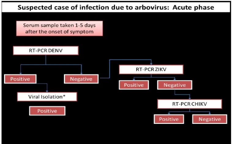
Algorithm for detecting zika virus (ZIKV) (Gourinat et al., 2015): This algorithm is addressed to laboratories with established capacity (molecular, antigenic and/or serological) to detect dengue (DENV), Zika (ZIKV) (Hayes, 2009), and chikungunya (CHIKV) as part of the differential diagnosis for arbor viruses. A BSL2 containment level is required to handle suspected samples.
Acknowledgement
We authors would like to thank our principal Dr. Mohan Kale, Head of department of pharmacology. Our college member like librarian, computer experts, and all other persons who help us in direct or indirect way to whom we fail to notice. Our sincere thanks to almighty God for their continuous monitoring of our work till its completion.
REFERENCES
Amman, B.R., Carroll, S.A., Reed, Z.D., Sealy, T.K., Balinandi, S., Swanepoel, R., Kemp, A., Erickson, B.R., Comer, J.A., Campbell, S., et al. 2012. Seasonal Pulses of Marburg Virus Circulation in Juvenile Rousettusaegyptiacus Bats Coincide with Periods of Increased Risk of Human Infection. PLoSPathog. 8, doi:10.1371/journal.ppat.1002877.
Bausch, D.G., Borchert, M., Grein, T., Roth, C., Swanepoel, R., Libande, M.L., Talarmin, A., Bertherat, E., Muyembe-Tamfum, J.J., Tugume, B., et al. 2003. Risk factors for Marburg hemorrhagic fever, Democratic Republic of the Congo. Emerg. Infect. Dis., 9, 1531–1537.
Brauburger, K., Hume, A.J., Muhlberger, E., Olejnik, J. 2012. Forty-Five Years of Marburg Virus Research. Viruses, 4, 1878–1927.
Brauburger, K., Hume, A.J., Muhlberger, E., Olejnik, J. 2012. Forty-Five Years of Marburg Virus Research. Viruses, 4, 1878–1927.
Briand S, Bertherat E, Cox P, et al. 2014. The International Ebola Emergency. N Engl J Med., 371:1180-3.
Carroll, S.A., Towner, J.S., Sealy, T.K., McMullan, L.K., Khristova, M.L., Burt, F.J., Swanepoel, R., Rollin, P.E., Nichol, S.T. 2013. Molecular evolution of viruses of the family Filoviridae based on 97 whole-genome sequences.
J. Virol., 87, 2608–2616.
Centro de operações de emergências em saúde pública sobre microcefalias. Monitoramento dos casos de microcefalias no brasil informe epidemiológico Nº 02/2015 – semana epidemiológica 47 (22 a 28/11/2015) (Internet). 2015 (cited 2015 Nov 30). Available from: http://portalsaude.saude. gov.br/images/pdf/2015/novembro/30/coes-microcefalias---informe-epidemiol--gico---se-47.pdf.
Daep CA, Munoz-Jordan JL, Eugenin EA. 2014. Flaviviruses, an expanding threat in public health: focus on dengue, West Nile, and Japanese encephalitis virus. J Neurovirol., 20(6):539-60.
Dick GW, Kitchen SF, Haddow AJ. Zika virus. I. 1952. Isolations and serological specificity. Trans R Soc Trop Med Hyg., 46(5):509-20.
Duffy MR, Chen TH, Hancock WT, Powers AM, Kool JL, Lanciotti RS, et al. Zika virus outbreak on Yap Island, Federated States of Micronesia. N Engl J Med. 2009 Jun 11;360(24):2536-43.
Enfissi A, Codrington J, Roosblad J, Kazanji M, Rousset D. Zika virus genome from the Americas. The Lancet, 387(10015):227-8.
European Centre for Disease Prevention and Control. Microcephaly in Brazil potentially linked to the Zika virus epidemic Stockholm: ECDC; 2015 (updated 2015 Nov 25). Available from: http://ecdc.europa.eu/en/publications/
Publications/zika-microcephaly-Brazil-rapid-risk-assessment-Nov-2015.pdf.
European Centre for Disease Prevention and Control. Rapid risk assessment: Zika virus infection outbreak, French Polynesia: ECDC; 2014 (updated 2014 Feb 14). Available from: http://ecdc.europa.eu/en/publications/_layouts/forms/ Publication_DispForm.aspx?List=4f55ad51-4aed-4d32-b960-af70113dbb90&ID=1025.
European Centre for Disease Prevention and Control. Zika virus epidemic in the Americas: potential association with microcephaly and Guillain-Barré syndrome Stockholm: ECDC; 2015 (updated 2015 Dec 10). Available from: http://ecdc.europa.eu/en/publications/Publications/zika- virus-americas-association-with-microcephaly-rapid-risk-assessment.pdf.
European Centre for Disease Prevention and Control. Zika virus infection outbreak, Brazil and the Pacific region: ECDC; 2014 (updated 2015 May 26). Available from: http://ecdc.europa.eu/en/publications/Publications/rapid- risk-assessment-Zika%20virus-south-america-Brazil-2015.pdf.
European Centre for Disease Prevention and Control. Zika virus infection (factsheet for health professionals) (Internet). Stockholm: ECDC; 2015 (cited 2015 18 May 2015). Available from:http://ecdc.europa.eu/en/health topics/zika_virus_infection/factsheet-healthprofessionals/ Pages/factsheet_health_professionals.aspx.
Its Emergence in the 20(th) Century. PLoS Negl Trop Dis., 8(1):e2636.
Feldmann H, Geisbert TW. 2011. Ebola haemorrhagic fever.
Lancet, 377:849-62.
Feldmann, H., Jones, S., Klenk, H.D., Schnittler, H.J. 2003. Ebola virus: From discovery to vaccine. Nat. Rev. Immunol., 3, 677–685.
Feldmann, H., Kiley, M.P. 1999. Classification, structure, and replication of filoviruses. Curr.Top.Microbiol.Immunol.,
235, 1–21.
Furuta Y, Gowen BB, Takahashi K, Shiraki K, Smee DF, Barnard DL. 2013. Favipiravir (T-705), a novel viral RNA polymerase inhibitor. Antiviral Res., 100:446-54.
Gourinat AC, O’Connor O, Calvez E, Goarant C, Dupont-Rouzeyrol M. 2015. Detection of Zika Virus in Urine. Emerging Infectious Diseases, 21:84-6. http://wwwnc. cdc.gov /eid/article /21/1/14-0894_article
Haddow AD, Schuh AJ, Yasuda CY, Kasper MR, Heang V, Huy R, et al. 2012. Genetic characterization of Zika virus strains: geographic expansion of the Asian lineage. PLoS Negl Trop Dis., 6(2):e1477.
Hawaii Department of health. Hawaii Department of health receives confirmation of Zika infection in baby born with microcephaly (News Release). Honolulu, 2016.
Hayes EB. 2009. Zika virus outside Africa. Emerg Infect Dis., 15(9):1347-50.
Hayes EB. 2009. Zika virus outside Africa. Emerging Infectious Diseases, 15:1347-50.
http://apps.who.int/iris/bitstream/10665/133833/1/roadmapsitr ep4_eng.pdf?ua=1
Kiley, M.P., Bowen, E.T., Eddy, G.A., Isaacson, M., Johnson, K.M., McCormick, J.B., Murphy, F.A., Pattyn, S.R., Peters, D., Prozesky, O.W., et al. 1982. Filoviridae: A taxonomic home for Marburg and Ebola viruses?
Intervirology, 18, 24–32.
Kuno G, Chang GJ, Tsuchiya KR, Karabatsos N, Cropp CB. 1998. Phylogeny of the genus Flavivirus. J Virol., 72(1):73-83.
Kuzmin, I.V., Niezgoda, M., Franka, R., Agwanda, B., Markotter, W., Breiman, R.F., Shieh, W.J., Zaki, S.R., Rupprecht, C.E. 2010. Marburg Virus in Fruit Bat, Kenya.
Emerg. Infect. Dis., 16, 352–354.
Leyssen P, De Clercq E, Neyts J. 2000. Perspectives for the treatment of infections with Flaviviridae. Clin Microbiol Rev., 13(1):67-82, table of contents.
Martinez-Pulgarin DF, Acevedo-Mendoza WF, Cardona-Ospina JA, Rodriguez-Morales AJ, Paniz-Mondolfi AE. 2015. A bibliometric analysis of global Zika research. Travel Med Infect Dis.
Melo OAS, Malinger G, Ximenes R, Szejnfeld PO, Alves Sampaio S, Bispo de Filippis AM. 2016. Zika virus intrauterine infection causes fetal brain abnormality and microcephaly: tip of the iceberg? Ultrasound Obstet Gynecol., 47(1):6-7.
Ministério da Saúde (Brazil). Microcefalia - Ministério da Saúde divulga boletim epidemiológico (Internet). Brasília: Ministério da Saúde; 2015 (updated 2015 Nov 17; cited 2015 Nov 17). Available from: http://portalsaude.saude. gov.br/index.php/cidadao/principal/agencia-saude/20805-ministerio-da-saude-divulga-boletim-epidemiologico Negredo, A., Palacios, G., Vazquez-Moron, S., Gonzalez, F.,
Dopazo, H., Molero, F., Juste, J., Quetglas, J., Savji, N., de la Cruz Martinez, M., et al. 2011. Discovery of an ebolavirus-like filovirus in europe. PLoS Pathog., 7, e1002304.
Nunes-Alves C. Ebola update. 2014. Nat Rev Microbiol., 12:656.
Oestereich L, Ludtke A, Wurr S, Rieger T, Munoz-Fontela C, Gunther S. 2014. Successful treatment of advanced Ebola virus infection with T-705 (favipiravir) in a small animal model. Antiviral Res., 105:17-21.
Pan American Health Organization, World Health Organization. Regional Office for the Americas. Epidemiological Alert: Neurological syndrome, congenital malformations, and Zika virus infection. Implications for public health in the Americas (Internet). Washington: World Health Organization; 2015 (updated 2015 Dec 1; cited 2015 Dec 1). Available from: http://www.paho.org/ hq/index.php?option=com_docman&task=doc_download& Itemid=&gid=32405&lang=en.
Pourrut, X., Kumulungui, B., Wittmann, T., Moussavou, G., Delicat, A., Yaba, P., Nkoghe, D., Gonzalez, J.P., Leroy, E.M. 2005. The natural history of Ebola virus in Africa.
Microbes Infect./Inst.Pasteur, 7, 1005–1014.
Pourrut, X., Souris, M., Towner, J.S., Rollin, P.E., Nichol, S.T., Gonzalez, J.P., Leroy, E. 2009. Large serological survey showing cocirculation of Ebola and Marburg viruses in Gabonese bat populations, and a high seroprevalence of both viruses in Rousettusaegyptiacus.
BMC Infect. Dis., 9, doi:10.1186/1471-2334-9-159
Sanchez, A., Geisbert, T.W., Feldmann, H. Filoviridae: Marburg and Ebola Viruses. In Fields virology; Knipe, D. M., Howley, P. M., Eds. 2007. Lippincott Williams and Wilkins: Philadelphia, PA,USA, 1279–1304.
Smither SJ, Eastaugh LS, Steward JA, Nelson M, Lenk RP, Lever MS. 2014. Post-exposure efficacy of oral T-705 (Favipiravir) against inhalational Ebola virus infection in a mouse model. Antiviral Res., 104:153-5.
Swanepoel, R., Smit, S.B., Rollin, P.E., Formenty, P., Leman, P.A., Kemp, A., Burt, F.J., Grobbelaar, A.A., Croft, J., Bausch, D.G., et al. 2007. Studies of reservoir hosts for Marburg virus. Emerg. Infect. Dis., 13, 1847–1851.
Taylor, D.J., Dittmar, K., Ballinger, M.J., Bruenn, J.A. 2011. Evolutionary maintenance of filovirus-like genes in bat genomes. BMC Evol.Biol., 11, 336.
Towner, J.S., Amman, B.R., Sealy, T.K., Carroll, S.A.R., Comer, J.A., Kemp, A., Swanepoel, R., Paddock, C.D., Balinandi, S., Khristova, M.L., et al. 2009. Isolation of Genetically Diverse Marburg Viruses from Egyptian Fruit Bats. PLoSPathog., 5, doi:10.1371/journal.ppat.1000536. Towner, J.S., Amman, B.R., Sealy, T.K., Carroll, S.A.R.,
Comer, J.A., Kemp, A., Swanepoel, R., Paddock, C.D., Balinandi, S., Khristova, M.L., et al. 2009. Isolation of Genetically Diverse Marburg Viruses from Egyptian Fruit Bats. PLoSPathog., 5, doi:10.1371/journal.ppat.1000536. Warren TK, Wells J, Panchal RG, et al. 2014. Protection
against filovirus diseases by a novel broad-spectrum nucleoside analogue BCX4430. Nature, 508:402-5. WHO. WHO statement on the first meeting of the
International Health Regulations (2005) (IHR 2005) Emergency Committee on Zika virus and observed increase in neurological disorders and neonatal malformations: WHO; 2016 (cited 2016 04022016). Available from: http://www.who.int/mediacentre/news/ statements/2016/1stemergencycommittee- zika/en/.
Yuan, J.F., Zhang, Y.J., Li, J.L., Zhang, Y.Z., Wang, L.F., Shi, Z.L. 2012. Serological evidence of ebolavirus infection in bats, China. Virol. J., 9, doi:10.1186/1743-422X-9-236.