ORIGINAL RESEARCH ARTICLE
A STUDY ON THE FAILURE OF TRANSPORT PROTEIN
*
Lee Donghee
Auckland International College, Grade 12
ARTICLE INFO ABSTRACT
Life is maintained by cell metabolism. The most important factor in cell metabolism is protein production and destruction. Once created, the protein must function and be ubiquitin immediately. When a three-dimensional protein which formed the helix structure and a folded structure occurs a deformation, it interferes with cell metabolism and even causes cell necrosis. In particular, modification of structure in the transport protein degrades transporting ability of the substrate, and inhibits the passage of metabolites. It is a misfolding that is the main pattern for the modification of the steric structure of proteins. Abnormal folding is the most serious problem in protein structure. Misfolding of proteins is the main cause of DNA mutations. The misfolded protein fails to bind to the substrate to be transported. Changing the amino acids involved in the folding structure is a fundamental problem. Altering the amino acid residues associated with the side chain is the cause of misfolding. There is a need for a strategy to respond to misfolded proteins in drug development.
*Corresponding author
Copyright ©2017,Lee Donghee.This is an open access article distributed under the Creative Commons Attribution License, which permits unrestricted use, distribution, and reproduction in any medium, provided the original work is properly cited.
INTRODUCTION
Hardly anything happens in the life of a cell that does not require proteins. They are the most versatile of macromolecules, each with its own inbuilt ability to carry out a cellular function. Some proteins aggregate to form relatively stiff filaments that help define the cell’s shape and hold organelles in position. Others span the cell membrane and form channels or pores through which ions and small molecules can move. Many others are enzymes that catalyze the thousands of chemical reactions needed to maintain life. Still others are signaling proteins that enable cells to coordinate their internal activities or to communicate with other cells. Protein is an important substance found in every cell in the human body. In fact, except for water, protein is the most abundant substance in your body. This protein is manufactured by your body utilizing the dietary protein you consume. It is used in many vital processes and thus needs to be consistently replaced. You can accomplish this by regularly consuming foods that contain protein.
Proteins are large, complex molecules that play many critical roles in the body. They do most of the work in cells and are required for the structure, function, and regulation of the body’s tissues and organs. Proteins are made up of hundreds or thousands of smaller units called amino acids, which are attached to one another in long chains. There are 20 different types of amino acids that can be combined to make a protein. The sequence of amino acids determines each protein’s unique 3-dimensional structure and its specific function. Proteins can be described according to their large range of functions in the body, listed in alphabetical order. Proteins are produced through the processes of DNA transcription and translation. In protein synthesis, DNA is first transcribed or copied into RNA. The resulting RNA transcript or messenger RNA (mRNA) is then translated to produce amino acids from the transcribed genetic code. Organelles called ribosomes and another RNA molecule called transfer RNA help to translate mRNA. The resulting amino acids are joined together through dehydration synthesis, a process in which a peptide bond is formed between the amino acids. A polypeptide chain is formed when a number of amino acids are linked together by peptide bonds.
ISSN: 2230-9926
International Journal of Development Research
Vol. 07, Issue, 09, pp.14991-14997, September,2017
Article History:
Received 18th June, 2017 Received in revised form 19th July, 2017
Accepted 24th August, 2017
Published online 30th September, 2017
Citation: Lee Donghee. 2017. “A Study on the Failure of Transport Protein”, International Journal of Development Research, 7, (09), 14991-14997.
ORIGINAL RESEARCH ARTICLE OPEN ACCESS
Keywords:
Amino Acids, Polypeptide Chain, Three-Dimensional Structure, Backbone, Main Chain, Side chain,
After several modifications, the polypeptide chain becomes a fully functioning protein. One or more polypeptide chains twisted into a 3-D structure form a protein. An amino acid is an organic molecule that, when linked together with other amino acids, forms a protein. Amino acids are essential to life because the proteins they form are involved in virtually all cell functions. Some proteins function as enzymes, some as antibodies, some as transport, while others provide structural support. Although there are hundreds of amino acids found in nature, proteins are constructed from a set of 20 All amino acids have the alpha carbon bonded to a hydrogen atom, carboxyl group, and amino group. The "R" group varies among amino acids and determines the differences between these protein monomers. The amino acid sequence of a protein is determined by the information found in the cellular genetic code. The genetic code is the sequence of nucleotide bases in nucleic acids (DNA and RNA) that code for amino acids. These gene codes not only determine the order of amino acids in a protein, but they also determine a protein's structure and function.
Source:https://bio.libretexts.org/LibreTexts/University_of_California_Davis/B IS_2A%3A_Introductory_Biology_(Facciotti)/Readings/2017_SUMMER_SE
[image:2.595.82.247.297.402.2]SSION_1/2017_SS1_Lecture_03
Figure 1. The Amino Acid Backbone
Amino acids can be classified into four general groups based on the properties of the "R" group in each amino acid. Amino acids can be polar, non-polar, positively charged, or negatively charged. Polar amino acids have "R" groups that are hydrophilic, meaning that they seek contact with aqueous solutions. Non-polar amino acids are the opposite (hydrophobic) in that they avoid contact with liquid. These interactions play a major role in protein folding and give proteins their 3-D structure. Below is a listing of the 20 amino acids grouped by their "R" group properties.
The non-polar amino acids are hydrophobic, while the remaining groups are hydrophilic. While amino acids are necessary for life, not all of them can be produced naturally in the body. Of the 20 amino acids, 11 can be produced naturally. These nonessential amino acids are alanine, arginine, asparagine, aspartate, cysteine, glutamate, glutamine, glycine, proline, serine, and tyrosine. With the exception of tyrosine, nonessential amino acids are synthesized from products or intermediates of crucial metabolic pathways. For example, alanine and aspartate are derived from substances produced during cellular respiration. Alanine is synthesized from pyruvate, a product of glycolysis. Aspartate is synthesized from oxaloacetate, an intermediate of the citric acid cycle. Six of the nonessential amino acids (arginine, cysteine, glutamine, glycine, proline, and tyrosine) are considered conditionally essential as dietary supplementation may be required during the course of an illness or in children. Amino acids that cannot be produced naturally are called essential amino acids.
They are histidine, isoleucine, leucine, lysine, methionine, phenylalanine, threonine, tryptophan, and valine. Essential amino acids must be acquired through diet. Common food sources for these amino acids include eggs, soy protein, and whitefish. Unlike humans, plants are capable of synthesizing all 20 amino acids. Understanding how proteins, which are polymers of amino acids, carry out different functions in a cell has been a problem of considerable interest (Madan Babu, 2016). Transport Proteins - are carrier proteins which move molecules from one place to another around the body. Examples include hemoglobin and cyto-chromes. Hemoglobin transports oxygen through the blood via red blood cells. Cyto-chromes operate in the electron transport chain as electron carrier proteins. The transport function of proteins is closely related to the stereo-structure of proteins. The three-dimensional protein structure must be balanced to allow the transport of substrates required for cell metabolism. It is because of the specificity of the steric structure that a protein can transport a substrate. When an abnormality occurs in the
three-dimensional structure, the carrying function is
significantly deteriorated.
Structure of Proteins
Alpha helices and beta sheets are linked by less-structured loop regions to form domains, which combine to form larger subunits and ultimately functional proteins. Proteins play countless roles throughout the biological world, from catalyzing chemical reactions to building the structures of all living things. Despite this wide range of functions all proteins are made out of the same twenty amino acids, but combined in different ways. The way these twenty amino acids are arranged dictates the folding of the protein into its unique final shape. Since protein function is based on the ability to recognize and bind to specific molecules, having the correct shape is critical for proteins to do their jobs correctly. The amino acid sequence largely determines the three-dimensional structure of a protein. The sequence of a protein encodes all information required for its fold and function, but we are not always able to decipher the function from sequence or structure alone
(Debnath Pal and David Eisenberg, 2005). The protein chain
Source: http://www.biologyreference.com/Po-Re/Protein-Structure.html
Figure 2. Levels of protein structure
The inset highlights the pattern of hydrogen bonds that stabilizes alpha helices. Many functional proteins fold into a compact globular shape, with many carbon-rich amino acids sheltered inside away from the surrounding water. The folded structure of hemoglobin includes a pocket to hold heme, which is the molecule that carries oxygen as it is transported throughout the body. The folding pattern of a polypeptide chain can be described in terms of the angles of rotation around the main chain bond. Chemical bonds have characteristic lengths, the peptide bond has partial double-bond character, meaning it is shorter, and rigid. Two or more polypeptide chains can come together to form one functional molecule with several subunits. The four subunits of hemoglobin cooperate so that the complex picks up and delivers more oxygen than is possible with single subunits. Protein function can be thought of on different interdependent levels and may be divided into three major categories: molecular function, biological process and cellular component
(David Lee et al. 2007).Molecular function describes activity
on the molecular level, such as catalysis, whereas biological process describes broader functions that are carried out by assemblies of molecular functions, such as a particular metabolic pathway. Cellular component describes the compartment or compartments of a cell in which the protein performs its function
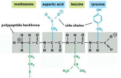
[image:3.595.61.260.531.659.2]Source: Essential Cell Biology, 4th Edition, Alberts et al. Garland Science.
Figure 3. A four-amino acid peptide
The primary sequence (or primary structure) of a protein is simply the linear sequence of amino acids in the polypeptide chain. Figure 3 above illustrates a peptide consisting of four amino acids. The full names and three-letter abbreviations for these amino acids are shown at the top and the bottom of the figure, respectively. If you were listing the primary sequence of this peptide using the three-letter abbreviations, it would be: Met-Asp-Leu-Tyr.
Detailed Functions of Transport Protein
There are two types of transport protein, one is proteins that directly transport substrates, the other one is proteins that help migration of substrates. Transferin is a glycoprotein with homologous N-terminal and C-terminal iron-binding domains. Transferrin is related to several other iron-binding proteins including lactoferrin, melanotransferin, and ovotransferrin. These molecules comprise the transferrin superfamily. All members of this superfamily have similar polypeptide folding patterns. The N-terminal and C-terminal domains of these proteins are globular moieties of about 330 amino acids. Each of these domains is divided into two subdomains, with the iron- and anion-binding sites found within the inter-subdomain cleft. The binding cleft opens with iron release, and closes with iron binding (Ching Ming Chung, 1984).Glucocorticoids
are released after hypothalamus-pituitary-adrenal axis
stimulation by stress and act both in the periphery and in the brain to bring about adaptive responses that are essential for life. Dysregulation of the stress response can precipitate psychiatric diseases, in particular depression. Recent genetic studies have suggested that the glucocorticoid carrier transcortin, also called corticosteroid-binding globulin (CBG),
may have an important role in stress response (Avery a.
structure Transporter proteins selectively bind to substrate in either its nonphosphorylated (inactive) or phosphorylated (active) form (Lutz Kummera, et al., 2012). When a phosphate group is attached to a protein, a structural change occurs. Serine, threonine, and tyrosine in the amino acids constituting the protein bind to the phosphate group. In order to carry out a unique transport function in the transport protein, it must bind to the target substrate. To this end, structural changes of the transport proteins must be accompanied. Phosphorylation refers to the activity of transporter proteins. Phosphorylation in transporter proteins means the conformational change through the activation, which is the key structural difference that occurs upon activation. Whereas the rigid phosphorylated activation loop remains in the same form when bound by transporter proteins, the more mobile unphosphorylated loop is pushed to a new position (Lutz Kummera, et al., 2012).
Regulation of enzyme activity by phosphorylation/
dephosphorylation is a regulation mechanism of universal enzymatic activity present in eukaryotes and is commonly observed in the process of hormone and other actions and cell response according to external environment. When the hydroxyl group of serine, threonine, tyrosine residues are phosphorylated, the enzyme protein changes in conformation, resulting in a change in enzyme activity. In addition, the phosphorylated enzyme protein returns to its original structure by being dephosphorylated. Phosphorylation is catalyzed by
enzymes collectively called protein kinases and
dephosphorylation is catalyzed by enzymes collectively. The cell responds to metabolism by receiving an external signal from the receptor on the cell membrane. When the receptor protein is activated, several proteins in the cytoplasm are sequentially phosphorylated to give a metabolic signal to the nuclear membrane. The cytoplasmic protein becomes active when the phosphate group is bound and becomes inactive when the phosphate group is separated. ROS are highly reactive, toxic oxygen moieties, such as hydroxyl radical, peroxyl radical, superoxide anion, and hydrogen peroxide. Collectively, ROS can lead to oxidation of proteins and DNA, peroxidation of lipids, and, ultimately, cell death. Protein carbonyls can result from oxidative cleavage of the protein backbone, direct oxidation of amino acids such as lysine, arginine, histidine, proline, glutamic acid, and threonine, or by the binding of aldehydes produced from lipid peroxidation. Direct oxidation of lysine, arginine, proline, and threonine residues may also yield carbonyl derivatives. In addition, carbonyl groups may be introduced into proteins by reactions with aldehydes (4-hydroxy-2-nonenal, malondialdehyde) produced during lipid peroxidation (Barbara S. Berlett and Earl R. Stadtman, 1997).
Carbonyl (CO) groups (aldehydes and ketones) are produced on protein side chains (especially of Pro, Arg, Lys, and Thr) when they are oxidised (Isabella Dalle-Donnea et al., 2003). Protein carbonyl content is actually the most general indicator and by far the most commonly used marker of protein oxidation, and accumulation of protein carbonyls has been observed in several human diseases including Alzheimer’s disease (AD), diabetes, inflammatory bowel disease (IBD), and arthritis (Isabella Dalle-Donnea et al., 2003). Active oxygen (ROS) is produced during mitochondrial oxygen metabolism. Active oxygen (ROS) migrates from the mitochondrial matrix to the cytoplasm and intervenes in the pathway of the proliferative signal. The ROS target is a protein tyrosine phosphatase (PTPs) that breaks down phosphate
groups in phosphate-coupled proteins and blocks the activity of the protein. Inactivation is induced by PTP oxidation. As a result, it continues to be activated for cell proliferation and develops into cancer cells (Jae Yeon Ahn, et al. 2006).Folding of newly synthesized polypeptides in the crowded cellular environment requires the assistance of so-called molecular chaperone proteins. Chaperones of the Hsp(heat shock protein)70 class and their partner proteins interact with nascent polypeptide chains on ribosomes and prevent their premature (mis)folding at least until a domain capable of forming a stable structure is synthesized (Hendrick and Hartl, 1995) Protein quality control constitutes the regulation of protein synthesis, folding, unfolding and turnover. It is mediated by chaperone and protease systems, together with cellular clearance mechanisms such as autophagy and lysosomal degradation. These quality control systems have an essential role in the life of cells, ensuring that proteins are correctly folded and functional at the right place and time (Helen Saibil, 2013). They are crucial for mitigating the deleterious effects of protein misfolding and aggregation, which, by unclear mechanisms, can cause cell death in neurodegeneration and other incurable protein misfolding diseases (Helen Saibil, 2013). Tertiary structures of proteins are characterised by hydrophobic cores (non polar amino acids) and hydrophilic surfaces (polar amino acids). Myoglobin is a protein found in muscle tissue that binds oxygen. Myoglobin is a monomeric heme protein found mainly in muscle tissue where it serves as an intracellular storage site for oxygen. During periods of oxygen deprivation, oxymyoglobin releases its bound oxygen which is then used for metabolic purposes. The tertiary structure of myoglobin is that of a typical water soluble globular protein. Its secondary structure is unusual in that it contains a very high proportion (75%) of α-helical secondary structure. A myoglobin polypeptide is comprised of 8 separate right handed a-helices, designated A through H, that are connected by short non-helical regions (as below Figure 4). Amino acid R-groups packed into the interior of the molecule are predominantly hydrophobic in character while those exposed on the surface of the molecule are generally hydrophilic, thus making the molecule relatively water soluble.
[image:4.595.356.515.528.642.2]Source: http://www.bioinfo.org.cn/ book/biochemistry /chapt07/bio2.htm
Figure 4. A myo-globin polypeptide
Mutations on Transporter Protein
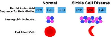
Mutations can change the phenotype and have a beneficial, deleterious or neutral effect on the fitness of the individual organism. Phenotypes and the corresponding mutations are then acted on by selection (natural or artificial) at the population level. Changes in proteins and DNA comprise single-point mutations (SNPs; single nucleotide polymorphisms), insertions–deletions, domain shuffling or copy number variations. These changes can affect diverse protein properties, such as stability, catalytic activity or the ability to interact with other molecules (Romain A.Studer, et al. 2013). Mutations can be the source of various diseases. In coding genes, mutations can cause a defect in the protein. A well-known case is sickle-cell anaemia, where the haemoglobin molecule is affected (as below Figure 5). Protein mutations in some cells can lead to deregulation and the emergence of cancer cells (Romain A.Studer, et al. 2013).
[image:5.595.70.257.275.338.2]Source: https://beyondthedish.wordpress.com/tag/sickle-cell-disease.
Figure 5. Changes to the Beta-Globin subunit of hemoglobin in sickle cell disease and the functional consequence for red blood
cells
Figure 5 above illustrates the basis of the most common version of sickle cell disease. Hemoglobin protein structure are altered in sickle cell disease, as shown in Figure 5. Normal red blood cells can easily travel through blood vessels, whereas sickle-shaped red blood cells get stuck. This is the basis of sickle cell anemia. The SERT(5-HTT) is also known as the "sodium-dependent Serotonin transporter. Its protein in humans is encoded by the "SLC6A4" gene. Serotonin transporter is called solute carrier family 6 (neurotransmitter transporter, serotonin), member 4. Mutations in SLC6A4 genes can alter the structure of Serotonin transporter the tree and it would be cause of depression. Point mutations in a protein sequence may result in a change or loss of the native structure, which in turn may cause a change or loss of function, and ultimately yields different phenotypes(Eric Feyfantet al. 2007). Amino acid residue replacements by point mutation often leave the backbone conformation almost unchanged. A single-point mutation in general does not trigger changes in many dihedral angles on the native structure of protein. Globular proteins are generally found in two states: a free unfolded state and a folded state, with the latter being functional. Each of them is associated with a Gibbs free energy value (ΔG), the unfolded state being more energetic than the folded state (ΔGunfold>ΔGfold). To move from one state to the other, there needs to be a transfer of energy. In their stable state, proteins are marginally stable, between −3 and −10 kcal·mol−1, and can tolerate some destabilization within a narrow range. This equilibrium is quite sensitive and any mutation that adds energy (i.e. more than +2 kcal·mol−1) to the folded state is likely to destabilize the structure and make the protein more likely to aggregate in its unfolded form, and this could be a factor in some diseases (Romain A. Studer et al. 2013). By contrast, a mutation that removes energy from the folded state (i.e. less than +2 kcal·mol−1) is likely to stabilize
the protein structure and make it too rigid and so non-functional in the case of enzymes (Romain A. Studer et al. 2013). The basic differences between the 20 natural amino acid residues are due to differences in their side-chain structures. It is necessary the contributions of side-chain–side-chain (s-s) and side-chain–backbone (s-b) interactions to the stabilization of folded protein structures. if one residue mutates, its interacting residue in the interacting partner will be under pressure to carry out a compensatory mutation for the interaction to be maintained (Velin Z. Spassov et al. 2007). Amino acid side chains are important intramolecular interactions in the structural realization of amino acid code(as
below Figure 6-1). Side chains may play the dominant role in amino acid side-chain packing and consequently in stabilizing protein native conformation. Among intramolecular side-chain interactions, the side-chain–backbone interaction is the dominant force for side-chain packing and for stabilizing the folded structure. The side-chain–backbone interactions outperform side-chain–side-chain interactions in differentiating native structure from the misfolded structures. The side-chain–backbone interactions show about a twice stronger effect on discriminating the decoy structures from the native states than s-s interactions and this is valid for both van der Waals and electrostatic contributions (Velin Z. Spassovet al. 2007)A protein in which many amino acids form a three-dimensional structure through peptide bonds, hydrogen bonds, and disulfide bonds has two patterns of helical structure and folding sheet.
The DNA of neuron cells continues to make abnormal proteins, and the proteins accumulate because they do not become ubiquitin in time. When the transport protein is misfolded as a mutant, like prion protein, its binding angle to the carrier (substrate) of the side-chain rotamers is changed and its carrying capacity is weakened. The sequence of side chains determines all that is unique about a particular protein, including its biological function and its specific three-dimensional structure. Each of the side groups has a certain "personality" which it contributes to this task. The hydrophobic residues provide a very strong driving force for folding, through the indirect effect of their ceasing to disrupt the water structure once they are buried. Bond angles are also essentially invariant, except perhaps for τ, the backbone N— Cα—C angle. The α-carbon is tetrahedral, which would give 110°, but there are indications from accurately refined protein structures that τ can sometimes stretch to larger values in order to accommodate other strains in the structure. Other bond angles are also now standardly treated as variable, which is certainly realistic at least up to variations of 2-3°.
[image:6.595.75.253.300.397.2]Source: http://chemistry.tutorvista.com/ biochemistry/ proteins.html
Figure 6-1. Backbone & Side Chain in Amino acids
Source :https://cnx.org/contents/WjXbYFJI@15/Representing-Proteins-in-Silic
Figure 6-2. The backbone dihedral angles & The side chain dihedral angles
The backbone dihedral angles are φ and ψ in sequence order on either side of the α-carbon, so that φ is the dihedral angle around the N—Cα bond and ψ around the Cα—C bond (Figure 6-2). The side chain dihedral angles are χ1, χ2, etc (Figure 6-2). The four atoms needed to define each dihedral angle are taken either along the main backbone or out the side chain, in sequence order: N, Cα, C, N define ψ and N, Cα, Cβ, Cγ define χ1. The dihedral angle is positive if the rear bond is clockwise from 0° and negative if it is counterclockwise. The structure of a protein in solution is stabilized by a network of hydrogen bonds formed by the polar atoms of the polypeptide chain.
There are three types of hydrogen bond in protein structures, which are main-chain to main-chain, side-chain to main-chain and side-chain to side-chain. These hydrogen bonds show such characteristic features of regular secondary structures observed in proteins, namely the α-helices, β-sheets and the sharp turns
(α-, β-, γ- and π-turns). In Neuron cell, helices of
transmembrane protein contain serine and/or threonine residues whose side chains form intrahelical H-bonds with upstream carbonyl oxygens(C=O)(Narayanan Eswar et al., 2000). Threonine side-chain is affected by the backbone dynamics of the amyloid precursor protein transmembrane helix. This helix consists of a N-terminal dimerization region and a C-terminal cleavage region, which is processed by γ-secretase to a series of products. Threonine mutations within this transmembrane helix are known to alter the cleavage pattern, which can lead to early-onset Alzheimer’s disease. Mutating threonine enhances the flexibility of this helix. The mutations of threonine residues reduce intrahelical amide H-bonding and H-bond lifetimes(Narayanan Eswaret al. 2000). In addition, the removal of side-chain/main-chain backbonding distorts the helix, which alters bending and rotation at a diglycine hinge connecting the dimerization and cleavage regions.
Conclusion
The most critical toxic in human diseases are misfolded proteins. But, it becomes metastable proteins with molecular chaperones such as Hsp90 which is essential for the blocking of disease progression. The three-dimensional structure of the misfolded cytotoxic monomer of the amyloidogenic human protein is characterized by the release of the C-terminal β-strand and perturbations of the A-B loop. The misfolded monomer, but not the wild-type protein, binds to human Hsp90. In the bound state, the Hsp90 dimer predominantly populates an open conformation, and the protein retains its globular structure. Proteins must have a unique stereo-structure to function properly. A folded and unfolded sheet structure is essential for protein structure formation. DNA mutations are the cause of protein misfolding. The light misfolded protein has the original shape with the help of the chaperone, but serious misfolding proteins cannot maintain their original shape and lead to disease. Chaperone proteins function to restore the structure of misfolded proteins. The activity of chaperone protein is the key to disease prevention. The major cause of the resistance of the target anticancer drug is the modification of the stereo-structure of the target protein associated with the therapeutic agent. The mutation of the DNA synthesizing the target protein should be confirmed first, and the therapeutic agent corresponding to the target protein whose stereo-structure is changed should be developed.
REFERENCES
Avery A. Sandberg and W. Roy Slaunwhite, JR. 1963. Transcortin: a corticosteroid-binding protein of plasma. v. in vitro inhibition of cortisol metabolism, Journal of Clinical Investigation, Vol. 42, No. 1, 19. NCTION
Barbara S. Berlett and Earl R. Stadtman. 1997. Protein Oxidation in Aging, Disease, and Oxidative Stress* THE JOURNAL OF BIOLOGICAL CHEMISTRY Vol. 272, No. 33, pp. 20313–20316.
David Lee, et al. 2007. Predicting protein function from
sequence and structure, molecular cell biology Volume 8 :
[image:6.595.94.201.456.592.2]Debnath Pal and David Eisenberg. 2005. Inference of Protein
Function from Protein Structure, Structure, Vol. 13, 121–
130, January, 2005, DOI 10.1016/j.str.2004.10.015.
Elodie M. Richard, et al. 2010. Plasma Transcortin Influences Endocrine and Behavioral Stress Responses in Mice, Endocrinology, 151(2): 649–659ND FUNCTION
Eric Feyfant, et al. 2007. Modeling mutations in protein structures, Protein Sci, 16(9): 2030–2041.
Hendrick JP, and Hartl FU. 1995. The role of molecular chaperones in protein folding. FASEB J. ;9(15):1559-69.
Helen Saibil. 2013. Chaperone machines for protein folding, unfolding and disaggregation. MOLECULAR CELL BIOLOGYVOLUME 14, 630-642.
Isabella Dalle-Donnea, et al. (2003). Protein carbonyl groups as biomarkers of oxidative stress, Clinica Chimica Acta 329, 23–38
Castilla, J. et al. 2005. Vertical Transmission of Bovine Spongiform Encephalopathy Prions Evaluated in a Transgenic Mouse Model, J Virol. 79(13): 8665–8668. Jacob Andersen, et al. 2014. Molecular Basis for Selective
Serotonin Reuptake Inhibition by the Antidepressant Agent Fluoxetine (Prozac) Mol Pharmacol 85:703–714. Jae Yeon Ahn, et al. 2006. Protein Phosphorylation as a
Regulatory Mechanism of Various Cellular Function, Cancer Prevention Research Vol. 11, No. 1.
Jacqueline Stöckli, et al. 2011. GLUT4 exocytosis, Journal of Cell Science 124 (24), 4147-4159.
Ju¨rgen A. Richt, and S. Mark Hall. (2008). BSE Case Associated with Prion Protein Gene Mutation,. PLoS Pathog 4(9)
Lutz Kummera, et al. 2012. Structural and functional analysis of phosphorylation-specific binders of the kinase ERK from designed ankyrin repeat protein libraries, PNAS, E2248–E2257.
M Ching Ming Chung. 1984. Function and Structure of Transferrin, biomedical education 12(4)
Madan Babu. M. 2016. The contribution of intrinsically disordered regions to protein function, cellular complexity, and human disease, Biochemical Society Transactions. 44 1185–1200 DOI: 10.1042/BST20160172.
Narayanan Eswar ,and C. 2000. Ramakrishnan Deterministic features of side-chain main-chain hydrogen bonds in globular protein structures, Protein Eng Des, 13 (4): 227-238.
Reshma P.Shetty, et al. 2003. Advantages of fine-grained side chain conformer libraries, Protein Engineering vol.16 no.12 pp.963-969.
Romain A. Studer, et al. 2013. Residue mutations and their impact on protein structure and function: detecting beneficial and pathogenic changes, Biochemical Journal Jan 09, 2013, 449 (3) 581-594.
Stephan Wilkens. 2015. Structure and mechanism of ABC transporters, F1000Prime Rep. 7: 14.