Summary
Heart malformations are common congenital defects in humans. Many congenital heart defects involve anomalies in cardiac septation or valve development, and understanding the developmental mechanisms that underlie the formation of cardiac septal and valvular tissues thus has important implications for the diagnosis, prevention and treatment of congenital heart disease. The development of heart septa and valves involves multiple types of progenitor cells that arise either within or outside the heart. Here, we review the morphogenetic events and genetic networks that regulate spatiotemporal interactions between the cells that give rise to septal and valvular tissues and hence partition the heart.
Key words: Signaling, Cardiac septation, Congenital heart disease, Heart development, Transcription, Valve development
Introduction
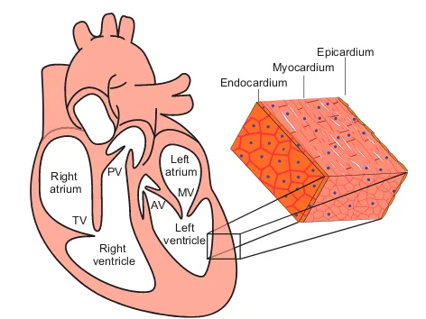
A mature mammalian heart has four valves and four chambers, with the wall of each chamber consisting of three tissue layers: endocardium, myocardium and epicardium (Fig. 1). The cardiac chambers and valves are organized such that they separate systemic from pulmonary circulation and ensure directional blood flow. The formation of these structures requires multiple cell types and complex morphogenetic processes, which often go awry in the developing human fetus. Heart malformations account for as many as 30% of embryos or fetuses lost before birth (Hoffman, 1995), and the incidence of heart defects in live births varies from 0.4% to 5% in different studies, depending on the severity of heart defects included in the statistics (Hoffman and Kaplan, 2002). On top of these statistics, another 2% of newborns have bicuspid aortic valves (BAVs; see Glossary, Box 1) or other defects (Hoffman and Kaplan, 2002), which may cause significant morbidity and mortality later in life (Brickner et al., 2000a). Congenital heart malformations, therefore, constitute an important medical issue challenging our society.
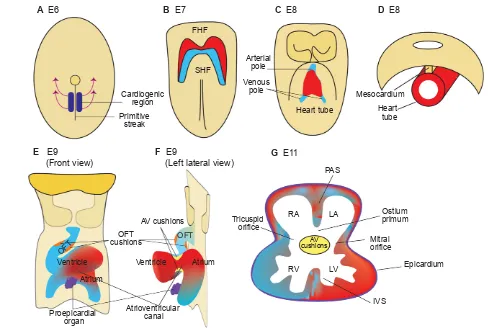
The heart of developing embryos originates from mesodermal cells located in the anterior part of the primitive streak (Lawson et al., 1991; Tam et al., 1997) (Fig. 2). During gastrulation, these cardiac mesodermal cells migrate from the streak to the splanchnic mesoderm underlying the head folds to form cardiac crescent (the first heart field, FHF; see Glossary, Box 1) (Abu-Issa and Kirby, 2007; Vincent and Buckingham, 2010) (Fig. 2A,B). As the embryo grows, the crescent of the FHF fuses in the ventral midline, forming a trough-like structure, which then closes dorsally to form a
primitive heart tube (Fig. 2C). The heart tube is suspended from the body wall by dorsal mesocardium (Fig. 2D), and the tube elongates on both the arterial and venous poles via the addition of progenitor cells originating from the secondary heart field (SHF; see Glossary, Box 1), which lies medially and posteriorly to the crescent (Kelly et al., 2001; Mjaatvedt et al., 2001; Waldo et al., 2001; Cai et al., 2003). Concurrent with heart tube elongation, the dorsal mesocardium dissolves except at the poles, liberating the majority of the heart tube and allowing it to undergo rightward looping. The looped heart tube, composed of an inner endocardial lining and an outer myocardial layer, is segmented into the atrium, the atrioventricular canal (AVC; see Glossary, Box 1), the ventricle and the outflow tract (OFT; see Glossary, Box 1) (Fig. 2E). In the lumen of the AVC and proximal OFT, local tissue swellings, termed endocardial cushions, are formed by the accumulation of abundant extracellular matrix (cardiac jelly) in between the endocardium and myocardium (Fig. 2F). These endocardial cushions are subsequently populated by mesenchymal cells that descend from the endocardium. In addition, within the lumen of the distal OFT, local tissue swellings (termed truncal cushions) arise and are later populated by mesenchymal cells originating from the neural crest. While the cushions are developing, a sheath of cells, which originate from the proepicardial organ, grows over the myocardium of the heart tube to form the outermost epicardial layer of the heart (Fig. 2E-G). Later in development, the atrial and ventricular chambers divide into two atria (left and right) and two ventricles (left and right), forming a prototypic four-chamber heart (Fig. 2G). Along with chamber septation, the AVC separates into left (mitral) and right (tricuspid) orifices, forming ventricular inlets that connect the respective atrium to the ventricle. The outflow tract divides into the left and right ventricular outlets that connect the left and right ventricle, respectively, to the aorta and pulmonary trunk. These septation events segregate the systemic from pulmonary circulation. In addition, the AVC endocardial cushions develop into atrioventricular (mitral and tricuspid) valves, whereas the OFT endocardial cushions give rise to semilunar (aortic and pulmonic) valves (Fig. 1). The formation of heart valves ensures that blood flows in one direction from the atria to ventricles and then to the arteries.
Multiple cells of distinct developmental origins contribute to the formation of a heart. Lineage tracing and clonal analyses in mice show the existence of two distinct myocardial lineages arising separately from the FHF and SHF. The FHF lineage contributes primarily to the myocardium of the left ventricle (Buckingham et al., 2005; Srivastava, 2006), whereas the SHF lineage contributes to the myocardium of the atria (Cai et al., 2003; Galli et al., 2008), right ventricle and outflow tract (Kelly et al., 2001; Cai et al., 2003; Zaffran et al., 2004; Verzi et al., 2005). By contrast, the epicardium arises from the proepicardial organ, which is located near the venous pole of the heart tube and originates from the coelomic mesenchyme of septum transversum (Männer et al., 2001) (Fig. 2E). Cells in the epicardium give rise to mesenchymal cells that Development 139, 3277-3299 (2012) doi:10.1242/dev.063495
© 2012. Published by The Company of Biologists Ltd
Partitioning the heart: mechanisms of cardiac septation and
valve development
Chien-Jung Lin1,*, Chieh-Yu Lin1,*, Chen-Hao Chen1, Bin Zhou2,‡and Ching-Pin Chang1,‡
1Division of Cardiovascular Medicine, Department of Medicine, Stanford Cardiovascular Institute, Stanford University, Stanford, California 94305, USA. 2Departments of Genetics, Medicine, and Pediatrics, Albert Einstein College of Medicine, Bronx, New York 10461, USA.
*These authors contributed equally to this work
‡Authors for correspondence (bin.zhou@einstein.yu.edu; chingpin@stanford.edu)
D
E
V
E
LO
P
M
E
N
migrate into the myocardium and differentiate into fibroblasts and coronary smooth muscle cells (Merki et al., 2005). The origin of endocardium, however, has been controversial: the endocardium may arise from the heart fields or vascular endothelial progenitors (Harris and Black, 2010; Vincent and Buckingham, 2010; Milgrom-Hoffman et al., 2011). The mesenchyme of cushions arises from two distinct origins. Endocardial cushions in the AVC and proximal OFT lumen derive their mesenchyme from the local endocardium that overlies the cushions (Eisenberg and Markwald, 1995), whereas the distal OFT cushions are populated by mesenchymal cells that migrate from the distant neural crest (Jiang et al., 2000).
Here, we review the interactions between these different progenitor cells and their derivatives that are essential for cardiac septation and valve development. We also highlight the key signaling pathways that are known to regulate cardiac septation and valve development.
Cardiac chamber septation and valve formation Septation of the primitive cardiac chambers, the AVC and the OFT is necessary for forming a four-chamber heart. The morphogenic events that direct cardiac septation and valve development are described below.
Atrioventricular septation
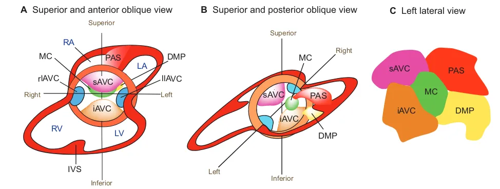
Four mesenchymal tissues are required for atrial and AVC septation: the superior and inferior atrioventricular (AV) endocardial cushions (atrioventricular cushions; see Glossary, Box 1), the mesenchymal cap (MC; see Glossary, Box 1), and the dorsal mesenchymal protrusion (DMP; see Glossary, Box 1) (Webb et al., 1998; Snarr et al., 2008) (Fig. 3). The AV cushions derive their mesenchyme from the endocardium through a cellular process called epithelial-to-mesenchymal transformation (EMT). During EMT, a subset of endocardial cells delaminates from the surface epithelium and transdifferentiates into mesenchymal cells, which migrate into the cardiac jelly and proliferate to cellularize the cushions (Eisenberg and Markwald, 1995). The MC, which envelops the growing edge of a muscular atrial septum, also arises
through EMT from the endocardium overlying the cap (Snarr et al., 2008). Conversely, the mesenchyme of DMP comes from the SHF, which gives rise to cells that migrate through the dorsal mesocardium and bulge into the atrial chamber as a mesenchymal protrusion (Snarr et al., 2007a; Snarr et al., 2008).
The mesenchyme of superior and inferior AV cushions fuses at the AV canal, dividing the canal into mitral and tricuspid orifices that form ventricular inlets (Fig. 2G, Fig. 3A,B). Meanwhile, a muscular septum (the primary atrial septum) grows from the atrial roof towards the AVC, with the MC at its leading edge. This muscular outgrowth partially septates the atrial chamber and leaves an opening (the ostium primum) between the MC and the AV canal (Fig. 2G). The MC then merges anteriorly with the AV cushions and posteriorly with the DMP to seal the ostium primum (Wessels et al., 1996; Schroeder et al., 2003; Wessels and Sedmera, 2003; Mommersteeg et al., 2006) (Fig. 3B,C). These mesenchymal tissues are later muscularized to form sturdy septum. While the ostium primum is closing, the upper margin of the primary atrial septum dissolves, creating a second opening (the ostium secundum) between the right and left atria. The ostium secundum is later sealed by a muscular septum (the secondary atrial septum), which is formed by part of the atrial roof that folds inward. The primary and secondary atrial septum then fuses to complete the septation of atrial chamber (Anderson et al., 2003a).
Within the ventricular chamber, an interventricular muscular septum emerges and grows superiorly to fuse with AV cushions, dividing the ventricular chamber into left and right ventricles (Fig. 2G, Fig. 3A) (Anderson et al., 2003a; Moorman et al., 2003). This muscular septum also connects with OFT cushions to separate the ventricular outlets. Abnormal chamber septation results in congenital heart diseases, including atrial septal defects (ASDs), ventricular septal defects (VSDs), and atrioventricular septal defects (AVSDs) (see Glossary, Box 1). These defects cause abnormal cardiac shunting and may lead to congestive heart failure (Brickner et al., 2000a).
Outflow tract septation
Septation of the cardiac OFT requires neural crest cells (NCCs) of neuroectodermal origin, as well as truncal and conal cushions situated in the distal and proximal OFT (Fig. 4A-C). Early in development, a group of NCCs delaminates from the neuroectodermal junction of rhombomeres 6-8 at the hindbrain (Kirby et al., 1983), traverses the pharyngeal arches and secondary heart field, and migrates into the distal OFT (Fig. 4A). The NCCs that reach the heart become the mesenchyme of truncal cushions (Fig. 4C). Subsequently, the mesenchymal truncal cushions (the right-superior and left-inferior cushions) fuse to form aortopulmonary septum, dividing the distal OFT into the aorta and pulmonary trunk (Jiang et al., 2000; Li et al., 2000) (Fig. 4D). By contrast, at the proximal OFT, the endocardium, through EMT, gives rise to the mesenchyme of conal cushions. The mesenchymal conal cushions (the right-posterior and left-anterior cushions) then merge to form a conal septum, separating the proximal OFT into the right and left ventricular outlets (Anderson et al., 2003b) (Fig. 4C,D). The ventricular outlets are aligned to the arteries by the connection of conal and truncal cushions and to the ventricles by the fusion of conal cushions with the interventricular septum. Misaligned or incomplete OFT septation leads to a variety of congenital heart defects, including overriding aorta (OA; see Glossary, Box 1), double-outlet right ventricle (DORV; see Glossary, Box 1), tetralogy of Fallot (TOF; see Glossary, Box 1), transposition of Endocardium
Myocardium Epicardium
TV PV Right atrium
Right ventricle
Left atrium
AV MV
[image:2.612.52.288.56.233.2]Left ventricle
Fig. 1. The structure of a mammalian heart.A mature mammalian heart contains four chambers (right atrium, left atrium, right ventricle, left ventricle) and four valves (pulmonary valve, PV; tricuspid valve, TV; atrial valve, AV; mitral valve, MV). The wall of each chamber consists of three tissue layers: endocardium, myocardium and epicardium.
D
E
V
E
LO
P
M
E
N
great arteries (TGA; see Glossary, Box 1) and persistent truncus arteriosus (PTA; see Glossary, Box 1) (Brickner et al., 2000b). These defects can cause mixing of arterial with venous blood, leading to cyanosis and/or heart failure.
Atrioventricular and semilunar valve development
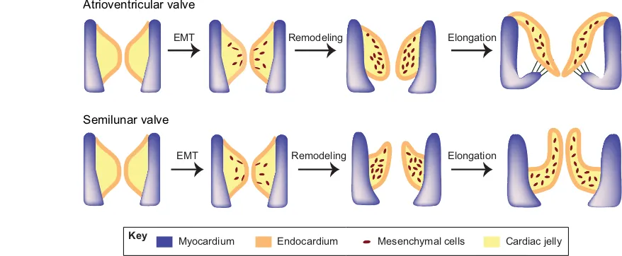
Heart valves develop at endocardial cushions of the AVC and OFT. Valve morphogenesis begins with the transformation of endocardial cells into mesenchymal cells through EMT (Fig. 5). Endocardial cushions with mesenchymal cells then elongate and remodel themselves to form primitive valves that gradually mature into thin valve leaflets. The elongation of valve leaflets is accomplished by a combination of cell proliferation at the growing edge and apoptosis at the base of the cushion (Hurle et al., 1980).
The mitral and tricuspid (atrioventricular) valves originate from AV endocardial cushions. The fusion and remodeling of superior and inferior AV cushions, while dividing the AVC, gives rise to the anterior mitral leaflet and the septal tricuspid leaflet (de Lange et al., 2004) (Fig. 6A,B). The left lateral AV cushion becomes the posterior mitral leaflet, whereas the right lateral cushion produces the anterior and posterior tricuspid leaflets. Failure of the superior and inferior cushions to fuse causes a cleft in the anterior mitral leaflet, resulting in leaky ‘cleft mitral valve’, a disease encountered in patients with Down syndrome (Fraisse et al., 2003). Tricuspid valve malformations also cause human disease, such as the Ebstein’s anomaly: the septal and often the posterior tricuspid leaflets are displaced into the right ventricle with the anterior leaflet becoming excessively large, thus causing tricuspid valve regurgitation or stenosis (see Glossary, Box 1) (Brickner et al., 2000b).
The aortic and pulmonic (semilunar) valves arise from the conotruncal and intercalated cushions at the OFT. The conotruncal cushions give rise to the right and left leaflets of semilunar valves (Fig. 4C, Fig. 6A,B) (Restivo et al., 2006; Okamoto et al., 2010). Conal cushions, capable of supporting EMT of the endocardium, probably have major contributions to the aforementioned valve leaflets, of which the mesenchymal tissues largely come from the endocardium (de Lange et al., 2004). Adjacent to the conotruncal cushions are two other distinct cushions – the right-posterior and the left-anterior intercalated cushions – that develop respectively into the posterior aortic and the anterior pulmonic leaflets (Fig. 4C, Fig. 6A,B) (Anderson et al., 2003b; Restivo et al., 2006). These semilunar valve leaflets also derive their mesenchyme primarily from the endocardium (de Lange et al., 2004). Semilunar valve malformations are common and occur in 2-3% of the population, causing valve regurgitation and/or stenosis (Brickner et al., 2000a).
Similarities between AVC and OFT septation
Morphogenesis of the AV and OFT cushions is similar. The analogous roles of these cushions in cardiac septation and valve formation are better appreciated by rotating the OFT 90° counter-clockwise to superimpose the corresponding OFT and AV cushions (Fig. 6A). The superior and inferior AV cushions fuse to divide the AVC, forming valve leaflets that flank the AVC septation site; comparably, the right-superior and left-inferior conal cushions merge to separate the OFT, bringing about valve leaflets that border the OFT septation (Fig. 6A,B). Conversely, the lateral AV cushions are similar in function to the intercalated OFT cushions in that they both lack direct contributions to AV/OFT septation and that they form valve leaflets that oppose the AV/OFT septation site (Fig. 6A,B). The AVC and OFT, therefore, have evolved similar strategies for lumen septation and valve formation, sharing many essential developmental genes and pathways (Fig. 7).
Box 1. Glossary
Atrial septal defect (ASD).A congenital heart defect resulting from incomplete atrial septation.
Atrioventricular canal (AVC).The junction between developing atria and ventricles.
Atrioventricular cushions.The four endocardial cushions located at the AV canal: superior, inferior, left-lateral and right-lateral cushions.
Atrioventricular septal defect (AVSD).A congenital heart defect resulting from incomplete septation of the atrioventricular canal. Bicuspid aortic valve (BAV).A congenital heart defect in which the aortic valve has only two cusps. The term BAV is also used broadly to describe any malformation of the aortic valve cusps. Dorsal mesenchymal protrusion (DMP).A mesenchymal tissue that protrudes into the atrial chamber through the dorsal mesocardium.
Double-outlet right ventricle (DORV).A congenital heart defect in which both aorta and pulmonary trunk arise from the right ventricle.
First heart field (FHF).A population of mesodermal cells that form the cardiac crescent located in splanchnic mesoderm underlying the head folds. Progenitors of the FHF give rise to myocardium of the left ventricle, part of the right ventricle and part of the atria. Interruption of the aortic arch (IAA).A congenital heart defect in which a segment of the aortic arch is occluded or absent. Mesenchymal cap (MC). A mesenchymal tissue that caps the growing (inferior) edge of the primary atrial septum.
Outflow tract (OFT).The outflow region of the embryonic heart that develops into the left and right ventricular outlets, as well as the aorta and pulmonary trunk.
Overriding aorta (OA).A congenital heart defect in which the aortic root connects with both the left and right ventricle and receives blood from both ventricles.
Patent ductus arteriosus (PDA).A congenital heart defect in which the ductus arteriosus fails to close after birth.
Persistent truncus arteriosus (PTA).A congenital heart defect in which the aorta fails to separate from the pulmonary trunk, resulting in a single arterial trunk that emerges from the ventricles. Pulmonary stenosis (PS).A congenital heart defect in which the pulmonary valve is malformed, causing narrowing of the pulmonary trunk and hindrance of blood flow.
Secondary heart field (SHF).A population of mesodermal cells located medially and posteriorly to the first heart field, then behind the heart tube, and extending into pharyngeal mesoderm as the embryo develops. Progenitor cells of the SHF give rise to myocardium of the right ventricle, cardiac outflow tract, and part of the left ventricle and atria.
Tetralogy of Fallot (TOF).A congenital heart defect characterized by right ventricular outflow tract obstruction, right ventricular hypertrophy, ventricular septal defect and overriding aorta. Total or partial anomalous pulmonary venous return (TAPVR or PAPVR). A congenital heart defect in which pulmonary veins are misconnected and drained into the systemic venous circulation. Transposition of the great arteries (TGA).A congenital heart defect in which the right ventricle connects to the aorta, and the left ventricle connects to the pulmonary trunk.
Tricuspid atresia (TA). A congenital heart defect in which the tricuspid valve is missing, hence blocking the blood flow from right atrium to right ventricle.
Ventricular septal defect (VSD). A congenital heart defect resulting from incomplete ventricular septation
D
E
V
E
LO
P
M
E
N
Cell lineages that contribute to septum formation and valve development
The endocardium, secondary heart field and neural crest contribute progenitor cells that give rise to septal tissues or valve leaflets. Besides direct lineage contributions, these progenitor cells of different origins interact with each other and with other cells in the heart to orchestrate cardiac septation and valve development.
Endocardium and EMT
EMT of the endocardium occurs only in the endocardial cushions and is regulated by many signaling factors secreted by the myocardium underlying the cushion. These EMT-regulating factors include bone morphogenetic proteins 2 and 4 (Bmp2, Bmp4), transforming growth factor 2 and 3 (TGF2, TGF3) and vascular endothelial growth factor (Vegf) (Fig. 7; Tables 1, 2). To react to myocardial signals and begin EMT, the endocardium at the cushion expresses receptors and effectors downstream of the myocardial signaling pathways, including Alk2 (Acvr1), Alk3
(Bmpr1a), Alk5 (Tgfbr1), Vegf-R, Notch1 and -catenin. Different from the chamber myocardium, the myocardium at the cushion is specified and programmed by genes, such as Tbx2, Bmp2, Nfatc2,
Nfatc3and Nfatc4, to suppress chamber-specific gene expression, produce EMT-regulating molecules, and deposit extracellular matrix to support EMT (Abedin et al., 2004; Chang, C. P. et al., 2004; Christoffels et al., 2004; Rivera-Feliciano and Tabin, 2006; Shirai et al., 2009). The endocardium at the cushion is also different from that in the cardiac chamber: the cushion endocardium expresses genes essential for septal and valvular development, such as those encoding Nfatc1 and Vegf receptors, in a temporal pattern different from that of the chamber endocardium (Chang, C. P. et al., 2004; Stankunas et al., 2010). Such distinct gene programming and disparate arrays of signaling factors and receptors at the endocardial cushion determines the regional specificity of EMT. Much less is known about the post-EMT events at the endocardial cushion: for example, how mesenchymal cells control the remodeling of valvular and septal tissues. Future efforts
A E6 B E7 C E8 D E8
E E9
(Front view)
SHF FHF
Heart tube
Mesocardium
Ventricle
Atrium OFT
Heart tube
F E9
(Left lateral view)
Ventricle Atrium AV cushions
Atrioventricular canal Proepicardial
organ
OFT OFT
cushions
RA LA
RV LV
AV cushions
G E11
Arterial pole
Venous pole Cardiogenic
region Primitive
streak
PAS
Tricuspid orifice
Mitral orifice
Ostium primum
IVS
[image:4.612.54.546.57.386.2]Epicardium
Fig. 2. The formation of a mouse heart.(A)Ventral view of a mouse embryo at E6. The heart originates from mesodermal cells in the primitive streak. During gastrulation, mesodermal cardiac progenitor cells migrate to the splanchnic mesoderm to form the cardiac crescent. (B)Ventral view at E7. One subset of cardiac progenitors forms a horseshoe-shaped cardiac crescent (the first heart field, FHF; red). Another subset of cardiac progenitor cells forms the secondary heart field (SHF; blue), which is located posteriorly and medially to the FHF. (C)Ventral view at E8. Cells in the FHF merge in the midline to form the heart tube, which then elongates on both arterial and venous poles via the addition of progenitor cells from the SHF. (D)Transverse section at E8. The developing heart tube is suspended from the body wall by the dorsal mesocardium, which later dissolves except at the poles of heart tube, allowing the tube to loop rightward. (E,F)Ventral (E) and left lateral (F) views at E9. The looped heart tube contains four anatomical segments: atrium, atrioventricular canal, ventricle and outflow tract (OFT). Within the AVC and OFT, AV cushions (yellow) and OFT cushions (orange) develop. The proepicardial organ (purple) houses epicardial progenitors that later migrate to the heart and give rise to the epicardium. (G)Transverse section at E11. At this stage, the heart is partially partitioned by the primitive atrial septum (PAS), interventricular septum (IVS) and atrioventricular cushions (AV cushions) into a prototypic four-chamber heart. The AVC is divided into tricuspid and mitral orifices, forming ventricular inlets that connect the respective atrium to the ventricle. The opening between the PAS and AVC is the ostium primum. RA, right atrium; LA, left atrium; RV, right ventricle; LV, left ventricle.
D
E
V
E
LO
P
M
E
N
to generate inducible and/or tissue-specific knockout mouse lines that specifically target cushion mesenchymal cells independent of their endocardial precursors will facilitate the investigation of post-EMT remodeling events at the cushion.
The secondary heart field
The SHF progenitor cells contribute to cardiac septation and valve development. SHF progenitors give rise to the DMP mesenchyme, which merges with AV cushions and becomes part of the atrial septum (Snarr et al., 2007b). SHF progenitors also give rise to the OFT myocardium (Verzi et al., 2005), which secretes signaling molecules that stimulate the conal endocardium to undergo EMT, an essential step for later development of the ventricular outlet septum and semilunar valve leaflets (Anderson et al., 2003b; de Lange et al., 2004). Besides promoting EMT, the SHF-derived OFT myocardium secretes chemotactic molecules, such as Sema3c, to attract NCCs into the OFT to form the aortopulmonary septum (Brown et al., 2001; Feiner et al., 2001; Toyofuku et al., 2008). Furthermore, at the base of the aorta and pulmonary trunk, the SHF gives rise to vascular smooth muscle cells (Cai et al., 2003; Verzi et al., 2005) to support the separation of these arteries from ventricular outlets.
Disruption of many signaling pathways in the SHF, using Mef2c- or Islet1-Cre lines (Cai et al., 2003; Verzi et al., 2005), results in abnormalities in OFT septation or semilunar valves. These pathways include Wnt/-catenin, BMP (Bmp4, Bmpr1a), Fgf8, Notch, Hedgehog/Smoothened, and calcineurin/Nfatc1 signaling (Fig. 7, Tables 1, 2). One major issue yet to be resolved is the actual action site(s) of these ‘SHF pathways’, i.e. whether they operate within the SHF or within SHF-derived tissues. Because of the overlap of many of the ‘SHF’ pathways with those functioning in cardiac tissues that are essential for EMT and cushion development, the outcomes of genetic manipulation in the SHF should be carefully interpreted and, in most cases, require further investigations. Nevertheless, calcineurin and Notch are known to operate in SHF progenitors to control OFT development. Deletion of calcineurin b1 (Cnb1; Ppp3r1 – Mouse Genome Informatics) in SHF progenitors causes cell apoptosis and
regression of conal cushions, resulting in absent semilunar valves (Lin et al., 2012). Conversely, Cnb1 deletion in the OFT myocardium does not cause semilunar valve defects (Lin et al., 2012), thus localizing the site of calcineurin action to SHF progenitors for semilunar valve development. Likewise, inhibition of the Notch pathway in SHF progenitors causes OFT septation defects (such as DORV and PTA), but a later inhibition of Notch in the myocardium does not produce such defects (High et al., 2007; High et al., 2009).
Besides OFT development, AV septation requires signaling in SHF progenitors. Embryos lacking Hedgehog signaling in SHF progenitors, but not those lacking Hedgehog signaling in the myocardium or endocardium, show AV septal defects and a failure of the SHF-derived DMP mesenchyme to protrude into the atrial chamber (Goddeeris et al., 2008). Signaling within the SHF progenitors before they differentiate into cardiac cells, therefore, is essential for both AV and OFT septation. Further studies will be needed to elucidate how SHF signaling specifies developmental functions of SHF-derived tissues and how SHF progenitors modulate the activities of NCCs as the latter cells traverse the SHF during their migration to the heart.
Neural crest cell lineage
Cardiac NCCs migrate from their original location in the hindbrain (rhombomeres 6-8) to the pharyngeal arches and then to the heart to form the aortopulmonary septum (Kirby et al., 1983). Migrating NCCs actively exchange signals with surrounding tissues, such as the pharyngeal arch and the OFT myocardium. The pharyngeal arch endothelium, through its endothelin-converting enzyme-1 (Ece1), produces the signaling molecule endothelin-1, which activates the endothelin receptor A on NCCs to modulate NCC activities for pharyngeal arch artery (PAA) and OFT development. Embryos lacking Ece1 or endothelin receptor A develop PAA defects, as well as septation defects such as VSD, OA, DORV, PTA or TGA (Clouthier et al., 1998; Yanagisawa et al., 1998a). The OFT myocardium secretes Sema3c (ligand) to attract NCCs, which express Plxna2 (receptor), and promotes them to migrate into the
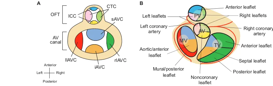
iAVC
DMP Superior
Inferior Left
Right
sAVC PAS
MC
rlAVC llAVC
RV
LV RA
PAS Superior
Inferior
Right Left
A Superior and anterior oblique view B Superior and posterior oblique view
sAVC
DMP PAS
iAVC MC
C Left lateral view
LA
iAVC sAVC
DMP
[image:5.612.52.539.57.241.2]IVS MC
Fig. 3. Endocardial cushion development.(A,B)Superior and anterior oblique view (A) and superior and posterior oblique view (B) of the heart. The superior and inferior atrioventricular cushions (sAVC and iAVC) are the two major cushions that develop in the central portion of the AVC. Two minor cushions, left and right lateral AV cushions (llAVC and rlAVC), form laterally at the AVC. The mesenchymal cap (MC) is a tissue that caps the leading edge of primary atrial septum (PAS) that grows from the atrial roof towards the AV canal. The dorsal mesenchymal protrusion (DMP) protrudes from the dorsal mesocardium into the atrial chamber. RA, right atrium; LA, left atrium; RV, right ventricle; LV, left ventricle; IVS, interventricular septum. (C)The composition of the atrial septum (left lateral view).
D
E
V
E
LO
P
M
E
N
OFT, where NCCs become the mesenchyme of truncal cushions. The truncal mesenchyme then fuses and differentiates to form a smooth muscle septum (aortopulmonary septum) that divides the aorta and pulmonary trunk. Knockout of Plxna2or Sema3cin mice impairs the migration of NCCs, leading to PAA defects and PTA (Brown et al., 2001; Feiner et al., 2001; Toyofuku et al., 2008).
Many other signaling pathways are necessary for NCCs to regulate OFT septation, including the Wnt/-catenin-Pitx2, Notch, TGF (Alk5), BMP (Alk2) and Hedgehog pathways (Fig. 7, Tables 1, 2). In contrast to the crucial roles of NCCs in OFT septation, most data suggest that NCCs do not have significant contribution to AV cushion development (Combs and Yutzey, 2009).
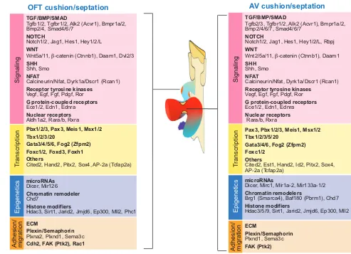
Molecular pathways that regulate septation and valve development
Many mouse genetic models have been established to demonstrate the function of genes involved in cardiac septation and valve
development (Fig. 7). These include genes that encode signaling molecules (Tables 1, 2), transcription factors (Table 3), chromatin or epigenetic regulators (Table 4), and cell adhesion/migration molecules (Table 4). Discussed below are some of the most well characterized pathways that are known to regulate cardiac septation and valve development.
TGF, BMP and SMAD pathways
TGFs were among the first signaling molecules to be implicated in the initiation of EMT (Brown et al., 1996; Ramsdell and Markwald, 1997; Boyer et al., 1999; Brown et al., 1999; Boyer and Runyan, 2001), and multiple TGFisoforms are expressed in the endocardial cushions of mouse embryos (Akhurst et al., 1990; Millan et al., 1991). These mammalian TGFisoforms are highly redundant; disruption of multiple TGFligands is often necessary to uncover their roles in heart development (Shull et al., 1992; Kaartinen et al., 1995; Sanford et al., 1997; Dünker and
Conotruncal curvature
T
runcal
Conal
A
B
CTC ICC
Truncal septum
Conal septum
C
RVOT LVOT AV
PV Aorta
Pulmonary trunk
Superior Right Left
Posterior
Anterior Inferior
Cardiac neural crest
OFT
D
ICC
[image:6.612.53.563.59.201.2]CTC
Fig. 4. Septation of the cardiac outflow tract.(A)Left lateral view of an E10 mouse embryo. The neural crest at rhombomere 6-8 gives rise to cells (blue) that migrate to and colonize the distal cardiac outflow tract (OFT). (B)The cardiac OFT contains conal (proximal) and truncal (distal) cushions. The boundary between the conal and truncal cushions is marked by an outer curvature of the OFT (the conotruncal curvature). (C)The conotruncal cushions (CTCs) and intercalated cushions (ICCs) develop within the OFT. These cushions occupy four quadrants of the OFT (shown in cross-section). The conotruncal cushions fuse to septate the OFT, as shown in D. (D)Fusion of the conotruncal cushions forms a spiral septum, the truncal part of which divides the OFT into aorta and pulmonary trunk, whereas the conal part septates the OFT into left and right ventricular outlets (LVOT, RVOT). The aortic valves (AV) and pulmonic valves (PV) develop at the conotruncal junction.
Semilunar valve
Myocardium Endocardium Cardiac jelly
EMT Remodeling
Mesenchymal cells Atrioventricular valve
EMT Remodeling Elongation
Elongation
Key
Fig. 5. Epithelial-to-mesenchymal transformation and valve elongation.Endocardial cells in the AV cushions and conal cushions undergo epithelial-to-mesenchymal transformation (EMT) and generate mesenchymal cells that populate the cushions. The mesenchymal cushions then remodel and elongate themselves to from primitive valves that mature into thin valve leaflets (shown here for the atrioventricular valves and the
semilunar valves).
D
E
V
E
LO
P
M
E
N
[image:6.612.58.497.512.693.2]Krieglstein, 2002). Regardless of the redundancy, Tgf2 is known to function downstream of the Notch1, Bmp2 and Tbx2 pathways to activate Wnt/-catenin signaling to promote EMT (Liebner et al., 2004; Timmerman et al., 2004; Shirai et al., 2009; Luna-Zurita et al., 2010).
BMPs have diverse roles in valve development and cardiac septation. Myocardial Bmp2 activates the expression of Has2 (hyaluronic acid synthetase 2) to produce the cushion extracellular matrix that is required for EMT (Rivera-Feliciano and Tabin, 2006). Also, myocardial Bmp2 signals the endocardial Bmp type 1A receptor (Bmpr1a) to induce the expression of Twist1, Msx1 and Msx2, which are essential for EMT (Ma et al., 2005). Bmp4 in the myocardium is necessary for cardiac septation. Absence of myocardial Bmp4 leads to ASD, VSD and PTA (Jiao et al., 2003; Liu et al., 2004; McCulley et al., 2008). Bmp6 and Bmp7, expressed in the myocardium and cushion/valve mesenchyme, are functionally redundant; mice with double knockout of these genes display hypocellular OFT cushions (Kim et al., 2001). Bmpr2, a receptor for Bmp2, Bmp4 and Bmp7, has different spatial roles. Mice with hypomorphic alleles of Bmpr2 exhibit interruption of the aortic arch (IAA; see Glossary, Box 1), PTA, and absent semilunar valves (Délot et al., 2003). Bmpr2 disruption in endothelial cells causes ASD, VSD, and hyperplastic semilunar and AV valve leaflets, whereas Bmpr2 deletion in myocardial cells or in NCCs leads to DORV or OA (Beppu et al., 2009).
SMAD proteins that transduce or modulate TGF/BMP signals are also essential for cardiac septation and valve formation. Loss of Smad4, the most common SMAD, in NCCs results in pharyngeal arch artery defects, OFT cushion hypoplasia and PTA (Jia et al., 2007). Smad4-null NCCs have increased apoptosis and reduced presence in the OFT, accompanied by a reduction in the Bmp4, Sema3c and Plxna2 signals that are necessary for OFT septation. By contrast, disruption of Smad6, which inhibits BMP signaling, causes hyperplasia of the AV and OFT cushions, leading to hyperplastic valves (Galvin et al., 2000).
Notch signaling
Notch signaling is necessary for EMT (Niessen and Karsan, 2008; MacGrogan et al., 2010), and mutation of Notch1 or its nuclear effector Rbpjk (Rbpj – Mouse Genome Informatics) causes EMT failure (Timmerman et al., 2004). Endocardial Notch1 induces the
expression of Tgf2 to activate the expression of Snail1 (Snai1) and Snail2 (Snai2), which repress VE-cadherin expression and hence disrupt cell-cell contact, allowing EMT to occur (Romano and Runyan, 2000; Timmerman et al., 2004; Luna-Zurita et al., 2010). EMT of the Notch1/Tgf2-primed endocardial cells requires myocardial Bmp2, the expression of which, however, is repressed by myocardial Notch1 (Luna-Zurita et al., 2010). These seemingly opposing effects of endocardial and myocardial Notch signaling suggest a complex tissue-specific role for Notch in orchestrating EMT of AV cushions.
In the SHF, inhibition of Notch decreases Fgf8 expression, reduces EMT of OFT cushions, and impairs NCC migration with consequent thickened, unequally sized semilunar valve leaflets (High et al., 2009; Jain et al., 2011). Such an EMT defect is rescued by exogenous Fgf8, suggesting that Notch functions through Fgf8 to activate EMT of OFT cushions (High et al., 2009). NOTCH1
mutations are observed in some families with a multi-generation history of BAV and/or calcific aortic stenosis (Garg et al., 2005; Garg, 2006; McKellar et al., 2007; McBride et al., 2008; Rusanescu et al., 2008). Because Notch1 is capable of suppressing Runx2, which promotes calcification (Garg et al., 2005), NOTCH1
mutations in humans might cause RUNX2 upregulation with consequent valve calcification.
Disruption of the SHF Notch in mice also causes septation defects, including ASD, VSD, DORV and PTA (High et al., 2009). In humans, JAG1 or NOTCH2 mutations are associated with Alagille syndrome (McDaniell et al., 2006; Warthen et al., 2006), an autosomal dominant disorder with abnormalities in multiple organs, including pulmonary stenosis and TOF. The Alagille heart phenotypes of pulmonary stenosis and TOF also occur in mice lacking Hey2, a Notch downstream target gene (Donovan et al., 2002).
The Wnt pathway
Wnt/-catenin signaling plays a major role in EMT and cardiac septation. For AV cushion development, -catenin functions downstream of Tgf2 in the endocardium to promote EMT of AV cushions (Liebner et al., 2004). Wnt2 also signals through -catenin to recruit SHF-derived mesenchymal cells into DMP. Mice lacking Wnt2 or -catenin in the SHF have reduced DMP mesenchyme, resulting in ASD and VSD (Lin et al., 2007; Tian et
Right
llAVC rlAVC
CTC
ICC
sAVC
iAVC
A B
AV canal OFT
Anterior
Left
Posterior
Mural/posterior leaflet
Septal leaflet
Posterior leaflet Anterior leaflet Left leaflets
Noncoronary leaflet
Anterior leaflet
Right leaflets
Right coronary artery Left coronary
artery
Aortic/anterior leaflet
TV MV
[image:7.612.54.494.63.202.2]AV PV
Fig. 6. Endocardial cushions and heart valve leaflets.(A)Schematic of endocardial cushions in the atrioventricular (AV) canal and the outflow tract (OFT). The figure is a superior view of the heart with atria removed. The cushions are color coded to correspond to their derived valve leaflets illustrated in B. CTC. conotruncal cushions; ICC. intercalated cushions; sAVC. superior AV cushion; iAVC. inferior AV cushion; rlAVC. right lateral AV cushion; llAVC. left lateral AV cushion. (B)Schematic (superior view) of atrioventricular and semilunar valve leaflets that develop from the
corresponding cushions color coded in A. PV, pulmonary valve; AV, aortic valve; TV, tricuspid valve; MV, mitral valve.
D
E
V
E
LO
P
M
E
N
al., 2010). For OFT development, -catenin has crucial roles in the SHF and in NCCs. In the SHF, -catenin is essential to prevent the development of abnormal pharyngeal arteries and PTA (Lin et al., 2007). In NCCs, Wnt/-catenin functions through Pitx2 to control OFT septation. Migrating NCCs that lack -catenin show reduced expression of Pitx2, disruption of which results in failure of NCC migration into the OFT, causing PTA, DORV or TGA (Kioussi et al., 2002).
The noncanonical Wnts, although not signaling through -catenin, are also essential for OFT septation; mice lacking Wnt5a or Wnt11 display TGA, DORV or PTA (Schleiffarth et al., 2007; Zhou et al., 2007).
Epidermal growth factor signaling
Epidermal growth factor (EGF) signaling is essential for AV and OFT development. EGF signaling between endocardial HB-EGF (Hbegf; ligand) and myocardial ErbB1 (Egfr; an EGF receptor), for example, suppresses cushion development. Mutations affecting HB-EGF, an endocardial ligand that directly binds ErbB1 and ErbB4, result in hyperproliferation of cushion mesenchymal cells and hyperplasia of AV and semilunar valves (Iwamoto et al., 2003; Jackson et al., 2003). Similar cushion and semilunar valve hyperplasia is observed in mice with mutations in ErbB1, which is present primarily in the myocardium (Chen et al., 2000; Jackson et
al., 2003; Sibilia et al., 2003). In contrast to myocardial ErbB1 signaling, ErbB2- or ErbB4-based signaling in the myocardium does not seem necessary for early cushion development. Mutations in ErbB2 or ErbB4, both present in the myocardium, have no apparent cushion defects at embryonic day (E) 10.5 before the mutant embryos die at E10-11 of severe hypotrabeculation (Gassmann et al., 1995; Lee et al., 1995).
EGF signaling within the mesenchyme promotes cushion development. Mutations in ErbB3, an EGF receptor present in the cushion mesenchyme (Meyer and Birchmeier, 1995), causes hypoplastic endocardial cushions (Erickson et al., 1997). Such mesenchymal ErbB3 signaling might be activated by endocardial Neuregulin 1 (Nrg1), which is a ligand that directly binds ErbB3 and ErbB4, because Nrg1 mutations, like ErbB3 mutations, cause cushion hypoplasia (Meyer and Birchmeier, 1995). The opposing effects of HB-EGF/ErbB1 and Nrg1/ErbB3 on the development of cushion mesenchyme suggest that a balance of signaling through ErbB1 and ErbB3 is essential to determine the extent of cushion and valve formation.
Tyrosine phosphorylation and activation of ErbB receptors is modulated by protein tyrosine phosphatases, including the phosphatase Shp2 encoded by Ptpn11(Neel et al., 2003). PTPN11
mutations are associated with Noonan syndrome, which is characterized by short stature, facial abnormalities,
Adhesion/ migration
OFT cushion/septation
microRNAs
Dicer, Mir126
Chromatin remodeler
Chd7
Histone modifiers
Hdac3, Sirt1, Jarid2, Jmjd6, Ep300, Mll2, Phc1
Pbx1/2/3, Pax3, Meis1, Msx1/2 Tbx1/2/3/20
Gata3/4/5/6, Fog2 (Zfpm2) Foxc1/2, Foxd3, Foxh1 Others
Cited2, Hand2, Pitx2, Sox4, AP-2a (Tcfap2a)
ECM
Plexin/Semaphorin
Plxna2, Plxnd1, Sema3c
Cdh2, FAK (Ptk2), Rac1
Signaling
Epigenetics
T
ranscription
AV cushion/septation
TGF/BMP/SMAD
Tgfb1/2, Tgfbr1/2, Alk2 (Acvr1), Bmpr1a/2, Bmp2/4, Smad4/6/7
NOTCH
Notch1/2, Jag1, Hes1, Hey1/2/L
WNT
Wnt5a/11, β-catenin (Ctnnb1), Daam1, Dvl2/3
SHH
Shh, Smo
NFAT
Calcineurin/Nfat, Dyrk1a/Dscr1 (Rcan1)
Receptor tyrosine kinases
Vegf, Egf, Fgf, Pdgf, Ror
G protein-coupled receptors
Ece1/2, Edn1, Ednra
Nuclear receptors
Aldh1a2, Rara/b, Rxra
TGF/BMP/SMAD
Tgfb2/3, Tgfbr1/2, Alk2 (Acvr1), Bmpr1a/2, Bmp2/4/6/7, Smad4/6/7
NOTCH
Notch1/2, Jag1, Hes1, Hey1/2/L, Rbpj
WNT
Wnt2/5a/11, β-catenin (Ctnnb1), Daam1
SHH
Shh, Smo
NFAT
Calcineurin/Nfat, Dyrk1a/Dscr1 (Rcan1)
Receptor tyrosine kinases
Vegf, Egf, Fgf, Pdgf, Ror
G protein-coupled receptors
Ece1/2, Edn1, Ednra
Nuclear receptors
Rara/b, Rxra
Pax3, Pbx1/2/3, Meis1, Msx1/2 Tbx1/2/3/5/20
Gata3/4/6, Fog2 (Zfpm2) Foxc1/2
Others
Cited2, Est1, Hand2, Id2, Pitx2, Sox4, AP-2a (Tcfap2a)
microRNAs
Dicer, Mirc1, Mir1a-2, Mir133a-1/2
Chromatin remodelers
Brg1 (Smarca4), Baf180 (Pbrm1), Chd7
Histone modifiers
Hdac3/5/9, Sirt1, Jarid2, Jmjd6, Ep300, Mll2
Signaling
T
ranscription
Epigenetics
Adhesion/ migration ECM
Plexin/Semaphorin
Plxnd1, Sema3c
[image:8.612.52.545.58.415.2]FAK (Ptk2)
Fig. 7. Genes and pathways essential for cardiac septation and valve development.Cushion and valve development, and hence septation, in the outflow tract (OFT) and the atrioventricular (AV) canal require similar molecular pathways. Factors required include those involved in signaling, transcription, epigenetics and cell adhesion/migration.
D
E
V
E
LO
P
M
E
N
myeloproliferative disease and heart malformations. The spectrum of heart defects in Noonan syndrome includes dysplastic/stenotic pulmonary valves, bicuspid/stenotic aortic valves, ASD or AVSD, and TOF (Tartaglia et al., 2001; Romano et al., 2010). Cardiac defects consistent with Noonan syndrome are present in mice bearing a Ptpn11 point mutation (D61G) and exhibiting hyperplastic cushions and large valves, AVSD and DORV (Araki et al., 2004). The Ptpn11 (D61G) mutation, possibly through activating ErbB/Erk, functions in the endocardium to enhance EMT and cause valve hyperplasia (Araki et al., 2009).
Calcineurin/NFAT signaling
The two distinct phases of valve development, EMT and valve elongation, are organized by sequential waves of nuclear factor of activated T cells (NFAT) signaling (Chang, C. P. et al., 2004). Myocardial Nfatc2, Nfatc3 and Nfatc4 first trigger EMT of the AV cushions by repressing the expression of a potent EMT inhibitor, VEGF-A. Subsequent to EMT, a second wave of NFAT signaling, directed by calcineurin and Nfatc1, occurs in the endocardium to promote valve remodeling and elongation. However, the mechanisms that control the transition from EMT to valve elongation phase are not entirely clear. The phase transition is likely to be facilitated by VEGF-A, which is upregulated along with Vegf-R2 (Kdr – Mouse Genome Informatics) at the transition window to terminate EMT (Dor et al., 2001; Dor et al., 2003; Chang, C. P. et al., 2004) as well as to help initiate AV valve elongation (Stankunas et al., 2010).
In the endocardium, calcineurin triggers the entry of Nfatc1 into the nucleus to activate target genes essential for valve elongation (de la Pompa et al., 1998; Ranger et al., 1998; Chang et al., Chang, C. P. et al., 2004; Wu et al., 2007; Zeini et al., 2009). Furthermore, to support the endocardial growth required for valve elongation, a subpopulation of Nfatc1-expressing endocardial cells does not undergo EMT but remains as a proliferative cell population (Wu et al., 2011). In the OFT, Nfatc1 keeps EMT of conal cushions in check to prevent excessive EMT and invasion of EMT-derived mesenchymal cells into truncal cushions that are occupied by NCC-derived mesenchyme (Wu et al., 2011). Nfatc1 thus delineates a boundary between EMT- and NCC-derived mesenchyme at the conotruncal junction, where the semilunar valves develop. Besides functioning in the endocardium, calcineurin-Nfatc1 signals in the SHF to maintain conal cushion development (Lin et al., 2012). Without SHF calcineurin or Nfatc1, the conal cushion mesenchyme displays enhanced apoptosis, resulting in failure of semilunar valve formation.
Calcineurin-NFAT signaling is counteracted by Dscr1 (Down syndrome critical region 1) and Dyrk1a (dual specificity tyrosine-phosphorylation-regulated kinase 1A). Dscr1 inhibits calcineurin activity to prevent nuclear entry of NFAT, whereas Dyrk1a promotes nuclear export of NFAT (Arron et al., 2006). Dscr1 and Dyrk1a thus synergistically inhibit NFAT signaling. The triplication of DSCR1 (RCAN1 – Human Gene Nomenclature Database) and DYRK1A genes in Trisomy 21 (Down syndrome) might, therefore, attenuate calcineurin-NFAT signals in multiple developmental tissues, leading to the endocardial cushion and valve defects, as well as other developmental phenotypes, associated with Down syndrome (Lange et al., 2004; Arron et al., 2006; Wu et al., 2007).
VEGF signaling
Regulation of valve development by VEGF signaling is a complex process. VEGF-A can function as an inhibitor of EMT (Dor et al., 2001; Dor et al., 2003; Chang, C. P. et al., 2004), a growth factor
for endothelial/endocardial cell proliferation (Fong et al., 1995; Shalaby et al., 1995; Olsson et al., 2006), and a promoter for valve elongation (Stankunas et al., 2010). Moreover, the expression of VEGF receptors is dynamically regulated during valve development (Stankunas et al., 2010). VEGF receptor 1 (Vegf-R1; Flt1 – Mouse Genome Informatics) is highly expressed in the early cushion endocardium, but its expression subsides after EMT, whereas VEGF receptor 2 (Vegf-R2) does not exhibit robust expression in the cushion endocardium until after EMT is complete. This distinct spatiotemporal expression of VEGF receptors correlates with their function in valve development. Vegf-R1 is essential for EMT of OFT cushions, whereas Vegf-R2 is required primarily for the elongation of AV valves after EMT (Stankunas et al., 2010).
Pax3, Pbx and Meis
Pax3 is a paired homeodomain transcription factor required for OFT septation (Epstein et al., 1991; Epstein, 1996; Conway et al., 1997). Pax3 is transiently expressed in premigratory NCCs and is quickly turned off before the emigration of those cells (Epstein et al., 2000; Chang et al., 2008). Deletion of Pax3 in early NCCs causes OFT septation defects (such as PTA), whereas a later deletion of Pax3in NCCs has no influence on OFT development (Olaopa et al., 2011). Pax3 thus functions within a short window to program NCCs for OFT septation.
The short burst of Pax3 in NCCs is absent in mice lacking Pbx1, a TALE homeodomain transcription factor (Chang et al., 2008). Pbx1-null embryos exhibit pharyngeal arch artery defects, VSD and PTA, accompanied by an absence of Pax3 expression and upregulation of Msx2 in premigratory NCCs (Chang et al., 2008).
Msx2encodes a homeodomain transcription factor and is repressed by Pax3 in NCCs to maintain OFT development (Kwang et al., 2002). Pax3, by contrast, is a direct transcriptional target of Pbx1 and Pbx’s co-factors – the Hox and Meis homeodomain proteins (Chang et al., 1995; Chang et al., 1997; Chang et al., 2008). Both Hox and Meis are required for heart development. Hoxa3knockout mice exhibit pharyngeal arch artery defects (such as patent ductus arteriosus; see Glossary, Box 1) and possible pulmonic stenosis (Chisaka and Capecchi, 1991), whereas Meis1-null mice show overriding aorta with VSD (Stankunas et al., 2008). The transcriptional cascade Pbx/Hox/Meis-Pax3-Msx2, composed of five different classes of homeodomain proteins, is therefore essential to program NCCs for the development of OFT.
Pbx1, the major Pbx gene, cooperates with two minor Pbx genes (Pbx2and Pbx3) to control OFT development (Chang et al., 2008; Stankunas et al., 2008). Mice with compound Pbx mutations develop a spectrum of OFT malformations, with the exact type of abnormalities determined by the Pbx genotype (Chang et al., 2008; Stankunas et al., 2008). The triple heterozygous mice (Pbx1+/–;2+/–;3+/–) have isolated BAV, whereas Pbx1+/–;2–/–mice display overriding aorta with BAV. By contrast, Pbx1+/–;2–/–;3+/–mice have TOF with small, malformed semilunar valves, yet Pbx1–/–mice exhibit PTA with abnormal valve leaflets. Such increasing OFT abnormalities, from isolated bicuspid valve to PTA, are a consequence of a decreasing dosage of major and minor Pbx genes. Also, mutations in the gene encoding a Pbx DNA-binding partner, Meis1, result in VSD and overriding aorta, defects that fall within the spectrum of OFT abnormalities caused by Pbx mutations. The genetic influence of a major gene (Pbx1), minor genes (Pbx2 and Pbx3) and an interacting gene (Meis1) demonstrates a multi-genetic origin of
congenital heart disease.
D
E
V
E
LO
P
M
E
N
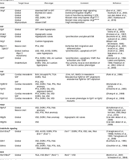
Table 1. Signaling molecules involved in cardiac septation and valve development
Gene Target tissue Phenotype Comment Reference
TGF/BMP/SMAD
Acvr1 (Alk2) Neural crest Endothelium
PTA, VSD, PAA anomaly ASD, VSD
Defective NCC migration
fEMT; fMsx1, Snail, pSmad1/2/5/8 (Kaartinen et al., 2004; Wang et al.,
2005)
Bmpr1a
(Alk3)
SHF
Endothelium
PTA, ASD, VSD
fAV cushion EMT
fTbx20, FIsl1 at OFT; fTbx2, Tbx3 in AV cushion (Ma et al., 2005;
Yang et al., 2006)
Bmpr2* Global
(hypomorph) Epiblast Endothelium Neural crest
PTA, IAA, VSD, no OFT valves
DORV, ASD, VSD Thick valves, ASD, VSD OA
fperiostin in OFT cushions (Délot et al., 2003;
Beppu et al., 2009)
Bmp2 Myocardium/
mesoderm
Hypoplastic AV/OFT cushions
fTbx2 and Has2 in cushion myocardium (Ma et al., 2005;
Rivera-Feliciano and Tabin, 2006)
Bmp4 SHF
Myocardium/ mesoderm Myocardium
PTA, VSD
PTA, VSD, PAA anomaly
DORV, ASD, AVSD
Semilunar valve hyperplasia
feHand, dHand, Sema3c, Pitx2; overlapping
functions of Bmp4/7 in OFT Hypomorph, fproliferation in AVC
(Jiao et al., 2003; Abedin et al., 2004; McCulley et al., 2008)
Bmp6/7 Global ASD, VSD Bmp6/7 are functionally redundant (Kim et al., 2001)
Chrd Global PTA, PAA anomaly fTbx1 and Fgf8 in mesoderm (Bachiller et al.,
2003)
Ltbp1 Global PTA, IAA, valve
hyperplasia
fperiostin (Todorovic et al.,
2011)
Smad4 Muscle
Neural crest
Endothelium
DORV, VSD
PTA, hypoplastic OFT cushion, PAA anomaly
fAV cushion cellularity
Fapoptosis; fBmp4, Sema3c, Plxna2
fproliferation, Fapoptosis in AV cushions
(Jia et al., 2007; Azhar et al., 2010; Song et al., 2011)
Smad6 Global Hyperplastic valves (Galvin et al., 2000)
Smad7 Global VSD, OFT misalignment F pSmad2/3 and apoptosis in AVC (Chen et al., 2009)
Tgfbr1 (Alk5) Neural crest Endothelium
PTA, PAA anomaly Hypoplastic AV cushion,
VSD
Fapoptosis in NCC
fproliferation in myocardium (Wang et al., 2006; Sridurongrit et
al., 2008)
Tgfb2 Global DORV, PTA, VSD, PAA
anomaly, thick valves
(Sanford et al., 1997; Bartram et al., 2001)
Tgfb2/3 Global VSD (Dünker and
Krieglstein, 2002)
Tgfbr2 Neural crest
Muscle
AV myocardium Ventricular
myocardium Endothelium
PTA, VSD, PAA anomaly ASD, VSD (Sm22Cre) DORV, VSD (cTntCre) Tricuspid valve defect PTA, OA, VSD, OFT valve
defect VSD, OA, DORV
Descending aorta defects
(Wurdak et al., 2005; Choudhary et al., 2006; Jiao et al., 2006; Langlois et al., 2010; Robson et al., 2010)
NOTCH
Hes1 Global VSD, OA (Rochais et al.,
2009)
Hey1/
HeyL
Global VSD, AV, OFT valve defects
Impaired EMT in AV cushion (Zhang and Fisher,
2007)
Hey2‡ Global ASD, VSD, OA, TOF, TA (Donovan et al.,
2002; Fischer et al., 2004)
Jag1§ SHF PTA, DORV, PS, VSD, ASD,
IAA
fPlxna2/Sema3c; fFgf8 and Bmp signaling (Krantz et al., 1999;
Eldadah et al., 2001; Warthen et al., 2006)
Notch¶ Neural crest
SHF
VSD, PTA, DORV, PAA anomaly, PS, valve defect
PTA, DORV, IAA, VSD, ASD, PS, TA, OFT valve defects
NCC differentiation defects
fFgf8 in SHF
(High and Epstein, 2007; High et al., 2009; Jain et al., 2011)
Notch1# Global Absent EMT fTgfb2, Tgfbr1/2/3 (Timmerman et al.,
2004; Garg et al., 2005; McKellar et al., 2007)
Table 1. Continued on next page
D
E
V
E
LO
P
M
E
N
Table 1. Continued
Gene Target tissue Phenotype Comment Reference
Notch2** Global ASD, VSD, PS (McCright et al.,
2002; McDaniell et al., 2006)
Psen1 Global VSD, DORV, PS (Nakajima et al.,
2004)
Rbpj Global
Twist2(+) tissue
Absent EMT VSD
(Timmerman et al.,
2004; Morimoto et al., 2010)
WNT
Ctnnb1
(-catenin) SHF
Neural crest Endothelium Pharyngeal
mesenchyme
PTA, ASD,VSD, PAA anomaly (Islet1Cre) PTA (Mef2cCre) PTA, TGA, DORV Hypoplastic AV cushions OA, DORV, PTA, VSD, ASD
Islet1 as a downstream target; Fapoptosis, fproliferation
fCyclinD2, Tgfb2, BMP4 fPitx2 in NCC
fEMT
FTbx1 and Fgf8; introduction of Fgf8+/– into mutant mice rescued the phenotype
(Kioussi et al., 2002; Liebner et al., 2004; Ai et al., 2007; Lin et al., 2007; Huh and Ornitz, 2010)
Daam1 Global DORV, VSD Affect cytoskeleton and sarcomeres (Li et al., 2011)
Dvl2 Global PTA, DORV, TGA fPitx2; Dvl2+/–;Pitx2+/–: PTA (Kioussi et al.,
2002)
Dvl3 Global PTA, DORV Dvl2+/–; Dvl3+/–: DORV, PTA, TGA
Dvl2+/–; Dvl3–/–: more severe phenotype
(Etheridge et al., 2008)
Wnt2 Global AVSD fEMT; fproliferation of DM/DMP (Tian et al., 2010)
Wnt5a‡‡ Global PTA, DORV, TGA, VSD,
IAA
fPlxna2 in NCC (Schleiffarth et al.,
2007; Person et al., 2010)
Wnt11 Global TGA, DORV, PTA, VSD fTgfb2 (Zhou et al., 2007;
Nagy et al., 2010)
SHH
Shh Global
Pharyngeal endoderm
PTA, PAA anomaly, ASD, VSD
PTA, AVSD, PAA anomaly
fNCC population; defective DMP
Shortening of OFT
(Washington Smoak et al., 2005; Goddeeris et al., 2007; Goddeeris et al., 2008)
Smo SHF
Neural crest
ASD, VSD, PAA anomaly, PTA, TGA (Islet1Cre) PTA, AVSD (Mef2cCre) PTA, PAA anomaly
fTbx1 in mesoderm; fNeuropilin2 in OFT; F
apoptosis in OFT
Defective DMP differentiation/migration Gain-of-function mutants also have PTA
(Lin et al., 2006; Goddeeris et al., 2007; Goddeeris et al., 2008)
NFAT
Nfatc1 Global Blunting of AV/OFT valves,
VSD
Rescued by endothelial Nfatc1 expression (de la Pompa et al., 1998; Ranger et al., 1998; Chang, C. P. et al., 2004; Zhou et al., 2005)
Nfatc2/3/4 Global fcushion mesenchyme Nfatc2/c3/c4 are functionally redundant (Chang, C. P. et al.,
2004)
Ppp3r2
(Calcineurin B1)
Endothelium SHF
Blunted AV/OFT valves Absent OFT valves
Phenocopies Nfatc1–/– embryos Fapoptosis in conal cushion
(Chang, C. P. et al., 2004; Lin et al., 2012)
Dyrk1a/
Dscr1§§
Global
(overexpressed)
Blunted valves Phenocopies Nfatc1–/– embryos (Arron et al., 2006)
For definitions, see Glossary, Box 1. PAA, pharyngeal arch artery. *BMPR2 mutations found in patients with ASD, VSD, AVSD, PDA, PAPVR. ‡HEY2 mutations in CHD or Alagille syndrome (ASD, VSD, TOF, PS). §JAG1 mutations in TOF or Alagille syndrome.
¶NOTCH mutations found in patients with BAV.
#NOTCH1 mutations found in patients with aortic valve anomaly. **NOTCH2 mutations in Alagille syndrome.
‡‡WNT5A mutations in Robinow Syndrome (ASD, VSD, TOF, PS). §§Duplication in Down Syndrome (ASD, VSD, AVSD, valve defects).
D
E
V
E
LO
P
M
E
N
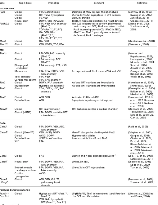
Table 2. Receptor tyrosine kinases, G protein-coupled receptors and nuclear receptors involved in cardiac septation and valve development
Gene Target tissue Phenotype Comment Reference
VEGF
Vegf Global
Global Global Global Global Myocardium
Aborted EMT in OFT Blunted AV valves VSD
VSD, DORV, TOF VSD
Absent EMT
sFlt to antagonize Vegf signaling Dominant-negative VegfR-2 Gain of function by deleting 3⬘UTR Mutant mice only express Vegf120/120 Mutant mice only express Vegf188/188 Vegf overexpression
(Dor et al., 2001; Stalmans et al., 2003; van den Akker et al., 2007; Stankunas et al., 2010)
EGF
Egfr Global OFT valve hyperplasia (Chen et al., 2000;
Sibilia et al., 2003)
Erbb3 Global Hypoplastic valves (Erickson et al., 1997)
Hbegf Global Hyperplastic valves Fproliferation and pSmad1/5/8 (Iwamoto et al., 2003;
Jackson et al., 2003)
Nrg1 Global Cushion hypoplasia (Meyer and
Birchmeier, 1995)
Ptpn11
(Sph2)*
Neural crest
Global (various
Ptpn11
mutations)
PTA, VSD
ASD, VSD, AVSD, DORV, valve hyperplasia
Defective NCC migration and differentiation
Fproliferation; fapoptosis of OFT
(Tartaglia et al., 2001; Araki et al., 2004; Araki et al., 2009; Nakamura et al., 2009)
Nf1‡ Global
Endothelium
PTA, DORV, VSD, AV cushion hyperplasia
DORV, VSD, AV cushion hyperplasia
Fproliferation; fapoptosis; FEMT; Ras
activation also FEMT
FRas activity; neural crest deletion of
Nf1 has no cardiac phenotype
(Brannan et al., 1994; Lakkis and Epstein, 1998; Friedman et al., 2002; Gitler et al., 2003)
FGF
Fgf8 Cardiac mesoderm
SHF
SHF
BAV, bicuspid PV, TGA DORV, TGA
PTA, VSD
fErm, Isl1, Mef2c in mesoderm
Mesodermal Fgf8 for OFT alignment; endodermal Fgf8 for OFT septation
(Park et al., 2006)
Fgf8/10 Cardiac mesoderm VSD, DORV, TGA, PTA, PAA
anomaly
(Watanabe et al., 2010)
Fgf15 Global PTA, DORV, OA, VSD,
alignment defect
(Vincentz et al., 2005)
Frs2 Cardiac mesoderm
SHF
PTA, DORV, OA, VSD DORV, OA, VSD
(Zhang et al., 2008)
Fgfr1/r2 Cardiac mesoderm PTA, DORV, OA, VSD Less severe phenotype in Fgfr1 or Fgfr2
mutants
(Zhang et al., 2008)
PDGF
Pgfra§ Global
Neural crest
VSD, DORV, PTA, PAA anomaly
VSD, PTA, PAA anomaly
(Schatteman et al., 1995; Tallquist and Soriano, 2003; Bleyl et al., 2010)
Pdgfb Global VSD, DORV, PAA anomaly Hypoplastic AV valves (Van den Akker et al.,
2008)
Pdgfrb Global VSD (Van den Akker et al.,
2008)
Endothelin signaling
Ece1/Ece2¶ Global VSD, AVSD, DORV, PTA
(Ece1–/–;Ece2–/–) Ece1
–/–: DORV, PTA, VSD, IAA, PAA (Yanagisawa et al., 1998b; Hofstra et al., 1999; Yanagisawa et al., 2000)
Edn1 Global VSD, DORV, PTA, PAA
anomaly
(Kurihara et al., 1995)
Ednra Global VSD, DORV, TGA, PTA, IAA,
PAA anomaly
(Clouthier et al., 1998)
ROR
[image:12.612.50.567.78.714.2]Ror1/Ror2# Global TGA, VSD (Ror1–/–;Ror2–/–) Ror2–/–: VSD (Nomi et al., 2001; Schwabe et al., 2004) Table 2. Continued on next page
D
E
V
E
LO
P
M
E
N
GATA factors
The zinc finger GATA transcription factors are essential for cardiac septation and valve formation. For example, mice with Gata4
hypomorphic alleles exhibit myocardial hypoplasia, DORV and AVSD (Crispino et al., 2001; Pu et al., 2004), and mice lacking endocardial Gata4 have EMT failure in AV cushions (Rivera-Feliciano et al., 2006). Gata4 cooperates with Smad4 to control AV septation and EMT (Moskowitz et al., 2011). Disruption of endocardial Smad4, like Gata4mutations, results in EMT failure in AV cushions. Gata4 and Smad4 synergistically activate the expression of Id2, a helix-loop-helix transcriptional repressor, to regulate AV septation (Moskowitz et al., 2011). Gata4 also interacts with Tbx5 to control AV cushion development. Gata4and Tbx5
double heterozygotes display thin myocardium as well as AVSD with a single atrioventricular valve (Maitra et al., 2009). GATA4
mutations are found in patients with septal defects (ASD or AVSD) or valve abnormalities (aortic regurgitation, mitral regurgitation and/or pulmonary stenosis) (Garg et al., 2003; Okubo et al., 2004; Sarkozy et al., 2005; Moskowitz et al., 2011). Interestingly, certain
GATA4 missense mutations (G303E and G296S) in humans are known to disrupt the binding of GATA4 to SMAD4 (Moskowitz et al., 2011) or to TBX5 (Maitra et al., 2009), suggesting a conserved function of the human GATA4-SMAD4 and GATA4-TBX5 complex for AV septation and valve development.
Gata5 is involved in aortic valve development. Gata5-null mice have reduced ventricular trabeculation and partially penetrant BAV (Laforest et al., 2011). Endocardial Gata5 regulates aortic valve formation possibly through Notch signaling and endothelial nitric oxide synthase Nos3 (Lee, T. C. et al., 2000; Laforest et al., 2011). Gata6 is essential for both AV and OFT development. Gata6 synergizes with its transcription target Wnt2 to regulate AV septation, and deletion of Gata6 in cardiac progenitor cells causes AV septation defects (Tian et al., 2010). By contrast, Gata6 regulates OFT septation through its activation of Sema3c and Plxna2 (Lepore et al., 2006; Kodo et al., 2009). Gata6 transcriptionally activates Sema3c in the OFT myocardium and Plxna2 in NCCs to orchestrate the migration of NCCs into the OFT. Deletion of Gata6 in the myocardium or NCCs causes Sema3c or Plxna2 downregulation, leading to pharyngeal arch artery defects and OFT abnormalities (PTA or DORV) (Lepore et al., 2006). GATA6 mutations that disrupt GATA6 nuclear localization (E486del) or abolish GATA6’s transcriptional activity on SEMA3Cand PLXNA2promoters (E486del and N466H) have been identified in patients with PTA (Kodo et al., 2009).
T-box genes
Tbx genes encode T-box transcription factors that regulate multiple developmental processes (Greulich et al., 2011). The absence of TBX1 is thought to be a major cause of 22q11 deletion syndrome (DiGeorge, velocardialfacial, and conotruncal face anomaly syndromes), which includes craniofacial abnormalities, pharyngeal arch artery defects and cardiac malformations (TOF, DORV and PTA). In mice, Tbx1germline mutations or tissue-specific mutations in the pharyngeal endoderm or mesoderm result in pharyngeal arch artery defects and cardiac abnormalities (PTA and VSD) (Jerome and Papaioannou, 2001; Merscher et al., 2001; Vitelli et al., 2002; Arnold et al., 2006; Zhang et al., 2006).
Tbx2 is essential for the developmental identity of myocardium at the AV and OFT cushions. Tbx2 is expressed in the cushion myocardium to repress the expression of chamber myocardium-specific genes (Habets et al., 2002; Christoffels et al., 2004; Harrelson et al., 2004). In Tbx2-null embryos, the cushion myocardium is partially turned into ventricular myocardium, resulting in hypoplastic endocardial cushions (Harrelson et al., 2004). Conversely, overexpression of Tbx2 in the myocardium of the heart tube inhibits cardiac chamber formation and chamber-specific gene expression (Christoffels et al., 2004). Such ectopic Tbx2 expression triggers excessive deposition of extracellular matrix in the ventricles and activates chamber myocardium to stimulate EMT (Shirai et al., 2009), rendering the chamber myocardium ‘cushion-like’. These changes are at least partly caused by ectopic activation by Tbx2 of the matrix-producing Has2 and the EMT-promoting Tgf2 (Shirai et al., 2009).
Tbx5 is essential for determining the left ventricle identity and interventricular boundary: the boundary between Tbx5-expressing left ventricle and non-Tbx5-expressing right ventricle determines the site of interventricular septation (Bruneau et al., 1999; Takeuchi et al., 2003). In mice, Tbx5 overexpression causes expansion of the left ventricle and a loss of interventricular septum, whereas in chick an extra interventricular septum forms at an ectopically induced boundary between Tbx5-positive and Tbx5-negative ventricles (Takeuchi et al., 2003). TBX5mutations are associated with Holt-Oram syndrome, which is characterized by upper limb and cardiac malformations (ASD, VSD) (Basson et al., 1997; Li et al., 1997). These limb and cardiac defects are seen in mice with a heterozygous
Tbx5mutation (Bruneau et al., 2001), and loss of endocardial Tbx5 causes excessive apoptosis in primary atrial septum, possibly through disruption of the Tbx5/Gata4-Nos3 pathway (Nadeau et al., 2010). Table 2. Continued
Gene Target tissue Phenotype Comment Reference
Nuclear receptors
Aldh1a2** Chimera PTA (Vermot et al., 2006;
Ryckebusch et al., 2008; Pavan et al., 2009)
Rara/Rarb Global
Cardiac mesoderm
PTA, PAA anomaly (RARa1–/–;RARb–/–) PTA, DORV, OA
Rara1–/– or Rarb–/–: no phenotype;
Rara1–/–; Rxra–/–: PTA, PAA anomaly (Lee et al., 1997; Jiang et al., 2002; Li et al., 2010)
Rxra Global PTA, DORV, VSD, AVSD Variable AV cushion defects (Gruber et al., 1996)
For definitions, see Glossary, Box 1. PAA, pharyngeal arch artery.
*PTPN11 mutation in Noonan syndrome (ASD, AVSD, TOF, OFT valve defects). ‡NF1 mutation in neurofibromatosis type I (PS, aortic coarctation).
§PDGFRA mutation in TAPVR patients.
¶ECE1/ECE2 mutation found in a patient with Hirschsprung disease and CHD. #ROR2 mutation found in Robinow syndrome.
**ALDH1A2 mutation found in TOF patients.