INTRODUCTION
During the development of the eye, in vertebrates and invertebrates, neural progenitors derive from multipotent and proliferative cells (reviewed by Chow and Lang, 2001; Dominguez and Casares, 2005). In the Drosophila eye primordium, the TALE-class homeodomain transcription factor Homothorax (Hth) is expressed in this multipotent population, where it is required to maintain these cells in a proliferative state and to prevent their premature differentiation (Pai et al., 1998; Pichaud and Casares, 2000; Bessa et al., 2002). The homologs of hth in vertebrates are the Meis and Prep (also known as Pknox) gene families (reviewed by Burglin, 1997; Moens and Selleri, 2006). Whereas the expression of Prep genes is widespread in mice and zebrafish, Meis genes show specific transcription patterns in vertebrates, including expression in the developing eye (Ferretti et al., 1999; Toresson et al., 2000; Waskiewicz et al., 2001; Maeda et al., 2002; Zhang et al., 2002; Hisa et al., 2004).
Recent work points to a role for Meis genes in eye development: Meis1 and Meis2 are upstream regulators of Pax6 in the developing lens in chicken and mouse (Zhang et al., 2002), and mouse embryos homozygous for a homeodomain-less Meis1 gene show eye malformations (Hisa et al., 2004). Still, the precise role(s) played by Meis genes during eye development remain(s) unknown. If the parallels in early eye development between flies and vertebrates hold true for Hth/Meis, Meis genes might be involved in stimulating
proliferation, or preventing premature differentiation in the optic primordium, or both. Here, we investigated these hypotheses in the zebrafish (Danio rerio).
MATERIALS AND METHODS
Probe preparation, in situ hybridization and immunolabeling Antisense RNA probes were prepared from cDNAs and labeled with digoxigenin. Specimens were fixed, hybridized and stained as described (Tena et al., 2007).
Fluorescent probes and antibodies
Propidium iodide (PI) was used as nuclear stain; FITC-phalloidin to mark filamentous actin; anti-Islet1 mouse monoclonal antibody labels GCL [36 hours post-fertilization (hpf)] and ganglion cell layer (GCL) plus inner nuclear layer (INL) (48-72hpf) (from DSHB, University of Iowa); rabbit anti-GFP (A11122, Molecular Probes), mouse anti-Myc (MMS 150P, Covance), mouse anti-cleaved Caspase 3 (Cell Signaling Technology). Fluorescent secondary antibodies were from Molecular Probes. Dissected eyes from stained embryos were imaged using a Leica-SP2 confocal system, and data processed with Adobe Photoshop.
In vitro RNA synthesis and microinjection of mRNA and morpholinos
cDNAs were linearized and transcribed as described (Tena et al., 2007). One- to two-cell-stage zebrafish embryos were injected in the yolk with mRNA and/or morpholino (MO) diluted in ~5 nl of injection solution (10% Phenol Red in DEPC-treated water).
MOs targeting the ATG region of meis1, meis2.2, meis3and meis4
mRNAs (see Fig. S1A in the supplementary material) were synthesized by GeneTools. We verified the target specificity of meis1-and meis2.2-MOs in
Xenopus laevisassays (see Fig. S1B in the supplementary material), and the biological specificity of the meis1-MO by testing its ability to reduce the rhombomere-3 expression of krox20 (also known as egr2– ZFIN) (see Figs S2 and S5 in the supplementary material).
As controls, we injected similar amounts (8-16 ng) of a control MO directed against the Xenopus tropicalis olig2gene that shows no match in the zebrafish genome (see Fig. S1 in the supplementary
meis1
regulates
cyclin D1
and
c-myc
expression, and controls
the proliferation of the multipotent cells in the early
developing zebrafish eye
José Bessa1,*, Maria J. Tavares1,*, Joana Santos1,*, Hiroshi Kikuta2, Mary Laplante2, Thomas S. Becker2, José Luis Gómez-Skarmeta1and Fernando Casares1,3,†
During eye development, retinal progenitors are drawn from a multipotent, proliferative cell population. In Drosophila the maintenance of this cell population requires the function of the TALE-homeodomain transcription factor Hth, although its
mechanisms of action are still unknown. Here we investigate whether members of the Meis gene family, the vertebrate homologs of
hth, are also involved in early stages of eye development in the zebrafish. We show that meis1 is initially expressed throughout the eye primordium. Later, meis1becomes repressed as neurogenesis is initiated, and its expression is confined to the ciliary margin, where the retinal stem population resides. Knocking down meis1 function through morpholino injection causes a delay in the G1-to-S phase transition of the eye cells, and results in severely reduced eyes. This role in cell cycle control is mediated by meis1 regulating cyclin D1 and
c-myc transcription. The forced maintenance of meis1 expression in cell clones is incompatible with the normal differentiation of the
meis1-expressing cells, which in turn tend to reside in undifferentiated regions of the retinal neuroepithelium, such as the ciliary margin. Together, these results implicate meis1as a positive cell cycle regulator in early retinal cells, and provide evidence of an evolutionary conserved function for Hth/Meis genes in the maintenance of the proliferative, multipotent cell state during early eye development.
KEY WORDS: meis1, Zebrafish, Cell cycle, Eye development, cyclin D1,c-myc(myca) Development 135, 799-803 (2008) doi:10.1242/dev.011932
1CABD, CSIC-Universidad Pablo de Olavide, 41013 Seville, Spain. 2SARS Institute, N-5008 Bergen, Norway. 3IBMC, Universidade do Porto, 4159-180 Oporto, Portugal.
*These authors contributed equally to this work †Author for correspondence (e-mail: [email protected])
Accepted 21 December 2007
D
E
V
E
LO
P
M
E
N
material). The meis3-MO, which has nine and seven mismatches with
meis1and meis2.2, respectively, also served as control MO in some experiments.
Eye phenotype measurements
The polygonal-lasso tool from Adobe Photoshop was used to measure in digital photographs taken with the same magnification, the eye surface area (in pixels) of control and morphant embryos. The volume of each eye was estimated considering it as a hemisphere of radius equal to the radius of a circle with that same area. Measurements from 20 eyes for each condition were compared using a 2test.
Plasmid constructs
I.M.A.G.E. cDNA clones, from the Lawrence Livermore National Laboratory Consortium, used were: ccnd1 (IMAGE IRALp962K2356Q), c-myc (IRBOp991F125D), meis1 (IRAKp961C08136Q), meis2.1 (IRBOp991C0733D), meis2.2 (IRBOp991D0437D), meis3 (IRALp962E1456Q) and meis4 (MPMGp609N1326Q). pCS2-ccnd1 was generated by inserting the full-length cDNA into EcoRI and XbaI sites of pCS2+. To generate GFP-meis1, MT-meis1, meis1-MT, MT-meis2.2, meis2.2-MT, MT-meis3, meis3-MT, MT-meis4 and meis4-MT constructs, we PCR amplified the corresponding Meis coding regions with the following primer pairs (5⬘-3⬘; EcoRI and XhoI sites underlined): GAATTCGATGGCGCAGAGGT and CTCGAGCATGTAGTGCCAC -TGTCCC for meis1; GAATTCGATGGCGCAAAGGTACGA and CTCGAGCATGTAGTGCCACTGGCC for meis2.2; GAATTCCATG -GATA AGAGGTATGAGGAGTT and CTCGAGGTGGGCATGTA -TGTCAA for meis3; and GAATTCCATGGCGCAACGGTACGA and CTCGAGCATGTAGTGCCACTGACTCTC for meis4.
The PCR fragments were subcloned into pGEMT-Easy (Promega) and sequenced. Meis cDNAs were cloned into pCS2 MT, pCS2p+MTC2 or pCS2eGFP (kindly provided by D. Turner, University of Michigan, USA) to generate N-terminal (Myc-meis) and C-terminal (meis-Myc) Myc-tagged meis or N-terminal GFP-tagged meis1(GFP-meis1), respectively. To generate the Tol2-GFP-meis1and Tol2-GFP constructs, we inserted the GFP-meis1and GFP fragments, respectively, into SalI and SspI sites of Tol2 (pT2KXIG).
Acridine Orange staining
Acridine Orange staining was performed as described (Perkins et al., 2005).
DNA content analysis and flow cytometry
Eyes dissected from 19hpf zebrafish embryos were disaggregated, and PI staining carried out as described (Langenau et al., 2003). DNA content was analyzed on a BD FACSAria and results processed with FloJo software (Tree Star). A 2test was used for statistical data analysis.
Induction of ectopic expression mosaics
The Tol2 transposon/transposase method of transgenesis (Kawakami et al., 2004) was used with minor modifications. Four- to 16-cell-stage zebrafish embryos were injected in the yolk with 5-12.5 pg of either Tol2-GFP-meis1 or Tol2-GFP constructs, plus 125 pg of transposase-encoding mRNA in a final volume of 5 nl of injection solution. Embryos were cultured at 28.5°C, staged and fixed. Anti-GFP antibody was used to detect the GFP- or GFP-meis1-expressing clones. A stack of confocal z -sections was obtained for each eye analyzed. Three-dimensional reconstruction of the stacks was used to determine the location of the clones.
RESULTS AND DISCUSSSION
meis1expression is restricted to the
undifferentiated and proliferating cells of the early zebrafish eye
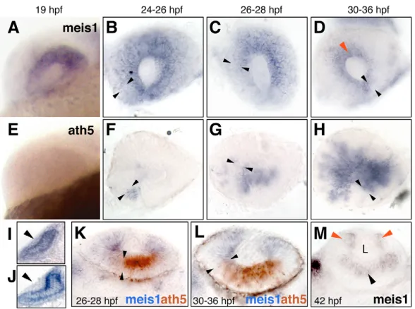
[image:2.612.52.346.520.739.2]Of all five zebrafish Meis genes (meis1, 2.1,2.2,3and 4.1), only meis1 and meis2.2 are expressed during early stages of eye development (Kudoh et al., 2001; Waskiewicz et al., 2001; Zerucha and Prince, 2001; Thisse and Thisse, 2005) (this work). meis1, as monitored by in situ hybridization, or by a YFP insertional reporter inserted close to meis1, was seen to be uniformly transcribed in the eye primordium from 15 to ~24hpf (Fig. 1A and see Fig. S3 in the supplementary material), a period in which all cells proliferate (Li et al., 2000). After this time, meis1 expression progressively retracted in the retina (Fig. 1B-D,K,L) as the neurogenic wave, marked by ath5 (also known as atoh7– ZFIN) expression, expands from antero-nasal to posterior-temporal positions (Fig. 1F-H) (Hu and Easter, 1999; Li et al., 2000; Masai et al., 2000). meis1 remained transiently expressed in the ciliary margin zone (CMZ), where the retinal stem population resides (Fig. 1D,M). meis2.2was also found to be expressed uniformly in early eye primordia, but its expression faded away by 20hpf (see Fig. S3 in the supplementary material). Similar to the situation found in chicken and mouse (Zhang et al., 2002), meis1was expressed in the prospective lens ectoderm, but was turned off as the lens placode started to thicken (Fig. 1I,J). Therefore, meis1 expression is associated with the undifferentiated, proliferative cells during the early development of the zebrafish eye. In addition, a new wave of Meis gene expression starts in postmitotic neurons at around 36-42hpf (Fig. 1M and see
Fig. 1. meis1retracts accompanying the ath5 wave and becomes restricted to the CMZ. (A-D) meis1and (E-H) ath5expression analyzed by single in situ hybridization. Developmental stages are indicated as hours post-fertilization (hpf) at 28.5°C. Lateral views of whole-mount (A,E) or dissected (B-D,F-H) eyes, with dorsal up and anterior to the left. The front of the ath5 domain is marked by black arrowheads. The red arrowhead in D points to meis1 expression in the prospective ciliary margin. (I-M) Transverse 40 m vibratome sections. (I,J) Dorsal is up. meis1is weakly expressed in the lens ectoderm before its thickening (I), but no signal is detected once the lens placode is formed (J). (K-M) Dorsal is to the left. meis1 and ath5 expression domains are complementary as shown by double in situ hybridization (K,L). Approximate limits of the ath5 signal are indicated by the black arrowheads. (M) At 42hpf, meis1 expression is detected by in situ hybridization in the ciliary margin (red arrowheads) and in the postmitotic ganglion cells (black arrowhead). L, lens.
D
E
V
E
LO
P
M
E
N
Fig. S3 in the supplementary material). Interestingly, at 4 days post-fertilization (dpf), meis2.2 expression had replaced meis1 at the CMZ.
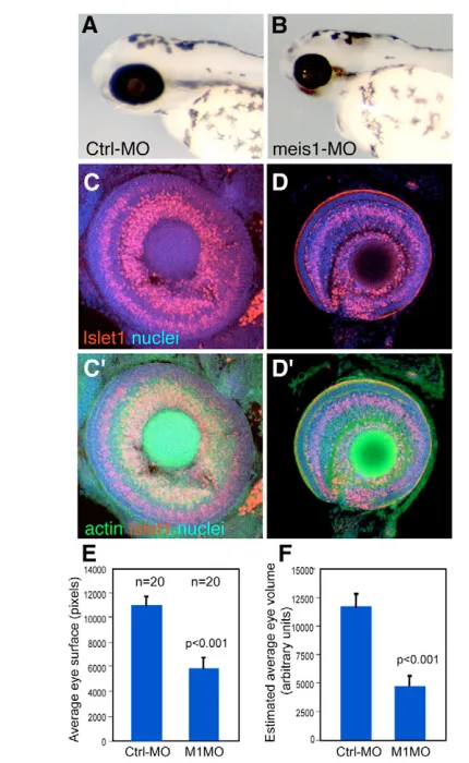
meis1is required to promote the G1-to-S transition of the eye primordium cells, and regulates the transcription of cyclin D1and c-myc The early expression pattern of meis1suggests that it plays a role in the proliferative/multipotent cells of the developing eye. To determine what role that is, we knocked down meis1function using meis1-specific morpholinos (meis1-MO). By the end of embryogenesis,meis1morphants were severely microphthalmic (>60% of embryos injected with 8 ng of meis1-MO, n=163) (Fig. 2A,B,E,F), with eyes containing fewer cells than controls (Fig. 2C,D). Despite this, meis1-morphant eyes showed apparently normal retinal lamination (Fig. 2C⬘,D⬘). The lens was normal or slightly reduced (Fig. 2D and data not shown). The co-expression of
meis2.2and meis1during optic vesicle stages suggested a possible functional redundancy between these two genes. Nevertheless, injection of meis2.2-MO (8 ng/embryo) caused only mild eye reductions in 22% of the treated embryos (n=299). Furthermore, co-injection of equivalent amounts of meis1-and meis2.2-MOs (4 ng of each MO/embryo) did not significantly enhance the penetrance or severity of the microphthalmia (29%, n=196). These results suggest that meis2.2does not have a major role during early stages of eye development in the zebrafish. The phenotype observed in meis1morphants does not appear to be due to an abnormal eye primordium specification. Although we found a slight decrease in pax6b expression in meis1-morphant eyes, as estimated by RT-PCR (see Fig. S4C,D in the supplementary material), the early expression of the eye selector genes pax6b and rx2 (Stigloher et al., 2006) seemed unaffected, when observed by in situ hybridization (see Fig. S4A,B in the supplementary material and data not shown). In agreement with this, we found that the expression of the Pax6 gene eyeless is independent of hth during the development of the Drosophilaeye (see Fig. S4E,F in the supplementary material).
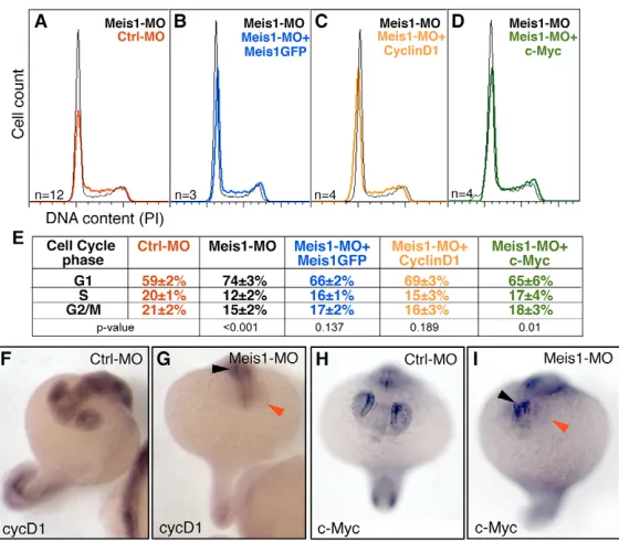
To further dissect the mechanisms underlying the observed microphthalmia, we assessed whether meis1controls the cell cycle. meis1-morphant eyes, at 19hpf, had a significantly higher percentage of cells in G1 phase than control embryos (Fig. 3A,E), indicating a requirement of meis1in promoting the G1–S transition. Viability of these cells was not compromised, as meis1-morphant eyes did not show a significant increase in the levels of active Caspase 3, or in the vital incorporation of Acridine Orange (not shown).
cyclin D1(ccnd1) and c-myc(also known as myca– ZFIN) are two major G1 regulators of the cell cycle in vertebrates (Levine and Green, 2004). During the development of the zebrafish eye, cyclin D1 and c-myc are first widely expressed in the optic vesicle, followed by a progressive restriction to the proliferating cells of the neural retina (Thisse and Thisse, 2005; Yamaguchi et al., 2005), a pattern that is reminiscent of that of meis1. In addition, recent work shows that cyclin D1is required for proliferation in the zebrafish developing retina, as cyclin D1 morphants are microphthalmic (Duffy et al., 2005). The similarity between the patterns of expression of meis1, cyclin D1 and c-myc, and the similar eye phenotypes of cyclin D1and meis1morphants, prompted us to ask whether cyclin D1and c-mycwere under meis1control. Indeed, meis1morphants showed a dramatic reduction of cyclin D1and c-mycexpression in the eye when compared with control-injected embryos (Fig. 3F-I and see Fig. S5 in the supplementary material). In addition, the co-injection of either cyclin D1or c-mycmRNAs partially rescued the cell cycle defects of meis1morphants to levels similar to those obtained by co-injection of GFP-meis1 mRNA (Fig. 3B-E). These results place cyclin D1 and c-myc functionally downstream of meis1in the control of cell cycle progression in the developing eye. Whether meis1 regulates the transcription of cyclin D1and c-mycdirectly or indirectly is unknown.
Maintenance of meis1expression is incompatible with cell differentiation
[image:3.612.54.269.268.618.2]In Drosophila, hth not only promotes proliferation in the eye primordium, but forced maintenance of its expression results in a delay or block of retinal differentiation (Pai et al., 1998; Pichaud and Casares, 2000; Bessa et al., 2002). Similarly, in the early zebrafish eye, meis1expression is found in undifferentiated cells but is turned off as neurogenesis advances (Fig. 1). To test whether maintaining meis1expression is incompatible with retinal differentiation, we analyzed the distribution of clones of cells expressing either GFP or Fig. 2. meis1is required for the growth of the eye primordium.
(A,B) Lateral views of representative 72hpf control-MO (A) and meis1 -MO (B) -injected fish. meis1morphants are microphthalmic.
(C,D) Confocal images of dissected eyes stained for propidium iodide (nuclei), rhodamine-phalloidin (filamentous actin) and Islet1, which labels GCL nuclei and some in the INL. The reduced eyes from meis1 morphants show apparently normal retina lamination (D,D⬘), but fewer cells than control eyes (C,C⬘). Area (E) and estimated volume (F) of control-MO and meis1-MO-injected embryos at 72hpf. meis1-morphant embryos show a significant (P<0.001) reduction in eye area
and volume (45% and 60%, respectively). n=20 for each condition.
D
E
V
E
LO
P
M
E
N
GFP-tagged-Meis1 in developing retinas, prior to and after the initiation of neuronal differentiation (Fig. 4 and see Fig. S6 in the supplementary material). Differentiation was followed using the GCL marker islet1. When analyzed between 24 and 30hpf, a stage at which most of the retina is undifferentiated, all GFP- and 80% of GFP-Meis1-expressing clones spanned the whole width of the neuroepithelium (n=57 and 46, respectively; Fig. 4A,D). Later in development, when retinal differentiation is ongoing and layering becomes apparent, most GFP clones appeared in the central retina
and contained both Islet1-expressing and non-expressing cells (90%) (Fig. 4B,C), whereas only a few (7%) were found in the CMZ (n=41). By contrast, of the GFP-Meis1 clones located in the central retina (72%, n=39), none contained Islet1-positive cells at this stage (Fig. 4E). In addition, a large portion of these Meis1-expressing clones (28%, n=39) was found in the CMZ (Fig. 4F). The fact that Meis1-expressing cells were always found in undifferentiated regions of the neuroepithelium, leads us to conclude that maintenance of meis1 expression in the first 48 hours of eye development is incompatible with neuronal differentiation.
Our results indicate that, during early eye development, meis1 shares two roles with its fly homolog, hth. First, meis1is required to maintain proliferation of the multipotent cells of the early eye, by promoting the G1-to-S transition of the cell cycle. Mechanistically, meis1regulates the transcription of at least two potent cell-cycle activators: cyclin D1and c-myc. Second, ath5 follows receding meis1 expression in a similar fashion as in Drosophila, where hthexpression retracts as the atonal-expressing differentiation wave advances. This finding is in accordance with a model in which the expression of meis1has to be downregulated to allow further differentiation of the fish retina, and agrees with our results that the sustained expression of meis1is incompatible with neural differentiation. Similar results have been obtained in chicken and mouse by Heine and co-workers (Heine et al., 2008). Although retinal lamination in meis1 morphants is not grossly affected, we cannot rule out specific effects on the specification and/or differentiation of specific retinal cell types, as meis1, together with meis2.1 and meis2.2, is redeployed in postmitotic cells of the ganglion cell and inner nuclear layers (Fig. 1M and see Fig. S3 in the supplementary material).
[image:4.612.54.334.58.303.2]The expression of meis1in the CMZ, and the fact that forcing meis1expression results in the localization of the expressing cells to the CMZ, suggest that meis1might function in specifying the retinal stem cells of the zebrafish. In this regard, it is interesting to note that meis1expression resembles that of Pax6, a previously described retinal progenitor transcription factor (Raymond et al., 2006) (reviewed by Amato et al., 2004). In Drosophila, previous results showed that hthand eyelessare co-expressed in the undifferentiated
Fig. 3. meis1is required for the G1–S transition and the expression of the G1–S regulators cyclin D1and c-myc.(A-D) Histograms displaying DNA content (cell cycle profile) of cells from dissected 19hpf eyes of meis1 -MO treated embryos relative to (A) controls (Crtl--MO), (B) meis1-MO+GFP-meis1mRNA, (C) meis1-MO+cyclin D1 mRNA, and (D) meis1-MO+c-mycmRNA injected embryos. Number of replicates (n) and Pvalues are indicated. meis1 knockdown induces a delay in G1, which is partially rescued by co-injection of GFP-meis1 (B), cyclin D1(C) and c-myc(D) mRNAs. (E) Average percentages are shown for G1, S and G2/M DNA content. The cell-cycle profiles of control morphants and of uninjected, wild-type embryos are indistinguishable (not shown). (F-I) In situ analysis of cyclin D1and c-myctranscription in control (F,H) and meis1 morphants (G,I) at 19hpf. meis1morphants show a dramatic reduction in cyclin D1 and c-mycin the eye (red arrowheads). cyclin D1and c-mycare still detected in other body regions (black arrowheads). Hybridization and reaction development were performed strictly in parallel. Representative embryos are shown.
Fig. 4. Clonal overerexpression of meis1in the developing eye prevents differentiation and results in cell sorting.Single optical sections from confocal z-stacks of GFP (A-C) or GFP-Meis1 (D-F) expressing clones induced genetically in developing eyes. GFP-meis1 signal is nuclear. At 24-30hpf, both clone types frequently span the whole width of the neuroepithelium (A,D). Confocal optical sections through the central retina (B,E) and z-sections (C,F) of 48hpf eyes. At this stage, GFP clones comprise both Islet1-expressing and non-expressing cells (B,C). By contrast, same-stage GFP-Meis1 clones in the central retina do not contain Islet1-positive cells (E). GFP-Meis1 clones are often located in the CMZ (F). The arrowheads (C,F) point to the
CMZ, and the retina and the lens (L) are outlined.
D
E
V
E
LO
P
M
E
N
[image:4.612.50.299.442.606.2]domain and that their products might directly interact in vivo (Bessa et al., 2002). All these results seem to indicate that a common molecular mechanism to maintain a multipotent stem-like state exists during eye development in vertebrates and invertebrates.
In addition to controlling several developmental processes, Meis genes are overexpressed in an increasing number of cancer types (Lawrence et al., 1999; Segal et al., 2004; Geerts et al., 2005; Dekel et al., 2006). Therefore, the identification of functional targets of the Meis genes involved in the maintenance of the undifferentiated and proliferative state during normal development, such as cyclin D1and c-myc, is likely to be instrumental in deciphering the mechanisms underlying Meis-associated tumors.
We are grateful to Dorothea Schulte for communicating results prior to publication. This work was supported by grants BMC2003-06248 and BFU2006-00349/BMC from the Spanish Ministry of Education and Science, co-funded by FEDER, to F.C. J.B., M.J.T. and J.S. are supported by the Fundação para Ciência e Tecnologia, Portugal. The CABD is institutionally supported by Junta de Andalucía.
Supplementary material
Supplementary material for this article is available at http://dev.biologists.org/cgi/content/full/135/5/799/DC1
References
Amato, M. A., Arnault, E. and Perron, M.(2004). Retinal stem cells in vertebrates: parallels and divergences. Int. J. Dev. Biol. 48, 993-1001. Bessa, J., Gebelein, B., Pichaud, F., Casares, F. and Mann, R. S.(2002).
Combinatorial control of Drosophila eye development by eyeless, homothorax, and teashirt. Genes Dev.16, 2415-2427.
Burglin, T. R.(1997). Analysis of TALE superclass homeobox genes (MEIS, PBC, KNOX, Iroquois, TGIF) reveals a novel domain conserved between plants and animals. Nucleic Acids Res. 25, 4173-4180.
Choe, S.-K., Vlachakis, N. and Sagerström, C. G.(2002). Meis family proteins are required for hindbrain development in the zebrafish. Development129, 585-595.
Chow, R. L. and Lang, R. A.(2001). Early eye development in vertebrates. Annu. Rev. Cell Dev. Biol. 17, 255-296.
Dekel, B., Metsuyanim, S., Schmidt-Ott, K. M., Fridman, E., Jacob-Hirsch, J., Simon, A., Pinthus, J., Mor, Y., Barasch, J., Amariglio, N. et al.(2006). Multiple imprinted and stemness genes provide a link between normal and tumor progenitor cells of the developing human kidney. Cancer Res.66, 6040-6049. Dominguez, M. and Casares, F.(2005). Organ specification-growth control
connection: new in-sights from the Drosophila eye-antennal disc. Dev. Dyn.232, 673-684.
Duffy, K. T., McAleer, M. F., Davidson, W. R., Kari, L., Kari, C., Liu, C. G., Farber, S. A., Cheng, K. C., Mest, J. R., Wickstrom, E. et al.(2005). Coordinate control of cell cycle regulatory genes in zebrafish development tested by cyclin D1 knockdown with morpholino phosphorodiamidates and hydroxyprolyl-phosphono peptide nucleic acids. Nucleic Acids Res. 33, 4914-4921.
Ferretti, E., Schulz, H., Talarico, D., Blasi, F. and Berthelsen, J.(1999). The PBX-regulating protein PREP1 is present in different PBX-complexed forms in mouse. Mech. Dev.83, 53-64.
Geerts, D., Revet, I., Jorritsma, G., Schilderink, N. and Versteeg, R.(2005). MEIS homeobox genes in neuroblastoma. Cancer Lett.228, 43-50. Heine, P., Dohle, E., Bumsted-O’Brien, K., Engelkamp, D. and Schulte, D.
(2008). Evidence for an evolutionary conserved role of homothorax/Meis during vertebrate retina development. Development135, 805-811.
Hisa, T., Spence, S. E., Rachel, R. A., Fujita, M., Nakamura, T., Ward, J. M., Devor-Henneman, D. E., Saiki, Y., Kutsuna, H., Tessarollo, L. et al.(2004). Hematopoietic, angiogenic and eye defects in Meis1 mutant animals. EMBO J.
23, 450-459.
Hu, M. and Easter, S. S.(1999). Retinal neurogenesis: the formation of the initial central patch of postmitotic cells. Dev. Biol. 207, 309-321.
Huang, H., Paliouras, M., Rambaldi, I., Lasko, P. and Featherstone, M.(2003). Nonmuscle myosin promotes cytoplasmic localization of PBX. Mol. Cell. Biol. 23, 3636-3645.
Kawakami, K., Takeda, H., Kawakami, N., Kobayashi, M., Matsuda, N. and Mishina, M.(2004). A transposon-mediated gene trap approach identifies developmentally regulated genes in zebrafish. Dev. Cell7, 133-144. Kudoh, T., Tsang, M., Hukriede, N. A., Chen, X., Dedekian, M., Clarke, C. J.,
Kiang, A., Schultz, S., Epstein, J. A., Toyama, R. et al.(2001). A gene expression screen in zebrafish embryogenesis. Genome Res. 11, 1979-1987. Langenau, D. M., Traver, D., Ferrando, A. A., Kutok, J. L., Aster, J. C., Kanki,
J. P., Lin, S., Prochownik, E., Trede, N. S., Zon, L. I. et al.(2003). Myc-induced T cell leukemia in transgenic zebrafish. Science299, 887-890. Laplante, M., Kikuta, H., Konig, M. and Becker, T. S.(2006). Enhancer
detection in the zebrafish using pseudotyped murine retroviruses. Methods39, 189-198.
Lawrence, H. J., Rozenfeld, S., Cruz, C., Matsukuma, K., Kwong, A., Komuves, L., Buchberg, A. M. and Largman, C.(1999). Frequent co-expression of the HOXA9 and MEIS1 homeobox genes in human myeloid leukemias. Leukemia13, 1993-1999.
Levine, E. M. and Green, E. S.(2004). Cell-intrinsic regulators of proliferation in vertebrate retinal progenitors. Semin. Cell Dev. Biol. 15, 63-74.
Li, Z., Hu, M., Ochocinska, M. J., Joseph, N. M. and Easter, S. S., Jr(2000). Modulation of cell proliferation in the embryonic retina of zebrafish (Danio rerio). Dev. Dyn.219, 391-401.
Maeda, R., Ishimura, A., Mood, K., Park, E. K., Buchberg, A. M. and Daar, I. O.(2002). Xpbx1b and Xmeis1b play a collaborative role in hindbrain and neural crest gene expression in Xenopus embryos. Proc. Natl. Acad. Sci. USA99, 5448-5453.
Masai, I., Stemple, D. L., Okamoto, H. and Wilson, S. W.(2000). Midline signals regulate retinal neurogenesis in zebrafish. Neuron27, 251-263. Moens, C. B. and Selleri, L.(2006). Hox cofactors in vertebrate development.
Dev. Biol. 291, 193-206.
Pai, C. Y., Kuo, T. S., Jaw, T. J., Kurant, E., Chen, C. T., Bessarab, D. A., Salzberg, A. and Sun, Y. H.(1998). The Homothorax homeoprotein activates the nuclear localization of another homeoprotein, extradenticle, and suppresses eye development in Drosophila. Genes Dev.12, 435-446.
Perkins, B. D., Nicholas, C. S., Baye, L. M., Link, B. A. and Dowling, J. E. (2005). dazed gene is necessary for late cell type development and retinal cell maintenance in the zebrafish retina. Dev. Dyn.233, 680-694.
Pichaud, F. and Casares, F.(2000). homothorax and iroquois-C genes are required for the establishment of territories within the developing eye disc.
Mech. Dev.96, 15-25.
Raymond, P. A., Barthel, L. K., Bernardos, R. L. and Perkowski, J. J.(2006). Molecular characterization of retinal stem cells and their niches in adult zebrafish. BMC Dev. Biol. 6, 36.
Segal, E., Friedman, N., Koller, D. and Regev, A.(2004). A module map showing conditional activity of expression modules in cancer. Nat. Genet. 36, 1090-1098.
Stigloher, C., Ninkovic, J., Laplante, M., Geling, A., Tannhauser, B., Topp, S., Kikuta, H., Becker, T. S., Houart, C. and Bally-Cuif, L.(2006). Segregation of telencephalic and eye-field identities inside the zebrafish forebrain territory is controlled by Rx3. Development133, 2925-2935.
Tena, J. J., Neto, A., de la Calle-Mustienes, E., Bras-Pereira, C., Casares, F. and Gomez-Skarmeta, J. L.(2007). Odd-skipped genes encode repressors that control kidney development. Dev. Biol. 301, 518-531.
Thisse, C. and Thisse, B.(2005). High throughput expression analysis of ZF-models consortium clones. ZFIN Direct Data Submission, http://zfin.org. Toresson, H., Parmar, M. and Campbell, K.(2000). Expression of Meis and Pbx
genes and their protein products in the developing telencephalon: implications for regional differentiation. Mech. Dev.94, 183-187.
Vlachakis, N., Choe, S. K. and Sagerstrom, C. G.(2001). Meis3 synergizes with Pbx4 and Hoxb1b in promoting hindbrain fates in the zebrafish. Development
128, 1299-1312.
Waskiewicz, A. J., Rikhof, H. A., Hernandez, R. E. and Moens, C. B.(2001). Zebrafish Meis functions to stabilize Pbx proteins and regulate hindbrain patterning. Development128, 4139-4151.
Yamaguchi, M., Tonou-Fujimori, N., Komori, A., Maeda, R., Nojima, Y., Li, H., Okamoto, H. and Masai, I.(2005). Histone deacetylase 1 regulates retinal neurogenesis in zebrafish by suppressing Wnt and Notch signaling pathways.
Development132, 3027-3043.
Zerucha, T. and Prince, V. E.(2001). Cloning and developmental expression of a zebrafish meis2 homeobox gene. Mech. Dev.102, 247-250.
Zhang, X., Friedman, A., Heaney, S., Purcell, P. and Maas, R. L.(2002). Meis homeoproteins directly regulate Pax6 during vertebrate lens morphogenesis.
Genes Dev.16, 2097-2107.