Peripheral
vestibular
dysfunction
in
patients
with
primary
ciliary
dyskinesia
J Rimmer1, M Patel2, K Agarwal2, C Hogg3, Q Arshad2, J Harcourt1.
1 Department of Otolaryngology andAudiological Medicine, Charing Cross Hospital
2 Division of Brain Sciences, Imperial College London, Charing Cross Hospital
3 Department ofRespiratory Medicine, Royal Brompton Hospital
Corresponding author: J Rimmer
Introduction
Primary ciliary dyskinesia (PCD) is a rare genetic defect of ciliary structure and/or motility (dyskinesia) with an incidence of approximately 1:15,000 [1]. Beating cilia located on the surfaces of cells of many tissues create localised fluid flow that regulates a plethora of developmental and physiological functions [2]. In PCD the defect is of motile cilia, found throughout the respiratory tract, including the Eustachian tubes. This results in the classical presentation of reduced mucociliary clearance from the upper and lower respiratory tract with subsequent chronic respiratory infections and eventual
bronchiectasis. The most common otological symptom of PCD is conductive hearing loss associated with otitis media [1,3], indicating the important role cilia have in the middle ear. PCD does affect cilia in other regions of the body as well, for example the Fallopian tubes and the tails of spermatozoa with resultant subfertility. Situs inversus (reversal of the internal organs) occurs in almost 50% of PCD patients, due to defective function of embryonic nodal cilia which leads to apparent randomisation of left‐right axis
development [4]. Patients with PCD may have associated ciliary defects such as retinitis pigmentosa; where such conditions co‐exist patients are considered to have syndromic ciliopathies of which PCD is a distinct entity.
their otolith organ [7,8]; otolith formation, positioning or nucleation might be regulated by ciliated cells known as kinocilia (tether cells) [9]. These kinocilia may serve to
distribute precursor particles and localise otolith deposition for seeding and positioning. The general hypothesis in humans is that kinocilium motility induces local fluid fluxes that propel otoconial precursor particles toward the base of the cilia, and in the direction of the growing stereocilia, ensuring that seeding particles are selectively drawn toward the anterior and posterior poles of the otic vesicle, and that otoconial formation is initiated only in these localized regions [7,10]. It remains unclear exactly where these cilia are located, but in zebrafish there are short motile and non‐motile cilia throughout the otic vesicle and longer motile cilia at the poles [7,11]; the same may be true in humans although mammalian otoconial development is still not well understood.
As there are widespread effects on ciliary function in PCD, we speculate that the PCD defect might affect kinocilia structure and/or motility, and subsequent position and/or orientation of the otoconia. Since the position and orientation of otoconia are deeply involved in signal mechanotransduction from sensory hair cells, an altered position or orientation of otoconia could impair mechanical transference between the otoconia and sensory hair cells, impairing full function from the saccule and utricle. With the
important role of ciliation for otolith development in zebrafish, we investigated whether PCD, a congenital disorder of ciliary function, is associated with impaired human
otoconial organ function, using routine clinical tests. The present pilot study is the first to assess vestibular function in PCD. We observed impaired saccular and utricular function in this patient cohort.
M
aterials
and
Methods
Ethical approval was sought and obtained from the National Research Ethics Service via the Joint Research Office at Imperial College Healthcare NHS Trust and the North London Research Ethics Committee 2 (protocol ID: JROHH0046; REC reference 10/H0701/96; ClinicalTrials.gov ID: NCT01246258).All procedures followed were in accordance with the ethical standards of the National Research Ethics Service, the NHS Research Governance Framework for Health and Social Care (2nd edition), and with the Helsinki Declaration.
Subjects
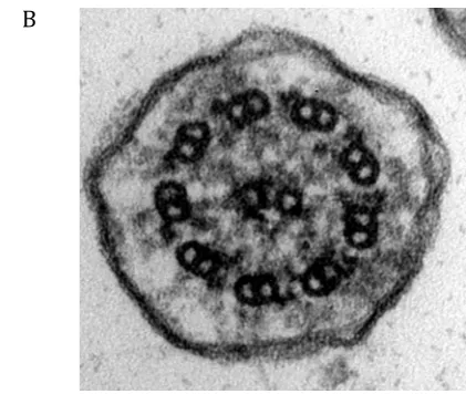
absent/abnormal outer dynein arms, compared to a normal cilium (Figure 1b). Subject 5’s normal electron microscopy (EM) appearances with dyskinetic ciliary movement are consistent with his known genetic mutation (DNAH11).
Audiometry
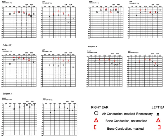
Hearing tests were air conduction, pure‐tone audiometry thresholds at 0.25, 0.5, 1, 2, 3, 4, 6 and 8 kHz. Subject 2 was not tested at 3 and 6 kHz. Headphones were used within a soundproof booth. An average from all thresholds was calculated for both ears.
Tympanometry
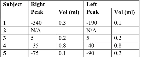
Tympanometry captured acoustic impedance from ‐400 to 200 daPa using a small tip inserted into the ear canal. Each ear was tested once during which participants were asked not to swallow or speak, yielding the peak pressure of tympanometric compliance and middle ear volume.
CervicalVestibularEvokedMyogenicPotential(cVEMP)
A custom system (Faldon & Buckwell) simultaneously delivered air‐conducted sound monaurally via audiological headphones and captured electromyographic (EMG) activity. Sound was delivered as 500Hz clicks at 110dB nHL (120dB SPL) of positive polarity, repeating 5 times per second. EMG signals were averaged over two hundred sweeps and rectified. Prior to electrode (Ag/AgCl, W‐60, Skintact UK) adhesion, skin was cleaned (NuPrep, Weaver Company, USA) and electrodes gelled (Spectra 360, Parker, USA). Resistance across electrodes was <5KΩ. The electrode montage comprised the right and left sternocleidomastoid (SCM) muscle bellies, the right and left ipsilateral mid‐clavical bones (reference) and sternum (ground).
Subjects lay on an examination bed, upper torso elevated with the backrest at 30° and lower body flat, perpendicular to the ground. For testing, they were asked to elevate their heads from the head rest and turn either to the left (right ear test) or to the right (left ear test) to increase tonic neck muscle activity. A biofeedback device (NeuroTrac Simplex unit, NeuroTrac, UK) regulated contractional strength (approximately 30 µV). The p13 was taken as the first distinctive peak in waveform and n23 as the first trough. VEMPs were scored in terms of presence or absence, p13 and n23 latency and
percentage amplitude asymmetry.
UtricularCentrifugation(UCF)
A full explanation of the protocol and theory for utricular centrifugation (UCF) has been published elsewhere [12‐14]. In brief, utricular function was measured by the subjective visual vertical (SVV) during unilateral centrifugation [15]. Testing was performed with a vertical axis rotation chair (Neurokinetics Inc, Pittsburugh, USA) capable of lateral chair translation for eccentric displacement, in total darkness and with the subject’s head, torso, legs and feet secured into the chair. Vertical head alignment was ensured with a vertically calibrated laser. Attached to the chair were two arms holding a thin wooden plaque 1m from participants, onto which a laser projected an illuminated red dot or line (160mm x 3mm). The line could be rotated about its centre using a wheel joy‐pad given to subjects who were instructed to position the line to their gravitational vertical. Upon doing so, the patient was asked to give a vocal cue that the line was vertical. The line angle was saved, and the line re‐set to a new angle. This was repeated over a 30 second period until the chair changed translation or after 7 SVV measurements. SVV was measured in the stationary position, during rotation centred on axis and during the left and right translations.
The UCF protocol was:
1. Chair stationary. SVV measured 7 times.
2. Chair accelerated at 3º/s2 up to a constant velocity of 400º/s over 120s and
maintained in the on‐axis position for 60s.
3. Translation from centre to the right laterally (4cm, 0.2cm/s) over 30s before SVV was measured for 30s.
4. Translation back to centre over 30s, and SVV measured for 30s.
5. Translation from centre to the left laterally (4cm, 0.2cm/s) over 30s, and SVV measured for 30s.
6. Translation back to the central position for 30s and decelerated at 2.5º/s to rest.
During earth‐centred centrifugation, displacing the head by 4 cm from the rotation axis stimulates the eccentrically positioned utricle unilaterally through the resultant
centrifugal force. The resulting utriculo‐ocular reflex (UOR) causes ocular counter‐roll in an opposite direction to the translation direction. At 400º/s the stimulated utricle is exposed to a centrifugal acceleration of ω2r=0.40 g, where ω is the angular velocity and r
sensation of 11.2° [14]. UCF was scored as median SVV angle and gain in each position and utricular percentage weakness. The normal range for static SVV is about 2° [13,16].
Electronystagmography(ENG)
Semi‐circular canal function was assessed by 90°/s step rotations (right‐ and leftward) in terms of gain (ratio of eye velocity to head velocity) and time constant (time taken for eye movement velocity to decay to 36.8% of the maximum velocity). The time constant is a technique based on eye movement response to an acceleration impulse generated by a cessation of rotational chair power following an epoch of constant velocity. The
vestibulo‐ocular reflex was also measured by sinusoidal rotations at 0.25Hz. Eye movements were captured with three small adhesive electrodes (Carefusion, Ag/AgCl) stuck to the outer canthi of both eyes and forehead.
Testing was performed in the same rotation chair as the UCF (Neurokinetics Inc, Pittsburgh, USA) and again in complete darkness. No visual cues were present.
Subjectivebalancefunctionandsymptoms
The Vertigo Symptom Scale‐short form (VSS) and Dizziness Handicap Inventory [17], both validated questionnaires, were completed by all subjects.
Results
Some subjects showed evidence of vestibular abnormality (Table 2).
Pure‐ToneAudiometry
As seen in Figure 2, a unilateral mild/moderate conductive hearing loss was present in 4 of the 5 subjects. The remaining subject (subject 5) had mild bilateral conductive
hearing loss.
Tympanometry
Table 3 shows that the stapedial reflex was reduced though not entirely eliminated. Tympanometry was available for all apart from subject 2. Otoscopic examination of this subject was unremarkable.
UCF weakness = 200 x Median L + Median C + Median R -3 x Median Static 3 x (Median L – Median R)
VestibularEvokedMyogenicPotentials
There were markedly reduced or unobtainable cVEMPs bilaterally in 3 of the 5 subjects (1, 2, and 4) and unilaterally in the remaining 2 subjects (Figure 3). The amplitude asymmetry in subject 3 was significant at 71.9%, but not in subject 5 (8.0%), probably owing to a small VEMP amplitude.
UtricularCentrifugation
No subject had a pathological UCF asymmetry (i.e. >100% weakness). Tilt perception was reduced unilaterally in subject 3 during left chair translation, as shown in Figure 4 by the closeness of the median tilt response to the dashed baseline mark. Subjects 1 and 4 showed small SVV responses bilaterally, represented by small gain values in table 2.
Electronystagmography
The vestibulo‐ocular reflex (VOR) at 0.25Hz sinusoidal rotation was normal in all subjects. The rotational nystagmic response of subject 3 was processed differently. Nystagmic decay in this participant was normal in both rotational directions. Rotation to the right yielded a time constant of 9s and to the left, 10s. Rotational time constants were borderline reduced bi‐directionally in subjects 1 and 4; both had unattainable VEMPs bilaterally. Subject 5 had a borderline reduced time constant on the right side, the same side as his unattainable VEMP. Rotational time constants were normal in subjects 2 and 3. Gains were typical.
Subjectivebalancefunctionandsymptoms
No evidence of vertigo or abnormal dizziness was found in any subject, as shown by entirely normal questionnaire scores.
Discussion
The goal of this study was to assess whether PCD, a congenital disorder of ciliary function, impairs otoconial organ function expressed through routine vestibular clinical tests. We show that saccular and utricular function is impaired in this small cohort of PCD patients. This finding suggests that ciliary structure and/or motility could
Congenital ciliary dysfunction in PCD commonly causes chronic respiratory infections and conductive hearing loss as result of otitis media. All subjects presented with a mild to moderate conductive hearing loss, but not the sensorineural loss that might be expected if motile cilia are required for normal development of the cochlea. A conductive hearing loss is frequently seen in PCD, commonly associated with otitis media with effusion [1]. In keeping with this, subject 3 had reduced tympanometric response and all except subject 4 had small middle ear volumes. Plausibly, hair cell function within the cochlea is preserved in PCD. We also observed an intact VOR in response to angular rotation (rVOR) in all 5 PCD subjects, confirming the presence of bilateral vestibular function. Upon rotational testing, however, the VOR time constant (which reflects vestibulo‐central processing [18] and is typically 13‐16s long [19]) was borderline reduced in subjects 1, 4 and 5 (Table 2), similar to that seen in vestibular disorders (such as acute vestibular neuritis [20]) and motion‐habituated professionals [21]. Perhaps the slight reduction in semicircular canal function points towards reciprocal inhibition to manage the otoconia‐semicircular canal mismatch [22,23].
Utricular function can be assessed by means of centrifugation (UCF) [23]. UCF has demonstrated utricular dysfunction in benign paroxysmal positional vertigo (BPPV) patients [24] and following intratympanic gentamicin therapy (causing ototoxic destruction of vestibular stereocilia and kinocilia) in 60‐70% of Meniere’s disease patients [25]. Subjects 1, 3 and 4 showed utricular abnormalities, subject 3 unilaterally on the left and subjects 1 and 4 with bilaterally reduced gain (below 0.3). In comparison, the gain in the 20 healthy subjects presented in Figure 4 was 0.7.
The cVEMP is an established test of saccular function [26]. Unobtainable or significantly reduced cVEMP responses were observed in all subjects. However, the conductive hearing loss present in these patients decreases cVEMP reliability [27] which may have affected our results. However, the conductive hearing loss seen in our patients was small or insignificant (Figure 2) which argues against unreliability. Still, bone‐conducted cVEMP as opposed to air‐conducted could provide better reliability [28].
Otoconial function, as measured by UCF (utricles) and VEMP (saccules), showed reduced function in this cohort of PCD patients compared to normal controls. The otoconial organs, arranged in the horizontal plane (utricle) and vertical plane (saccule), respond to linear acceleration and gravity, serving a functional role in the maintenance of
subjects experienced no obvious postural impairment. Recently, unilateral ablation of vestibular function in tadpoles, resulting in disequilibrium in descending vestibulospinal information, led to an altered assembly of adult spinal locomotor circuitry and normal swimming behaviour after metamorphosis, in contrast to frogs lesioned after
metamorphosis who never recovered [29]. These findings suggest adaptive
compensation for vestibular dysfunction in the developmental stages. Similarly, subjects with chronic congenital nystagmus or chronic progressive external ophthalmoplegia are typically symptom free [30].
Limitations
A major factor worthy of discussion is our rotational protocol. We have chosen to use a step rotation at 90°/s to maintain consistency with our previous studies [20,31] and others of the velocity storage mechanism [32]. One measure of the velocity storage system is the vestibular velocity storage mechanism. We have chosen this measure because the time constant can reflect vestibular impairment by disease [20,33],
intervention [31] and drug effects [21,34‐36]. However, it should be pointed out that the time constant can habituate which lends itself to poor repeatability. The tests were only performed once for rightward and leftward rotations to prevent a habituation effect. Furthermore, sinusoidal rotation at 0.25Hz is limited compared to the range of
frequencies available where the phase could produce a more reliable measure of canal dysfunction than gain. Importantly though, we showed larger human abnormalities in otoconial function than semicircular canal function in PCD, which is consistent with previous theories on ciliary motility and otolith seeding, positioning [9] in zebrafish.
Conclusion
References
1. Rimmer J. Congenital problems of mucociliary clearance: primary ciliary
dyskinesia. Rhinology 2012;50:353‐9.
2. Roy S. The motile cilium in development and disease: emerging new insights.
Bioessays 2009;31:694‐9.
3. Badano JL, Mitsuma N, Beales PL, Katsanis N. The ciliopathies: An emerging class
of human genetic disorders. AnnuRevGenomHumGenet 2006;7:125‐8.
4. Castleman VH, Romio L, Chodhari R, et al. Mutations in radial spoke head protein
genes RSPH9 and RSPH4A cause primary ciliary dyskinesia with central‐
microtubular‐pair abnormalities. AmJHumGenet 2009;84:197‐209.
5. Anniko M. Cytodifferentiation of cochlear hair cells. AmJOtolaryngol
1983;4:375‐88.
6. Bisgrove BW, Yost HJ. The roles of cilia in developmental disorders and disease.
Development 2006;133:4131‐43.
7. Colantonio JR, Vermot J, Wu D, et al. The dynein regulatory complex is required
for ciliary motility and otolith biogenesis in the inner ear. Nature 2009;457:205‐
9.
8. Riley BB, Zhu C, Janetopoulos C, Aufderheide KJ. A critical period of ear
development controlled by distinct populations of ciliated cells in the zebrafish.
DevBiol 1997;191:191‐201.
9. Stooke‐Vaughan GA, Huang P, Hammond KL, Schier AF, Whitfield TT. The role of
hair cells, cilia and ciliary motility in otolith formation in the zebrafish otic
vesicle. Development 2012;139:1777‐87.
10. Wirschell M, Olbrich H, Werner C, et al. The nexin‐dynein regulatory complex
subunit DRC1 is essential for motile cilia function in algae and humans. Nat
11. Wu D, Freund JB, Fraser SE, Vermot J. Mechanistic basis of otolith formation
during teleost inner ear development. DevCell 2011;20:271‐8.
12. Clarke AH, Schonfeld U, Helling K. Unilateral examination of utricle and saccule
function. JVestibRes 2003;13:215‐25.
13. Akin FW, Murnane OD, Pearson A, Byrd S, Kelly KJ. Normative data for the
subjective visual vertical test during centrifugation. JAmAcadAudiol
2011;22:460‐8.
14. Weerts AP, De Meyer G, Pauwels G, et al. Pharmaceutical countermeasures have
opposite effects on the utricles and semicircular canals in man. AudiolNeurootol
2012;17:235‐42.
15. Janky KL, Shepard NT. Unilateral centrifugation: utricular assessment and
protocol comparison. OtolNeurotol 2011;32:116‐21.
16. Janssen M, Lauvenberg M, van der Ven W, Bloebaum T, Kingma H. Perception
threshold for tilt. OtolNeurotol 2011;32:818‐25.
17. Morris AE, Lutman ME, Yardley L. Measuring outcome from vestibular
rehabilitation, part II: refinement and validation of a new self‐report measure.
IntJAudiol 2009;48:24‐37.
18. Ramat S, Bertolini G. Estimating the time constants of the rVOR. A model‐based
study. AnnNYAcadSci 2009;1164:140‐6.
19. Raphan T, Matsuo V, Cohen B. Velocity storage in the vestibulo‐ocular reflex arc
(VOR). ExpBrainRes 1979;35:229‐48.
20. Cousins S, Kaski D, Cutfield N, et al. Vestibular perception following acute
unilateral vestibular lesions. PLoSOne 2013;8:e61862.
21. Cohen B, Dai M, Raphan T. The critical role of velocity storage in production of
22. Deutschlander A, Bense S, Stephan T, Schwaiger M, Brandt T, Dieterich M.
Sensory system interactions during simultaneous vestibular and visual
stimulation in PET. HumBrainMapp 2002;16:92‐103.
23. Schonfeld U, Clarke AH. A clinical study of the subjective visual vertical during
unilateral centrifugation and static tilt. ActaOtolaryngol 2011;131:1040‐50.
24. von Brevern M, Schmidt T, Schonfeld U, Lempert T, Clarke AH. Utricular
dysfunction in patients with benign paroxysmal positional vertigo. OtolNeurotol
2006;27:92‐6.
25. Helling K, Schonfeld U, Clarke AH. Treatment of Meniere's disease by low‐dosage
intratympanic gentamicin application: effect on otolith function. Laryngoscope
2007;117:2244‐50.
26. Colebatch JG, Halmagyi GM, Skuse NF. Myogenic potentials generated by a click‐
evoked vestibulocollic reflex. JNeurolNeurosurgPsychiatry 1994;57:190‐7.
27. Zhou G, Poe D, Gopen Q. Clinical use of vestibular evoked myogenic potentials in
the evaluation of patients with air‐bone gaps. OtolNeurotol 2012;33:1368‐74.
28. Yang TL, Young YH. Comparison of tone burst and tapping evocation of myogenic
potentials in patients with chronic otitis media. EarHear 2003;24:191‐4.
29. Beyeler A, Rao G, Ladepeche L, Jacques A, Simmers J, Le Ray D. Vestibular lesion‐
induced developmental plasticity in spinal locomotor networks during Xenopus
laevis metamorphosis. PLoSOne 2013;8:e71013.
30. Seemungal BM, Masaoutis P, Green DA, Plant GT, Bronstein AM. Symptomatic
recovery in Miller Fisher syndrome parallels vestibular‐perceptual and not
vestibular‐ocular reflex function. FrontNeurol 2011;2:2.
31. Arshad Q, Nigmatullina Y, Bronstein AM. Handedness‐related cortical
32. Bertolini G, Ramat S, Laurens J, et al. Velocity storage contribution to vestibular
self‐motion perception in healthy human subjects. JNeurophysiol 2011;105:209‐
23.
33. Okada T, Grunfeld E, Shallo‐Hoffmann J, Bronstein AM. Vestibular perception of
angular velocity in normal subjects and in patients with congenital nystagmus.
Brain 1999;122:1293‐303.
34. Cohen B, Dai M, Yakushin SB, Raphan T. Baclofen, motion sickness susceptibility
and the neural basis for velocity storage. ProgBrainRes 2008;171:543‐53.
35. Shaikh AG, Marti S, Tarnutzer AA, et al. Effects of 4‐aminopyridine on nystagmus
and vestibulo‐ocular reflex in ataxia‐telangiectasia. JNeurol 2013;260:2728‐35.
36. Shaikh AG, Palla A, Marti S, et al. Role of cerebellum in motion perception and
Figures
[image:13.595.344.555.96.274.2]A B
Figure 1a:Electron micrograph showing transverse section of a cilium lacking outer
dynein arms (absence indicated by red arrows); where present outer dynein arms
appear shortened (x 100,000) Figure 1b: Electron micrograph showing transverse
Figure 2: Pure‐tone audiograms from the five PCD subjects
RIGHT
FREQUENCY (Hz) LEFTFREQUENCY (Hz)
-10 0 10 20 30 40 50 60 70 80 90 100 110 120 130 140
125 250 500 1000 2000
3000 4000 6000 8000 -10 0 10 20 30 40 50 60 70 80 90 100 110 120 130 140
125 250 500 1000 2000
3000 4000
6000 8000
x x x x
x x x
x
Subject 1
RIGHT
FREQUENCY (Hz) LEFTFREQUENCY (Hz)
-10 0 10 20 30 40 50 60 70 80 90 100 110 120 130 140 -10 0 10 20 30 40 50 60 70 80 90 100 110 120 130 140
125 250 500 1000 2000
3000 4000
6000 8000
125 250 500 1000 2000
3000 4000 6000 8000 -10 0 10 20 30 40 50 60 70 80 90 100 110 120 130 140 -10 0 10 20 30 40 50 60 70 80 90 100 110 120 130 140
125 250 500 1000 2000
3000 4000
6000 8000
125 250 500 1000 2000
3000 4000
6000 8000
x x x x
x x x
x
Subject 1
RIGHT
FREQUENCY (Hz) LEFTFREQUENCY (Hz)
-10 0 10 20 30 40 50 60 70 80 90 100 110 120 130 140
125 250 500 1000 2000
3000 4000 6000 8000 -10 0 10 20 30 40 50 60 70 80 90 100 110 120 130 140
125 250 500 1000 2000
3000 4000
6000 8000
x x x x x
x
Subject 2
RIGHT
FREQUENCY (Hz) LEFTFREQUENCY (Hz)
-10 0 10 20 30 40 50 60 70 80 90 100 110 120 130 140 -10 0 10 20 30 40 50 60 70 80 90 100 110 120 130 140
125 250 500 1000 2000
3000 4000
6000 8000
125 250 500 1000 2000
3000 4000 6000 8000 -10 0 10 20 30 40 50 60 70 80 90 100 110 120 130 140 -10 0 10 20 30 40 50 60 70 80 90 100 110 120 130 140
125 250 500 1000 2000
3000 4000
6000 8000
125 250 500 1000 2000
3000 4000
6000 8000
x x x x x
x
Subject 2
RIGHT
FREQUENCY (Hz) LEFTFREQUENCY (Hz)
-10 0 10 20 30 40 50 60 70 80 90 100 110 120 130 140
125 250 500 1000 2000
3000 4000 6000 8000 -10 0 10 20 30 40 50 60 70 80 90 100 110 120 130 140
125 250 500 1000 2000
3000 4000
6000 8000
x x
x x x
x x x
Subject 3
RIGHT
FREQUENCY (Hz) LEFTFREQUENCY (Hz)
-10 0 10 20 30 40 50 60 70 80 90 100 110 120 130 140 -10 0 10 20 30 40 50 60 70 80 90 100 110 120 130 140
125 250 500 1000 2000
3000 4000
6000 8000
125 250 500 1000 2000
3000 4000 6000 8000 -10 0 10 20 30 40 50 60 70 80 90 100 110 120 130 140 -10 0 10 20 30 40 50 60 70 80 90 100 110 120 130 140
125 250 500 1000 2000
3000 4000
6000 8000
125 250 500 1000 2000
3000 4000
6000 8000
x x
x x x
x x x
Subject 3
RIGHT
FREQUENCY (Hz) LEFTFREQUENCY (Hz)
-10 0 10 20 30 40 50 60 70 80 90 100 110 120 130 140
125 250 500 1000 2000
3000 4000 6000 8000 -10 0 10 20 30 40 50 60 70 80 90 100 110 120 130 140
125 250 500 1000 2000
3000 4000
6000 8000
x
x x x
x x x x Subject 4 RIGHT
FREQUENCY (Hz) LEFTFREQUENCY (Hz)
-10 0 10 20 30 40 50 60 70 80 90 100 110 120 130 140 -10 0 10 20 30 40 50 60 70 80 90 100 110 120 130 140
125 250 500 1000 2000
3000 4000
6000 8000
125 250 500 1000 2000
3000 4000 6000 8000 -10 0 10 20 30 40 50 60 70 80 90 100 110 120 130 140 -10 0 10 20 30 40 50 60 70 80 90 100 110 120 130 140
125 250 500 1000 2000
3000 4000
6000 8000
125 250 500 1000 2000
3000 4000
6000 8000
x
x x x
x x x x Subject 4 RIGHT
FREQUENCY (Hz) LEFTFREQUENCY (Hz)
-10 0 10 20 30 40 50 60 70 80 90 100 110 120 130 140
125 250 500 1000 2000
3000 4000 6000 8000 -10 0 10 20 30 40 50 60 70 80 90 100 110 120 130 140
125 250 500 1000 2000
3000 4000
6000 8000
x x x x x
x x x
Subject 5
RIGHT
FREQUENCY (Hz) LEFTFREQUENCY (Hz)
-10 0 10 20 30 40 50 60 70 80 90 100 110 120 130 140 -10 0 10 20 30 40 50 60 70 80 90 100 110 120 130 140
125 250 500 1000 2000
3000 4000
6000 8000
125 250 500 1000 2000
3000 4000 6000 8000 -10 0 10 20 30 40 50 60 70 80 90 100 110 120 130 140 -10 0 10 20 30 40 50 60 70 80 90 100 110 120 130 140
125 250 500 1000 2000
3000 4000
6000 8000
125 250 500 1000 2000
3000 4000
6000 8000
x x x x x
x x x
Subject 5
x
RIGHT EAR LEFT EAR
Air Conduction, masked if necessary
Bone Conduction, not masked
Bone Conduction, masked
x
RIGHT EAR LEFT EAR
Air Conduction, masked if necessary
Bone Conduction, not masked
Figure 3:VEMPs of the 5 PCD subjects and of a healthy normal subject. Reductions in
VEMP responses are clearly visible. Only the right VEMP in subject 3 is in keeping with a
normal response.
RIGHT LEFT
Normal
Subject 4
Subject 5 Subject 3 Subject 2 Subject 1
Figure 4:Median UCF SVV responses (degrees) to stationary (S), centre centrifugation
and right and left chair translations in all five PCD subjects and a healthy group (20
control subjects, minimum‐maximum range given by the bar and mean (circle)). The
dashed line indicates the subject’s static vertical perception. Subject 3 showed evidence
of reduced tilt perception on left chair translation.
Centre Right Left S
Centre Right Left S
Centre Right Left S Subject 1 -5 -4 -3 -2 -1 0 1 2 3 4 5 Subject 2 -6 -4 -2 0 2 4 6 8 Subject 3 -40 -35 -30 -25 -20 -15 -10 -5 0 5 10
Centre Right Left S
Centre Right Left S Subject 1 -5 -4 -3 -2 -1 0 1 2 3 4 5 Subject 2 -6 -4 -2 0 2 4 6 8 Subject 3 -40 -35 -30 -25 -20 -15 -10 -5 0 5 10 Subject 4 -6 -5 -4 -3 -2 -1 0 1 2 3 Subject 5 -10 -5 0 5 10 15 Healthy group 10 20 0 -10 -20
Centre Right Left S Centre Right Left S
Centre Right Left S Centre Right Left S
Centre Right Left S Centre Right Left S
Centre Right Left S
Centre Right Left S Centre Right Left S
Tables
Patient Age Ciliary function (LM) Defect (EM) Symptoms Situs
1
28
Unknown
Unknown
URT
LRT
Normal
2
28
Static
ODA
URT
LRT
Normal
3
29
Unknown
Unknown
URT
LRT
Inversus
4
18
Static
ODA
URT
LRT
Normal
5
18
Dyskinetic with
reduced beat frequency
Normal EM URT
LRT
Inversus
Table 1:
Patient details (LM=light microscopy, EM=electron microscopy,
ODA=outer dynein arm, URT=upper respiratory tract, LRT=lower respiratory tract)
Table 2:
Pure tone thresholds (averaged across all frequencies),
VEMP, UCF and
ENG results in all participants. (A=absent, P=present, L=left, R=right,
Asym=asymmetry, TC=time constant, N/A=not available)
Subject PTA (dB) VEMP
UCF
ENG
R
L
R
L
R
L
Gain Asym
Gain TC Gain TC
1
24.4 42.5
A
A
0.36 6 – L
0.76 5
0.67 5.2
2
11.6 36.7
A
A
0.49 7 – L
0.45 14.2 0.4
10.7
3
1.25 16.9
P
A
1.64 81 – L
N/A 9
N/A 10
4
9.4 20
A
A
0.30 23 – L
0.55 5.9 0.49 3.6
[image:17.595.84.515.464.581.2]Table 3:
Tympanometric results from the five PCD subjects
Subject Right
Left
Peak
Vol (ml) Peak
Vol (ml)
1
-340
0.3
-190
0.1
2
N/A
N/A
3
5
0.2
5
0.2
4
-35
0.8
-40
0.8