Biology Dissertations Department of Biology
8-31-2008
Regulation of Connexin40 Gap Junctions
Thomas Vinaya SheelaREGULATION OF CONNEXIN40 GAP JUNCTIONS
by
THOMAS V. SHEELA
Under the Direction of Charles F. Louis
ABSTRACT
Gap junctions provide direct electrical and biochemical communication between
cardiomyocytes in the heart. Connexin40 (Cx40) is the major connexin in the atria of the
heart and little is known regarding its regulation. Thus, the goal was to investigate the
regulation of Cx40 in both physiological and pathophysiological conditions. The first
objective of this thesis was to determine whether Cx40 gap junctions were regulated by
β-adrenergic receptor activation. Cx40 has previously been shown to be acutely activated
by cAMP, this cAMP-induced increase in Cx40-mediated cell-to-cell dye transfer has
been shown to be effected through the β-adrenergic receptor-adenylyl cyclase- Protein
Kinase A (PKA) pathway in Cx40-transfected HeLa cells. The second objective of this
thesis was to determine whether Cx40 gap junctions were regulated by intracellular Ca2+
concentration ([Ca2+]i ). [Ca2+]i was increased by addition of the ionophore ionomycin
and elevating extracellular calcium [Ca2+]o from 1.8 mM to 21.8 mM. This resulted in an
elevation of [Ca2+]i and effected an inhibition of Cx40-mediated cell-to-cell dye transfer
Cx40-mediated cell-to-cell dye transfer that was shown to be Calmodulin dependent.
The main conclusions of this thesis were: (1) β-adrenergic receptor activation increases
Cx40-mediated cell-to-cell dye transfer which requires the activation of PKA; (2) A
sustained elevation in [Ca2+]i causes a partial inhibition of Cx40 gap junction-mediated
cell-to-cell dye transfer which was Ca2+-and Calmodulin dependent; (3) Chemical
ischemia causes a partial inhibition of Cx40 gap junction-mediated cell-to-cell dye
transfer which was shown to be Calmodulin-dependent.
Index words: Connexin40, Atria, β-adrenergic receptor, Intracellular calcium, Chemical
REGULATION OF CONNEXIN40 GAP JUNCTIONS
by
THOMAS V. SHEELA
A Thesis Submitted in Partial Fulfillment of the Requirements for the Degree of
Doctor of Philosophy
In the College of Arts and Sciences
Georgia State University
REGULATION OF CONNEXIN40 GAP JUNCTIONS by
THOMAS V. SHEELA
Major Professor: Charles F. Louis Committee: Delon Barfuss Vincent Rehder Jenny Yang
Electronic Version Approved:
ACKNOWLEDGEMENT
I would like to express my deepest appreciation to many people, especially my advisor, Dr. Charles Louis, who read my numerous revisions and helped make some
sense of it. Also thanks to my committee members, Dr. Delon Barfuss, Dr. Vincent
Rehder and Dr. Jenny Yang who offered guidance and support. Thanks to American
Heart Association for awarding me a Pre-doctoral fellowship. I wish to specially thank
my spouse for unwavering support and understanding during themany hours I dedicated
to achieving this milestone in my life and career. And finally, thanks to my parents, and
numerous friends who endured this long process with me, always offering support and
Table of Contents Chapter One: Introduction
List of Tables………... vi
List of Figures……….. vii
Cell-cell junctions……… 2
Tight junctions……… 2
Adherens junctions……… 3
Gap junctions……… 4
Connexins……… 6
Structure……… 6
Formation……… 7
Metabolism……… 9
Function……… 10
Gap junctions in diseases………. 12
Cardiac gap junctions……… 15
Cardiac morphology and function……… 15
Connexin expression and function in the heart……… 16
Gap junction mutations in cardiac diseases……… 19
Significance of Connexin40 in the heart……… 21
Thesis objectives……… 24
References……… 29
Figures……… 38
Chapter Two: Beta-adrenergic regulation of Cx40 gap junctions Introduction……… 44 Methods……… 48 Results……… 52 Discussion……… 67 References……… 75 Figures……… 80
Chapter Three: Regulation of Cx40 gap junctions by Intracellular Calcium Introduction……… 121
Methods……… 124
Chapter Four: Regulation of Cx40 gap junctions during Ischemia
Introduction……… 162
Methods……… 164
Results……… 167
Discussion……… 172
References……… 176
Figures……… 178
Chapter Five: Regulation of Connexin43 hemichannels Introduction……… 193
Methods……… 196
Results……… 198
Discussion……… 205
References……… 209
Figures……… 212
Chapter Six: Conclusions and Future Research Conclusions and Future Research…..……… 232
References……… 242
List of Tables
2.1. Several putative Cx40 consensus phosphorylation sequences ……… 81 2.2. Several receptors, receptor ligands, activators and inhibitors of various
1.1. A model of cell-cell junction……… 39 1.2. A model of gap junction proteins with all the protein……… 41
Chapter 2
2.1. Expression of Cx40 in HeLa cells transfected with rat Cx40………… 85 2.2. Cell-to-cell dye transfer in Cx40-transfected HeLa cells ……… 88 2.3. Cell-to-cell transfer of Alexa Fluor 594 dye in Cx40-transfected HeLa cells……….
90
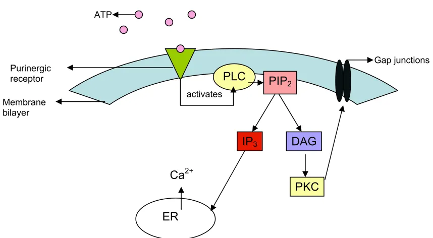
2.4. Cx40 amino acid sequence with the putative transmembrane regions… 92 2.5. Schematic diagram of thePurinergic receptor pathway……… 94 2.6. Effect of the purinergic receptor agonist ATP on Cx43 mediated
cell-to-cell dye transfer……… 96
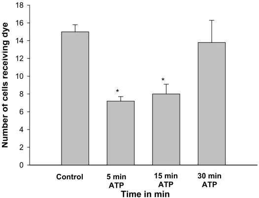
2.7. Effect of the purinergic receptor agonist ATP on Cx40 mediated cell-to-cell dye transfer ………
98
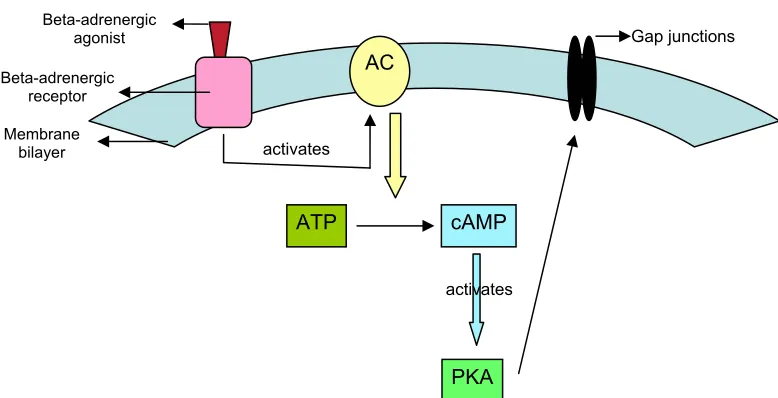
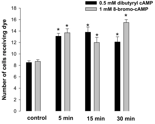
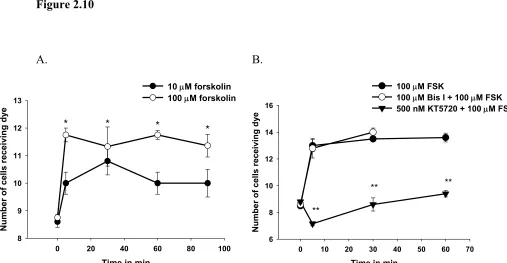
2.8. Schematic diagram of theβ-adrenergic receptor pathway ……… 100 2.9. Effect of cAMP analogs on Cx40 mediated cell-to-cell dye transfer… 102 2.10.Effect of forskolin on Cx40 mediated cell-to-cell dye transfer……… 104 2.11. Effect of the α-adrenergic agonist metaraminol on Cx40 mediated
cell-to-cell dye transfer ……… 106
2.12. Effect of the β-adrenergic agonist isoproterenol on Cx40 mediated cell-to-cell dye transfer ………..
108
2.13. Western blots of β-adrenergic receptor subtypes in untransfected or Cx40-transfected HeLa cells ………
110
2.14. Effect of the the β-1 adrenergic agonist dobutamine on Cx40
mediated cell-to-cell dye transfer ……… 112
2.15. Effect of the β-2 adrenergic receptor agonist salbutamol on Cx40 mediated cell-to-cell dye transfer ………
114
2.16. Effect of the β-3 adrenergic agonist BRL 37344 on Cx40 mediated
cell-to-cell dye transfer……… 116
2.17. Putative consensus phosphorylation sequences in Cx45 ……… 118
3.6. Cx40 amino acid sequence with the possible Calmodulin binding region……….
155
3.7. Effect of a Calmodulin inhibitor on the Ca2+-dependent inhibition of Cx40-mediated cell-to-cell dye transfer ………
157
3.8. Effect of a transient increase in [Ca2+]i on Cx40-mediated cell-to-cell
dye transfer ……… 159
Chapter 4
4.1. Effect of chemical ischemia on intracellular ATP concentration in Cx40-transfected HeLa cells ………
179
4.2. Effect of chemical ischemia on Cx40-mediated cell-to-cell dye transfer
at 37oC ……… 181
4.3. Effect of temperature on Fura-2 fluorescence……… 183 4.4. Effect of chemical ischemia on [Ca2+]i in Cx40-transfected HeLa cells
at 20οC……… 185
4.5. Effect of chemical ischemia on Cx40-mediated cell-to-cell dye transfer at 20ºC………
187
4.6. Effect of the Calmodulin inhibitor calmidazolium on the chemical ischemia-dependent decrease in Cx40-mediated cell-to-cell dye transfer at
37oC………
189
Chapter 5
5.1. Expression of Cx43 in HeLa cells transfected with Cx43. Western
Immunoblot of Cx43……….. 213
5.2. Effect of reduced extracellular [Ca2+] on Cx43 hemichannel-mediated dye uptake………
215
5.3. Effect of dye concentration on Cx43 hemichannel-mediated dye uptake………
217
5.4. Effect of time of incubation with dye on Cx43 hemichannel-mediated dye uptake………
219
5.5. Effect of temperature on Cx43 hemichannel-mediated dye uptake…… 221 5.6. Effect of ATP on Cx43 hemichannel-mediated dye uptake……… 223 5.7. Effect of a PKC inhibitor on the ATP-dependent inhibition of Cx43
hemichannels……….. 225
5.8. Effect of reduced [Ca2+]o on Cx43 hemichannel-mediated dye uptake. 227
5.9. Effect of reduced extracellular [Ca2+] on hemichannel-mediated dye
CHAPTER 1
Cell-cell junctions
In the process of building a multicellular organism, cells are organized to form
tissues that adhere to each other by specialized structures or junctions which are critical
for the development and function of these tissues. Through development, and also as
tissues are maintained in a mature organism, cells express informational and junctional
molecules on their surface, so that cells recognize and adhere to like cells. In mammalian
cells these cell-to-cell junctions are classified into three types of junctions: tight
junctions, adherens junctions and gap junctions.
Tight junctions
Epithelial and endothelial cells have junctional complexes on their apical domains
that form seals between adjacent cells called tight junctions. These are important in
tissues such as the bladder, intestines and glands where they form a barrier separating two
fluid compartments and allowing controlled transport of ions and solutes between these
compartments through the paracellular pathway. Freeze fracture studies show that tight
junctions form a network of parallel strands between the lateral faces of two opposing
cells called zonula occludens (Staehelin, 1973).
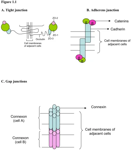
Tight junctions are composed of membrane proteins that include claudins,
occludins, zonula occludens-1 (ZO-1), ZO-2 and ZO-3. The extracellular loops of
kDa (Furuse et al., 1993). They contain four transmembrane domains with two
extracellular loops and one intracellular loop, with the amino and carboxy termini in the
cytoplasm. Like occludins, claudins are integral membrane proteins with four
transmembrane domains, two extracellular loops, one intracellular loop, and their N- and
C-termini in the cytoplasm (Furuse et al., 1998). There are 24 members in the claudin
family with molecular masses ranging from 20 – 27 kDa (Gonzalez-Mariscal et al.,
2003). Zonula occludens are peripheral membrane proteins in the vicinity of tight
junctions in the plasma membrane. They bind to the C-termini of occludins and claudins
forming an interface between these proteins and the F-actin filaments (Anderson and
Itallie, 1995). There are three types of zonula occluding proteins ZO-1, ZO-2 and ZO-3
with molecular masses of 200, 160 and 100 kDa respectively.
Adherens junctions
Adherens junctions are protein complexes found at cell-cell junctions of epithelial
cells; their main function is to anchor cells by connecting the actin cytoskeleton of
neighboring cells through direct interaction; they are also responsible for contact
inhibition of cell growth and cell signaling. They can appear as bands encircling the
apical pole of epithelial cells called zonula adherens or as spots of attachment to the
binding sites are located, a single transmembrane domain, and a relatively short
cytoplasmic C-terminal tail (two domains) (Fig.1.1B). Cadherins are classified as type I
and type II; Type I includes N- (Neural), E- (Epithelial), P-(placental) and R
(Retinal)-cadherins to name a few depending on the tissue they are expressed in; type II comprises
cadherin-5 through -12. Cadherins are anchored to the microfilament network of the
cytoskeleton via a complex composed of multiple intracellular proteins including α- and
β-catenin. β-catenin binds directly to the carboxy-terminal cytoplasmic domain of E-cadherin (Nagafuchi and Takeichi, 1988; Nagafuchi and Takeichi, 1989; Ozawa et al.,
1990).
Gap junctions
In tissues, cells are connected to each other by means of intercellular channels
called gap junctions. Gap junctions connect the cytoplasms of neighboring cells allowing
the passage of ions (electrical coupling) and small molecules (biochemical coupling)
between them. Gap junction proteins are classified into three classes: innexins,
pannexins and connexins.
Innexins
In invertebrates, cell-to-cell communication occurs via gap junction proteins
called innexins (Phelan et al., 1998a). Innexins associate to form hexameric subunits
forming a hemichannel in the plasma membrane with hemichannels from adjacent cells
docking to form intercellular channels. Each innexin subunit is comprised of four
Shak-B from Drosophila (Phelan et al., 1998b) and Inx-3 from C.elegans (Landesman et
al., 1999). Innexins are known to form homotypic junctions (both hemichannels
composed of a single type of innexin), heterotypic junctions (each hemichannel is
composed of a unique but different innexin) (Jacobs et al., 2000) and heteromeric
junctions (both hemichannels are composed of mixtures of different innexins) (Stebbings
et al., 2000). Innexins have been shown to form functional intercellular channels that are
voltage (Landesman et al., 1999; Phelan et al., 1998b) and pH sensitive (Landesman et
al., 1999).
Pannexins
Recently, the human genome database was searched for orthologs of the innexin
gene family yielding three members of a new family of vertebrate gap junction proteins
termed pannexins. These proteins appear to be highly conserved in worms, mollusks,
insects and mammals (Panchin et al., 2000), and their sequences show significant
homology to the innexins. Like the innexins, pannexins contain four transmembrane
domains, two extracellular loops, and one intracellular loop with both the N- and
C-termini in the cytoplasm (Fig.1.2B). This suggests that in vertebrates, gap junction
proteins are classified into two families, the well studied connexins and much less studied
homomeric and heteromeric gap junctions (Bruzzone et al., 2003). Panx1 protein has a
molecular mass of ~ 48 kDa. Panx1 hemichannels have been shown to mediate the
release of ATP from erythrocytes during low oxygen conditions (Locovei et al., 2006).
The function of Panx 2 and 3 are not yet understood.
Connexins
Structure:
In mammals, gap junctions are composed of protein subunits called connexins
which form intercellular channels (Fig.1.1C). Connexin gap junctions are composed of
two hemichannels termed a connexon (Musil & Goodenough, 1993; Laird, 1996), with
each connexon hemichannel comprised of six connexin subunits forming a channel with a
central pore (Musil & Goodenough, 1993; Laird, 1996) (Fig.1.1C). Gap junctions formed
from two hemichannels each containing the same connexin protein are called homotypic
gap junctions, while those composed of hemichannels containing different connexins are
called heterotypic gap junctions. When both hemichannels in a gap junction are
composed of a single type of connexin it is called a homomeric gap junction, when
composed of different connexins it is called a heteromeric gap junction. Each connexin
consists of four transmembrane domains with their amino (N-) and carboxy (C-) termini
in the cytoplasm (Fig.1.2C). In most connexins the amino acid sequences in the two
extracellular loops are highly conserved while the sequences of the intracellular loops
vary. The major difference between the different connexins lies in the length and amino
that the center-to-center distance of a gap junction is ~ 9-10 nm, the central pore diameter
is 2 nm, a depth of 1 nm and width of 3.8 nm (Hoh et al., 1993; Lal et al., 1995; Revel
and Karnovsky, 1967). Twenty highly related human connexin genes have been
identified (Willecke et al., 2002) with their nomenclature based on their expected
molecular weight in kilodaltons, so that the major connexin (Cx), a protein of 43 kDa, is
referred to as Cx43. A problem with this system is that homologous proteins in different
species may have different molecular weights or different connexin proteins in different
species may have identical molecular weights. An alternative nomenclature to eliminate
this problem was developed based on connexin sequence homology (Kumar and Gilula,
1992) in which connexins are divided into 3 classes α, β and γ, with different members of each class being given different number eg., α1 for rodent Cx43 and β1 for rodent Cx32 etc.
Formation:
Gap junctions are formed when six connexin subunits oligomerize to form a
connexon, with a connexon from one cell pairing with a connexon of the neighboring cell
to form a gap junction channel. Large numbers of these gap junctions aggregate in the
plasma membrane to form gap junctional plaques. Like other membrane proteins,
secretory pathway, suggesting that gap junction formation occurrs via this pathway (Laird
et al., 1995). Studies in which connexins were synthesized using translation competent
cell lysates supplemented with pancreatic ER-derived microsomes have demonstrated
that connexins are co-translationally integrated into the ER membrane in a
signal-recognition particle-dependent manner (Falk et al., 1994; Zhang et al., 1996). The
organization of the connexin topology also occurs in the ER (Falk and Gilula, 1998). A
combination of protease protection assays and antibody binding studies on isolated liver
and heart gap junction membranes established that Cx32 and Cx43 pass through the
membrane four times forming two extracellular loops and a cytoplasmic loop with both
the amino and carboxy termini exposed to the cytoplasm (Milks et al., 1988).
Following their synthesis, connexins oligomerize to form connexons, with the
location of connexin oligomerization known to be connexin-type specific (Diez et al.,
1999; Sarma et al., 2001). Cx32 oligomerization is known to occur in the ER or the
ER/golgi intermediate compartment, Cx43 oligomerization occurs in the trans-golgi
network (Martin et al., 2001; Musil and Goodenough, 1993), and Cx26 oligomerization
occurs in the plasma membrane (integrated directly into the plasma membranes in a
posttranslational manner) (Evans, 2002). In heteromeric connexons (connexons
comprised of two different connexins) an oligomeric intermediate is observed, for
instance in Cx26/Cx32 heteromeric connexons where oligomerization is observed in a
Golgi membrane fraction (Diez et al., 1999).
et al., 1974) where they dock with a connexon from the neighboring cell to form a gap
junction.
Metabolism:
Connexins are short lived as their half-lives are known to be in the range of ~ 1 -
5 hrs (Fallon and Goodenough, 1981). Pulse chase studies have demonstrated that Cx43
has a half-life of ~ 1-2 hr (Beardslee et al., 1998; Laird DW, 1991). Gap junction
internalization occurs via structures called “annular gap junctions” (Jordan et al., 2001),
double membrane vesicles containing entire gap junctions. Degradation of annular gap
junctions is known to occur either in lysosomes or proteosomes. Indeed Rahman, Carlile
et al. (1993) have demonstrated the presence of Cx32 in lysosomes at levels comparable
to the Golgi apparatus in rat liver (Rahman et al., 1993). It has be demonstrated that
inhibiting lysosomal enzymes results in the intracellular accumulation of Cx43 in
BICR-M1Rk cells, suggesting that like Cx32, Cx43 gap junctions are also degraded in
lysosomes. Other studies have demonstrated that specific inhibitors of proteosomal
enzymes prolonged the half-life of Cx43 in BWEM cells (Laing and Beyer, 1995),
suggesting that Cx43 gap junctions can be degraded in either lysosomes or proteosomes.
The preference for one degradativepathway over the other may be cell type dependent
Function:
Gap junctions are regulated by various factors that include transjunctional
voltage, intracellular pH, phosphorylation, intracellular Ca2+, and more recently it has
been recognized that associated proteins may also play this role.
Transjunctional voltage: Gap junctions are known to be regulated by
transjunctional voltage and most gap junctions close when large voltages (~ 60-80 mV)
are applied. Different connexins exhibit voltage gating to different degrees, for example
Cx43, the major connexin ubiquitously expressed in mammals, has a half-maximal
inactivation of ± 60 mV (Moreno et al., 1994); Cx40 which is expressed in the atria,
conduction system and endothelium of the heart has a half maximal inactivation of ± 50
mV (Beblo et al., 1995); Cx45 which is expressed in the cardiac conduction system has a
half-maximal inactivation of ± 20 mV (Moreno et al., 1995).
Intracellular pH: Gap junctions are regulated by intracellular pH and in general
gap junctions close in response to intracellular acidosis. Different connexins have
variable degrees of sensitivity to intracellular acidification, for instance the pKa for Cx43
is 6.71±0.03 (Stergiopoulos et al., 1999), while the pKa for Cx50 is 7.17 ± 0.03 (more
basic than the pKa for Cx32) (Stergiopoulos et al., 1999). It is believed that for Cx43 the
regulatory region for pH gating is within the intracellular loop and carboxy-terminus
domains of the connexin described as “particle-receptor” or “ball and chain” model
(Homma et al., 1998).
trafficking, channel function, and turnover (Herve and Sarrouilhe, 2002). Connexins are
differentially regulated by phosphorylation. For example phosphorylation of Cx32 (Saez
et al., 1990) or Cx40 (Van-Rijen et al., 2000) by c-AMP analog, 8-bromo cAMP resulted
in an increase in gap junction permeability, whereas phosphorylation of Cx43 by PKC
resulted in a decrease in gap junction permeability (Lampe et al., 2000). Lin,
Warn-Cramer et al. (2001) have provided evidence for the interaction of Cx43 with the SH2 and
SH3 domains of v-src, which is associated with the phosphorylation of Cx43 at Y247 and
Y265 residues in the carboxyl terminal domain (Lin et al., 2001). This interaction of
Cx43 with v-src is known to disrupt gap junctional communication.
Intracellular Ca2+: Gap junctions are sensitive to Ca2+ and are known to close at
elevated intracellular [Ca2+] (~ micromolar concentrations) levels. Ca2+ does not appear
to directly bind to connexins but this Ca2+ effect on gap junction gating is mediated by
the Ca2+-binding protein Calmodulin (CaM) (Peracchia, 2004). It has been postulated
that CaM serves as a mediator for this Ca2+- mediated effect on Cx32 (Hertzberg and
Eldik, 1987) and Cx43. Evidence for the direct interaction of CaM with connexins was
provided by immunofluorescence studies, where CaM co-localized with Cx32 in HeLa
cells stably transfected with Cx32 (Peracchia et al., 2000) and with Cx37 and Cx43 in
tight junction proteins ZO-1(Toyofuku et al., 1998), cytoskeletal proteins such as the α
and β-tubulins (Giepmans et al., 2001), as well as the adherens junction proteins cadherin, β-catenin (Ai et al., 2000), and the occludins (Kojima et al., 2001). The C-terminus of Cx43 is known to interact with the second PDZ domain of ZO-1. This
interaction is speculated to have a potential role in the trafficking of Cx43 to the
intercalated discs of the heart, thus facilitating the formation of Cx43 gap junctions
(Toyofuku et al., 1998). GST binding assays and immunolocalization studies have shown
that Cx43 binds to α and β-tubulin, indicating that the connexins interact with the cytoskeletal proteins α and β-tubulin (Giepmans et al., 2001). Co-immunoprecipitation and immunofluorescence studies have provided evidence for the direct association of
Cx32 with the occludins and claudin-1 in cultured hepatocytes, indicating that connexins
directly associate with tight junction proteins (Kojima et al., 2001). Finally, there is
evidence that connexins interact with signaling proteins such as src (Lin et al., 2001).
Gap junctions in diseases
Gap junction-mediated communication is important for various cellular functions
such as regulation of cell growth, differentiation, and development. Gap junctions effect
multiple cellular functions including the intercellular movement of ions, metabolites,
transmitting signals such as second messengers between cells to regulate cell growth and
development, the flow of current between cells to maintain electrical communication,
spectrum of human diseases, such as in demyelinating neuropathies/peripheral
neuropathy, various skin disorders, cataracts, deafness, oculodentodigital dysplasia
(ODDD) and cardiac diseases to name a few (Chang et al., 2003; Wei et al., 2004).
Peripheral neuropathies:
Cx32 is expressed in both the peripheral nervous system (located in the paranodal
loops and Schmidt-Lantermann incisures in myelinating Schwann cells) and central
nervous system (oligodendrocytes and its processes) (Scherer et al., 1995). Mutation in
the Cx32 protein is associated with an X-linked form of Charcot-Marie-Tooth (CMTX)
disease (Bergoffen et al., 1993). CMTX is a demyelinating syndrome associated with
progressive degeneration of peripheral nerves brought on by a defect in Schwann cells.
Over 200 unique Cx32 mutations have been identified in CMTX patients (Nelis et al.,
1999).
Skin disorders:
Cx26, Cx30, Cx31 and Cx43 are the major connexins expressed in the epidermis
and hair follicles where their main function is to regulate keratinocyte growth and
differentiation (Choudhry et al., 1997). Cx26 mutations are associated with skin
disorders such as palmoplantar keratoderma, which is a group of disorders characterized
genodermatosis characterized by persistent plaque-like or generalized hyperkeratosis and
transient red patches of variable size, shape, and location (Richard et al., 2003).
Cataracts:
Cx43, Cx46 and Cx50 are expressed in human eye lens. Cx43 is expressed in the
lens epithelium, while the fiber cells express Cx46 and Cx50. The main function of these
lens connexins is to maintain homeostatis (movement of fluid, ions, nutrients and
metabolites) and lens growth (White, 2002). Point mutations in Cx46 and Cx50 genes
are known to cause zonular pulverulent (dust-like appearance) cataracts (Mackay et al.,
1997; Shiels et al., 1998). Cx50 knockout mice developed microphthalmia (small eyes),
suggesting that Cx50 is responsible for lens growth and development (Rong et al., 2002),
while Cx46 knockout mice developed severe cataracts suggesting that Cx46 is
responsible for the coupling of central fibers to the peripheral cells, and this coupling is
essential for fiber cell homeostasis and maintaining lens clarity (Gong et al., 1998).
Deafness:
Mutations in at least 4 connexin genes are known to be associated with deafness
namely, Cx26, Cx30, Cx30.3, and Cx31 (Kelsell et al., 2001). Cx26 mutations are
known to result in autosomal-recessive (DFNB1) and dominant (DFNA3) forms of
genetic hearing impairment (White, 2000). Knockout mouse studies have also revealed
the role of Cx26 and Cx30 in hearing. Cx26 is responsible for cochlear function and cell
survival in the sensory epithelium of the inner ear (Cohen-Salmon et al., 2002), while
Cardiac Morphology and function
The heart is a muscular organ in vertebrates, responsible for pumping blood
through the blood vessels to and from various tissues providing metabolic substrates and
removing metabolic products from tissues in the body by repeated rhythmic contractions.
The heart consists of 4 cavities/chambers; the upper 2 cavities comprise the right and left
atria and the lower 2 cavities the right and left ventricles, each cavity has a valve to
prevent backflow of blood. The atria and ventricles are enclosed by 4 layers, the
pericardium, which is the outermost layer, the epicardium, myocardium and
endocardium. The myocardium forms the bulk of the mass of the heart wall and is
composed of muscular tissue. Energy, nutrients and oxygenated blood are provided to the
working myocardium by blood vessels called coronary arteries. The myocardium is
capable of organized contraction and relaxation propelling blood throughout the body,
delivering metabolic substrates to and transferring metabolic products from each organ.
Contraction occurs when the myocardium is excited by electrical impulses that are
carried through the conduction system. These impulses control heart rate and rhythm.
The electrical impulse originates in the sinus node (SA), which is located at the upper
portion of the right atrium then travels to the atrioventricular node which is located near
discs that separate individual cells from one another at their ends. Intercalated discs
contain specialized intercellular channels between cardiac muscle cells composed of gap
junctions. Gap junctions allow the cell-to-cell flow of electrical current (electrical
communication) and the movement of small molecules (intercellular communication)
between cardiomyocytes. Propagation of electrical impulses between adjacent
cardiomyocytes is accomplished via gap junctions. Any alteration in gap junction
function affects electrical conduction, thereby affecting heart rate and rhythm indicating
the critical role of gap junctions in cardiac function.
Connexin expression and function in the heart
Cell-to-cell action potential propagation in the heart is mediated through gap
junction channels that are located in regions called “intercalated discs” found at lateral
cell-to-cell borders in cardiomyocytes. The 3 major connexins in the heart are Cx40,
Cx43 and Cx45, with Cx43 expressed at much higher levels in the heart compared to the
other connexins (Van-Kempen et al., 1996). Cx40 is most abundant in the atria (upper
chambers of the heart) and atrioventricular conduction system (conducting link between
the atria and ventricles) (Delorme et al., 1995; Van-Kempen et al., 1996) whereas Cx45 is
restricted to the atrioventricular conduction system.
Connexin40: Cx40 is the major atrial connexin together with Cx43, it is also
expressed in the ventricular conduction system along with Cx45 (Beyer et al., 1992) and
in lower levels in the sinoatrial node (SAN) and atrioventricular nodes (AVN) (Kwong et
dependent protein kinase (Van-Rijen et al., 2000). Thus, in HeLa cells transfected with
human Cx40, a single open channel conductance of 120 pS, and 2 substates of 30 and 80
pS were observed (Van Rijen HVM, 2000). Furthermore, in oocytes transfected with
mouse Cx40, CO2-induced cytosolic acidification decreased the voltage gating sensitivity
of these junctions (Peracchia et al., 2004), indicating Cx40 is also regulated by
intracellular pH. Cx40 gap junctions are somewhat cation selective and Cx40-transfected
N2A cells are only slightly permeable to 2',7'-dichlorofluorescein and also to the more
polar 6-carboxyfluorescein dye (Beblo et al., 1995). Cx40 is a phospho-protein and
contains potential consensus phosphorylation sites for protein kinases including casein
kinase II, PKA and PKC. The effect of these protein kinases on Cx40 gap junction
conductance or permeability is still unknown and therefore a focus of this study.
Connexin43: Cx43 is the major connexin expressed throughout the heart
including the atria, ventricles and conduction system, except in the sinoatrial and
atrioventricular nodes (Davis et al., 1995). The human Cx43 protein sequence consists of
382 amino acids. Cx43 gap junctions are known to be regulated by transjunctional
voltage, intracellular pH, intracellular Ca2+ and protein kinase catalyzed phosphorylation.
In Cx43-transfected SKHep1 cells, Cx43 channels are regulated by transjunctional
Cx43 is a phospho-protein and its amino acid sequence contains putative phosphorylation
consensus sequences for PKC, PKA, PKG and MAP kinases in its carboxy-terminus
(Kemp and Pearson, 1990). Direct activation of PKC with the phorbol ester TPA is
known to catalyze the phosphorylation of Cx43 on S368, S372 (Shah et al., 2002), and
S262 residues (Doble et al., 2004; Lampe et al., 2000). Activation of PKC with TPA or
PKG with 8-bromo cGMP decreased both electrical (gap junctional conductance) and
biochemical communication (cell-to-cell dye transfer) in Cx43-transfected SKHep1 cells
(Kwak et al., 1995). Activation of PKA with 8-bromo cAMP did not affect
Cx43-mediated gap junction conductance or dye permeability in these cells (Kwak et al., 1995).
Activation of MAP kinase by epidermal growth factor in rat liver epithelial cells
decreased Cx43-mediated gap junctional conductance that was associated with the
phosphorylation of Cx43 at residues Ser255, 279 and 282 (Lau et al., 1992), indicating
Cx43 gap junctions are regulated by MAP kinase, PKC and PKG but not PKA.
Connexin45: Cx45 is expressed mainly in the ventricular conduction system
(Coppen et al., 1998), in the sinoatrial (Coppen et al., 1999), and atrioventricular nodes; it
is also expressed at very low levels in the atria and ventricles. Cx45 gap junctions are
known to be regulated by transjunctional voltage, intracellular pH, and phosphorylation
catalyzed by various protein kinases (PKA and PKC). Cx45 gap junctions are highly
sensitive to transjunctional voltage with half maximal activation at ±20 mV (Moreno et
al., 1995) and unitary conductances of approximately 20 and 40 pS (van-Veen et al.,
was completely inhibited when intracellular pH was decreased from 7.0 to 6.3, indicating
that Cx45 gap junctions like Cx40 and Cx43 are also highly sensitive to intracellular pH
(Hermans et al., 1995). Cx45 is a phospho-protein that contains putative phosphorylation
consensus sequences for PKA and PKC (Kemp and Pearson, 1990; Kennely and Krebs,
1991). The conductance of Cx45 gap junctions in SKHep1 cells increased upon
activation of PKC with TPA and elicited a third conductance state of 16 pS. Although
Cx45 electrical communication was increased by PKC activation, it did not affect
Cx45-mediated biochemical communication (dye permeability) (Kwak et al., 1995). In
contrast, activation of PKA with 8-bromo cAMP resulted in a decrease in Cx45-mediated
gap junctional conductance (van-Veen et al., 2000) suggesting that Cx45 gap junctions
are regulated by both PKA and PKC.
Gap junction mutations in Cardiac diseases
Gap junctions are responsible for electrical and intercellular communication in the
heart. Studies using connexin knockout mice have revealed the function of the individual
cardiac connexins. Thus, Cx43 knockout mice die shortly after birth (Reaume et al.,
1995). Tissue-specific Cx43 knockout mice (heart) are known to develop normal heart
a rare autosomal dominant congenital disorder mainly affecting the development of the
face, eyes, skeletal system, heart, dentition (Kelly et al., 2006) and congenital heart
malformation (Chen et al., 2005). Connexin43 mutations are associated with abnormal
heart morphogenesis that includes atrial septal defect (ASD), ventricular septal defect
(VSD) and other cardiac defects (Xie et al., 2005). Cx43 mutations are thought to be
associated with congenital heart malformation. Thus, when fetuses with congenital heart
malformation were examined for Cx43 mutations, several amino acid substitutions were
found that included Arg239Trp, Ser251Thr, Ala253Pro, Pro283Leu and Thr290Asn
(Chen et al., 2005), suggesting that Cx43 mutations are associated with several cardiac
diseases. Reduction in gap junction number or expression is known to be associated with
a number of pathological conditions. For example there is reduction in Cx43 expression
following myocardial ischemia and hypertrophy in the heart, suggesting that this
reduction in Cx43 content may play an important role in the development of hypertrophy
(Peters et al., 1993). During end-stage ischemic heart disease there is a remodeling of
gap junctions, and Cx43 gap junction distribution is altered from the lateral cell-cell
borders (intercalated discs) to the transverse cell-cell borders (Smith et al., 1991a),
suggesting that alteration in gap junction distribution is a response to the development of
cardiac diseases.
In studies with Cx40 knock-out mice (mice lacking Cx40), there is an increased
incidence of death, reduced velocity of impulse conduction in both the atria and
where P waves are absent in electrocardiogram suggesting the absence of atrial activity
(Hauer et al., 2006).
Cx45 knockout mice die during embryogenesis due to abnormalities of vascular
development and cardiogenesis (Kruger et al., 2000), suggesting an important role of
Cx45 in the development of these organs.
Significance of Connexin40 in the heart
In the heart, Cx40 is most abundant in the atria and atrioventricular conduction
system, with lower levels of expression in the ventricles (Vozzi et al., 1999a). Cx40
together with Cx43 is expressed in vascular smooth muscle, endothelial cells, and a
subpopulation of cardiomyocytes in the vertebrate cardiovascular system (Little et al.,
1995).
There is evidence that Cx40, together with Cx43 and Cx45 is required for normal
cardiac rhythm (Hagendorff et al., 1999),(Kumai et al., 2000), (Kruger et al., 2000),
(Tamaddon et al., 2000). Thus, studies of Cx40 deficient mice show an increased
incidence of death between embryonic day (ED) 11.5 and embryonic day 13.5, and fewer
than 50% of the mice survive after birth. These mice that do survive show reduced
myocardial hypertrophy and atrioventricular septal defects between the two bridging
valvar leaflets (Kirchhoff et al., 2000).
Cx40 deficiency in the heart is also associated with disturbances of sinoatrial
electrophysiology, intra-atrialpulse propagation and a prolongation of the PQ interval
and QRSduration. This results in spontaneous atrioventricular block (impaired electrical
conduction in the AV node and His-Purkinje system) indicating a decreased AV
conduction capacity (Hagendorff et al., 1999). These results point to the critical role that
Cx40 plays in electrical conduction in the heart. Any alteration in Cx40 function will
clearly impact normal cardiac muscle performance.
In the atria, gap junctions are mainly comprised of Cx40 and Cx43 proteins.
However, several studies have demonstrated that Cx40 and Cx43 do not form heterotypic
gap junctions (Bruzzone et al., 1993; Elfgang et al., 1995; Haubrich et al., 1996),
suggesting that gap junctions formed by these connexins are regulated independently of
each other in this region of the heart. Cx43 is a well studied connexin and its regulation
has been studied under both physiological and pathophysiological conditions, whereas
Cx40 regulation is still not well characterized.
Although Cx40 has been shown to be regulated by transjunctional voltage
(Bruzzone R, 1993; Beblo DA, 1995), intracellular pH (Peracchia et al., 2004)
(Stergiopoulos et al., 1999) and 8-bromo-cAMP (Van-Rijen et al., 2000), its regulation
by intracellular [Ca2+], purinergic and adrenergic receptor agonists and ischemia as
Thesis objectives
The goal of this thesis was to examine the regulation of Cx40 gap junctions in
both physiological and pathophysiological conditions. This thesis has four main
objectives: To study the regulation of
1. Cx40 gap junctions by β-adrenergic receptor activation 2. Cx40 gap junctions by intracellular [Ca2+]
3. Cx40 gap junctions by ischemia
4. Cx43 hemichannels by purinergic receptor activation
Cardiomyocytes express at least three different connexins, namely Cx40, Cx43
and Cx45 and therefore are not an optimal cell type for determining the functional
properties of individual cardiac connexins. Thus, to study the regulation of gap junctions
comprised of Cx40 alone, studies were conducted using this connexin stably transfected
in communication-deficient HeLa cells. Cell-to-cell communication mediated by Cx40
gap junctions was determined by measuring the level of cell-to-cell transfer of the
fluorescent dye Alexa Fluor 594.
Beta-adrenergic regulation of Cx40 gap junctions
Catecholamines play an important role in cardiac physiology and
pathophysiology. Sympathetic nerve activation is known to release the catecholamines
epinephrine and norepinephrine into the blood stream affecting both contraction and
relaxation of the heart through α- and β- adrenergic receptors. The human heart
Cx40-mediated cell-to-cell dye transfer can be regulated via β-adrenergic receptor-dependent activation of Protein Kinase A as would occur in the intact heart. The first
objective of this thesis will be to determine whether Cx40 gap junctions are regulated by
β-adrenergic receptor activation and the signaling pathway/s involved in the β-adrenergic receptor-mediated effects on Cx40 gap junctions. Cx40-transfected HeLa cells will be
incubated with β- adrenergic receptor agonists for various times and Cx40 gap junction function detected by measuring the extent of cell-to-cell transfers of Alexa Fluor 594 dye.
Components of the signal transduction pathway and the receptor subtypes will then be
characterized through a pharmacological approach using selective agonists for the
receptor subtypes and inhibitors for various protein kinases.
Regulation of Connexin40 Gap junctions by Intracellular Ca2+
During myocardial ischemia there is accumulation of intracellular Ca2+
concentration [Ca2+]i in cardiomyocytes (Watts et al., 1980) that may be due to the influx
of Ca2+ from the extracellular space via the Na+/Ca2+ exchanger or release from the
sacroplasmic reticulum (during ischemia there is increase in intracellular [H+] along with
[Ca2+] (Lazdunski et al., 1985). This increased [Ca2+]i is known to be associated with a
decrease in gap junction-mediated electrical (gap junction conductance) and biochemical
transfected HeLa cells. Although Cx40 is most abundantly expressed in the atria, its
regulation by [Ca2+]i is unknown. Therefore the second objective of this thesis will be to
examine the possible effect of [Ca2+]i on Cx40 gap junctions and the mechanisms
underlying any action of [Ca2+]i on Cx40 gap junction permeability. Intracellular Ca2+
will be measured ratiometrically (340 nm /380 nm) with Fura-2. [Ca2+]i will be elevated
in Cx40-transfected HeLa cells by adding ionomycin (2 µM) to permeabilze the plasma
membrane to Ca2+, and then elevating extracellular Ca2+ ([Ca2+]
o) from 1.8 to 21.8 mM
by adding CaCl2 to the bathing buffer which elevates [Ca2+]i from nano molar to micro
molar. Cx40 gap junction function will be detected by measuring the level of cell-to-cell
transfer of Alexa Fluor 594 dye. The mechanism/s responsible for the[Ca2+]i-dependent
changes in Cx40 gap junction permeability will be determined using inhibitors of [Ca2+]i
-dependent protein kinases and Ca2+-binding proteins.
Regulation of Connexin40 Gap junctions by Ischemia
Myocardial ischemia is known to cause arrhythmias which lead to sudden cardiac death
(Janse and Wit, 1989). During ischemia there is a significant decrease in cellular ATP and
oxygen concentration that is associated with inhibition of cardiac gap junction-mediated
electrical conduction (Beardslee et al., 2000). One of the important mechanisms responsible for
the myocardial ischemia-induced origin of arrhythmias is the uncoupling of gap junctions (Groot
and Coronel, 2004). Other studies have also shown that gap junction-mediated electrical
conduction was inhibited during ischemia in cardiomyocytes (Beardslee et al., 2000). Since
is most abundantly expressed in the atria, it remains to be determined whether it is affected by
ischemia. Therefore the third objective of this thesis will be to determine whether Cx40 gap
junction permeability is affected by ischemia. Chemical ischemia will be induced by inhibiting
the electron transport chain with sodium cyanide and glycolysis with iodoacetate and
2-deoxyglucose.
Regulation of Cx43 hemichannels
Hemichannels are “half” gap junctions, composed of 6 connexin subunits that
connect the interior of the cell to its exterior space. Evidence for a physiologic role for
non-junctional connexin hemichannels in vivo was first provided by electrical studies on
horizontal cells of fish retina (DeVries and Schwartz, 1992; Malchow et al., 1993).
Cardiomyocytes are known to express functional hemichannels in the heart (Kondo et al.,
2000) and Kondo and colleagues (John et al., 1999) (Kondo et al., 2000) have examined
the presence of functional hemichannels in isolated cardiomyocytes. In isolated
cardiomyocytes hemichannel-mediated conductance and calcein dye uptake was shown
to be increased in the presence of reduced extracellular Ca2+ concentration or metabolic
inhibition in the presence of normal extracellular Ca2+ concentration (John et al., 1999).
These results suggested that functional hemichannels existed in the heart. The presence
Valiunas and Weingart, 2000), metabolic state (Contreras et al., 2002; John et al., 1999),
membrane potential (Trexler et al., 1996; Valiunas and Weingart, 2000), Protein Kinase
C (Li, 1996), and Ca2+-dependent volume regulation (Quist et al., 2000). Many of the
properties of these hemichannels are similar to those of the corresponding gap junction
channels (Trexler et al., 1996).
ATP is released with norepinephrine from sympathetic nerve terminals
(Burnstock, 1995), from endothelial cells during vascular injury (Born and Kratzer,
1984), and from cardiomyocytes during hypoxia (Forrester and Williams, 1977). Little
information is known about the subcellular ionic changes which accompany purinergic
receptor activation (caused by ATP) of cardiac myocytes, even under physiological
conditions. It has been shown previously in our laboratory that Cx43-mediated
cell-to-cell dye transfer was transiently inhibited by ATP via activation of PKC in
Cx43-transfected HeLa cells. Since cardiomyocytes are known to express Cx43 hemichannels
(Kondo et al., 2000) it is important to understand the effect of agonists such as ATP on
Cx43 hemichannels. Therefore the fourth objective of this thesis will be to determine
whether like Cx43 gap junctions, Cx43 hemichannels are regulated by agonists such as
Ai, Z., Fischer, A., Spray, D., Brown, A. and Fishman, G. (2000). Wnt-1 regulation of connexin43 in cardiac myocytes. J Clin Invest.105, 161-171.
Anderson, J. and Itallie, C. V. (1995). Tight junctions and the molecular basis for regulation of paracellular permeability. Am J Physiol.269, G467-475.
Baranova, A., Ivanov, D., Petrash, N., Pestova, A., Skoblov, M., Kelmanson, I., Shagin, D., Nazarenko, S., Geraymovych, E., Litvin, O. et al. (2004). The
mammalian pannexin family is homologous to the invertebrate innexin gap junction proteins. Genomics.83, 706-716.
Beardslee, M., Laing, J., Beyer, E. and Saffitz, J. (1998). Rapid turnover of connexin43 in the adult rat heart. Circ Res.83, 629-635.
Beardslee, M., Lerner, D., Tadros, P., Laing, J., Beyer, E., Yamada, K., Kleber, A., Schuessler, R. and Saffitz, J. (2000). Dephosphorylation and intracellular redistribution of ventricular connexin43 during electrical uncoupling induced by ischemia. Circ Res.87, 656-662.
Beblo, D., Wang, H., Beyer, E., Westphale, E. and Veenstra, R. (1995). Unique conductance, gating, and selective permeability properties of gap junction channels formed by connexin40. Circ Res.77, 813-22.
Bergoffen, J., Scherer, S., Wang, S., Scott, M., Bone, L., Paul, D., Chen, K., Lensch, M., Chance, P. and Fischbeck, K. (1993). Connexin mutations in X-linked Charcot-Marie-Tooth disease. Science.262, 2039-2042.
Beyer, E., Reed, K., Westphale, E., Kanter, H. and Larson, D. (1992).
Molecular cloning and expression of rat connexin40, a gap junction protein expressed in vascular smooth muscle. J Membr Biol.127, 69-76.
Born, G. and Kratzer, M. (1984). Source and concentration of extracellular adenosine triphosphate during haemostasis in rats, rabbits and man. J Physiol.354, 419-429.
Bruzzone, R., Haefliger, J., Gimlich, R. and Paul, D. (1993). Connexin40, a component of gap junctions in vascular endothelium, is restricted in its ability to interact with other connexins. Mol Biol Cell.4, 7-20.
Bruzzone, R., Hormuzdi, S., Barbe, M., Herb, A. and Monyer, H. (2003). Pannexins, a family of gap junction proteins expressed in brain. Proc Natl Acad Sci U S A.100, 13644-13649.
Burnstock, G. (1995). Noradrenaline and ATP: cotransmitters and neuromodulators. J Physiol Pharmacol.46, 365-384.
Choudhry, R., Pitts, J. and Hodgins, M. (1997). Changing patterns of gap junctional intercellular communication and connexin distribution in mouse epidermis and hair follicles during embryonic development. Dev Dyn.210, 417-430.
Cohen-Salmon, M., Ott, T., Michel, V., Hardelin, J., Perfettini, I., Eybalin, M., Wu, T., Marcus, D., Wangemann, P., Willecke, K. et al. (2002). Targeted ablation of connexin26 in the inner ear epithelial gap junction network causes hearing impairment and cell death. Curr Biol.12, 1106-1111.
Coppen, S., Dupont, E., Rothery, S. and Severs, N. (1998). Connexin45 expression is preferentially associated with the ventricular conduction system in mouse and rat heart. Circ Res.82, 232-243.
Coppen, S., Kodama, I., Boyett, M., Dobrzynski, H., Takagishi, Y., Honjo, H., Yeh, H. and Severs, N. (1999). Connexin45, a major connexin of the rabbit sinoatrial node, is co-expressed with connexin43 in a restricted zone at the nodal-crista terminalis border. J Histochem Cytochem.47, 907-918.
Crow, J., Atkinson, M. and Johnson, R. (1994). Micromolar levels of intracellular calcium reduce gap junctional permeability in lens cultures. Invest Ophthalmol Vis Sci.35, 3332-3341.
Davis, L., Rodefeld, M., Green, K., Beyer, E. and Saffitz, J. (1995). Gap junction protein phenotypes of the human heart and conduction system. J Cardiovasc Electrophysiol.6, 813-822.
DeVries, S. and Schwartz, E. (1992). Hemi-gap-junction channels in solitary horizontal cells of the catfish retina. J Physiol.445, 201-230.
Diez, J., Ahmad, S. and Evans, W. (1999). Assembly of heteromeric connexons in guinea-pig liver en route to the Golgi apparatus, plasma membrane and gap junctions.
Eur J Biochem.262, 142-148.
Doble, B., Dang, X., Ping, P., Fandrich, R., Nickel, B., Jin, Y., Cattini, P. and Kardami, E. (2004). Phosphorylation of serine 262 in the gap junction protein connexin-43 regulates DNA synthesis in cell-cell contact forming cardiomyocytes. J Cell Sci.117, 507-514.
Duffy, H., Delmar, M. and Spray, D. (2002). Formation of the gap junction nexus: binding partners for connexins. J Physiol Paris.96, 243-249.
Elfgang, C., Eckert, R., Lichtenberg-Frate, H., Butterweck, A., Traub, O., Klein, R., Hulser, D. and Willecke, K. (1995). Specific permeability and selective formation of gap junction channels in connexin-transfected HeLa cells. J Cell Biol.129, 805-817.
Evans, S. A. W. (2002). Post-translational integration and oligomerization of connexin 26 in plasma membranes and evidence of formation of membrane pores: implications for the assembly of gap junctions. Biochem J.365, 693-699.
Falk, M. and Gilula, N. (1998). Connexin membrane protein biosynthesis is influenced by polypeptide positioning within the translocon and signal peptidase access. J Biol Chem.273, 7856-7864.
isolated adult heart cells in response to hypoxia. J Physiol.268, 371-390.
Furuse, M., Fujita, K., Hiiragi, T., Fujimoto, K. and Tsukita, S. (1998). Claudin-1 and -2: novel integral membrane proteins localizing at tight junctions with no sequence similarity to occludin. J Cell Biol.141, 1539-1550.
Furuse, M., Hirase, T., Itoh, M., Nagafuchi, A., Yonemura, S., Tsukita, S. and Tsukita, S. (1993). Occludin: a novel integral membrane protein localizing at tight junctions. J Cell Biol.123, 1777-1788.
Giepmans, B., Verlaan, I. and Moolenaar, W. (2001). Connexin-43 interactions with ZO-1 and alpha- and beta-tubulin. Cell Commun Adhes.8, 219-223.
Gong, X., Baldo, G., Kumar, N., Gilula, N. and Mathias, R. (1998). Gap junctional coupling in lenses lacking alpha3 connexin. Proc Natl Acad Sci U S A.95, 15303-15308.
Gonzalez-Mariscal, L., Betanzos, A., Nava, P. and Jaramillo, B. (2003). Tight junction proteins. Prog Biophys Mol Biol.81, 1-44.
Groot, J. D. and Coronel, R. (2004). Acute ischemia-induced gap junctional uncoupling and arrhythmogenesis. Cardiovasc Res.62, 323-334.
Guerrier, A., Fonlupt, P., Morand, I., Rabilloud, R., Audebet, C.,
Krutovskikh, V., Gros, D., Rousset, B. and Munari-Silem, Y. (1995). Gap junctions and cell polarity: connexin32 and connexin43 expressed in polarized thyroid epithelial cells assemble into separate gap junctions, which are located in distinct regions of the lateral plasma membrane domain. J Cell Sci.108, 2609-2617.
Gumbiner, B. (1987). Structure, biochemistry, and assembly of epithelial tight junctions. Am J Physiol.253, C749-758.
Gutstein, D., Morley, G., Tamaddon, H., Vaidya, D., Schneider, M., Chen, J., Chien, K., Stuhlmann, H. and Fishman, G. (2001). Conduction slowing and sudden arrhythmic death in mice with cardiac-restricted inactivation of connexin43. Circ Res.88, 333-339.
Hagendorff, A., Schumacher, B., Kirchhoff, S., Lüderitz, B. and Willecke, K. (1999). Conduction Disturbances and Increased Atrial Vulnerability in Connexin40-Deficient Mice Analyzed by Transesophageal Stimulation. Circulation99, 1508-1515.
Haubrich, S., Schwarz, H., Bukauskas, F., Lichtenberg-Frate, H., Traub, O., Weingart, R. and Willecke, K. (1996). Incompatibility of connexin 40 and 43
Hemichannels in gap junctions between mammalian cells is determined by intracellular domains. Mol Biol Cell.7, 1995-2006.
Hauer, R., Groenewegen, W., Firouzi, M., Ramanna, H. and Jongsma, H. (2006). Cx40 polymorphism in human atrial fibrillation. Adv Cardiol.42, 284-291.
Herve, J. and Sarrouilhe, D. (2002). Modulation of junctional communication by phosphorylation: protein phosphatases, the missing link in the chain. Biol Cell.94, 423-432.
Hoh, J., Sosinsky, G., Revel, J. and Hansma, P. (1993). Structure of the extracellular surface of the gap junction by atomic force microscopy. Biophys J.65, 149-163.
Homma, N., Alvarado, J., Coombs, W., Stergiopoulos, K., Taffet, S., Lau, A. and Delmar, M. (1998). A particle-receptor model for the insulin-induced closure of connexin43 channels. Circ Res.83, 27-32.
Jacobs, K., Todman, M., Davies, J. and Bacon, J. (2000). Heterotypic gap junctions formed from Drosophila innexins - a model for synaptic rectification. Eur J Neurosci12, 13.
Janse, M. and Wit, A. (1989). Electrophysiological mechanisms of ventricular arrhythmias resulting from myocardial ischemia and infarction. Physiol Rev.69, 1049-169.
John, S., Kondo, R., Wang, S., Goldhaber, J. and Weiss, J. (1999). Connexin-43 hemichannels opened by metabolic inhibition. J Biol Chem.274, 236-240.
Johnson, R., Hammer, M., Sheridan, J. and Revel, J. (1974). Gap junction formation between reaggregated Novikoff hepatoma cells. Proc Natl Acad Sci U S A.71, 4536-4540.
Jordan, K., Chodock, R., Hand, A. and Laird, D. (2001). The origin of annular junctions: a mechanism of gap junction internalization. J Cell Sci.114, 763-773.
Kelly, S., Ratajczak, P., Keller, M., Purcell, S., Griffin, T. and Richard, G. (2006). A novel GJA 1 mutation in oculo-dento-digital dysplasia with curly hair and hyperkeratosis. Eur J Dermatol.16, 241-245.
Kelsell, D., Di, W. and Houseman, M. (2001). Connexin mutations in skin disease and hearing loss. Am J Hum Genet.68, 559-568.
Kemp, B. and Pearson, R. (1990). Protein kinase recognition sequence motifs.
Trends Biochem Sci.15, 342-346.
Kennely, P. and Krebs, E. (1991). Consensus sequences as substrate specificity determinants for protein kinases and protein phosphatases. Biol Chem.266, 15555– 15558.
Kirchhoff, S., Kim, J., Hagendorff, A., Thonnissen, E., Kruger, O., Lamers, W. and Willecke, K. (2000). Abnormal cardiac conduction and morphogenesis in connexin40 and connexin43 double-deficient mice. Circ Res.87, 399-405.
Kirchhoff, S., Nelles, E., Hagendorff, A., Kruger, O., Traub, O. and Willecke, K. (1998). Reduced cardiac conduction velocity and predisposition to arrhythmias in connexin40-deficient mice. Curr Biol.8, 299-302.
Kojima, T., Kokai, Y., Chiba, H., Yamamoto, M., Mochizuki, Y. and Sawada, N. (2001). Cx32 but not Cx26 is associated with tight junctions in primary cultures of rat hepatocytes. Exp Cell Res.263, 193-201.
K., Traub, O., Lamers, W. and Willecke, K. (2000). Defective vascular development in connexin 45-deficient mice. Development.127, 4179-93.
Kumai, M., Nishii, K., Nakamura, K., Takeda, N., Suzuki, M. and Shibata, Y. (2000). Loss of connexin45 causes a cushion defect in early cardiogenesis.
Development.127, 3501-12.
Kumar, N. and Gilula, N. (1992). Molecular biology and genetics of gap junction channels. Semin Cell Biol.3, 3-16.
Kwak, B., Hermans, M., Jonge, H. D., Lohmann, S., Jongsma, H. and
Chanson, M. (1995). Differential regulation of distinct types of gap junction channels by similar phosphorylating conditions. Mol Biol Cell.6, 1707-1719.
Kwak, B., Kempen, M. v., Theveniau-Ruissy, M., Gros, D. and Jongsma, H. (1999). Connexin expression in cultured neonatal rat myocytes reflects the pattern of the intact ventricle. Cardiovasc Res.44, 370-380.
Kwong, K., Schuessler, R., Green, K., Laing, J., Beyer, E., Boineau, J. and Saffitz, J. (1998). Differential expression of gap junction proteins in the canine sinus node. Circ Res.82, 604-612.
Laing, J. and Beyer, E. (1995). The gap junction protein connexin43 is degraded via the ubiquitin proteasome pathway. J Biol Chem.270, 26399-26403.
Laird, D., Castillo, M. and Kasprzak, L. (1995). Gap junction turnover, intracellular trafficking, and phosphorylation of connexin43 in brefeldin A-treated rat mammary tumor cells. J Cell Biol.131, 1193-1203.
Laird DW, P. K., Revel JP. (1991). Turnover and phosphorylation dynamics of connexin43 gap junction protein in cultured cardiac myocytes. Biochem J.273, 67-72.
Lal, R., John, S., Laird, D. and Arnsdorf, M. (1995). Heart gap junction preparations reveal hemiplaques by atomic force microscopy. Am J Physiol.268, C968-977.
Lampe, P., TenBroek, E., Burt, J., Kurata, W., Johnson, R. and Lau, A. (2000). Phosphorylation of connexin43 on serine368 by protein kinase C regulates gap junctional communication. J Cell Biol.149, 1503-12.
Landesman, Y., White, T., Starich, T., Shaw, J., Goodenough, D. and Paul, D. (1999). Innexin-3 forms connexin-like intercellular channels. J Cell Sci.112, 2391-2396.
Lau, A., Kanemitsu, M., Kurata, W., Danesh, S. and Boynton, A. (1992). Epidermal growth factor disrupts gap-junctional communication and induces
phosphorylation of connexin43 on serine. Mol Biol Cell.3, 865-874.
Lin, R., Warn-Cramer, B., Kurata, W. and Lau, A. (2001). v-Src-mediated phosphorylation of connexin43 on tyrosine disrupts gap junctional communication in mammalian cells. Cell Commun Adhes.8, 265-269.
Little, T., Beyer, E. and Duling, B. (1995). Connexin 43 and connexin 40 gap junctional proteins are present in arteriolar smooth muscle and endothelium in vivo. Am J Physiol.268, H729-39.
Locovei, S., Bao, L. and Dahl, G. (2006). Pannexin 1 in erythrocytes: function without a gap. Proc Natl Acad Sci U S A.103, 7655-7659.
Mackay, D., Ionides, A., Berry, V., Moore, A., Bhattacharya, S. and Shiels, A. (1997). A new locus for dominant "zonular pulverulent" cataract, on chromosome 13. Am J Hum Genet.60, 1474-1478.
Maestrini, E., Korge, B., Ocana-Sierra, J., Calzolari, E., Cambiaghi, S., Scudder, P., Hovnanian, A., Monaco, A. and Munro, C. (1999). A missense mutation in connexin26, D66H, causes mutilating keratoderma with sensorineural deafness (Vohwinkel's syndrome) in three unrelated families. Hum Mol Genet.8, 1237-1243.
Malchow, R., Qian, H. and Ripps, H. (1993). Evidence for hemi-gap junctional channels in isolated horizontal cells of the skate retina. J Neurosci Res.35, 237-245.
Martin, P., Blundell, G., Ahmad, S., Errington, R. and Evans, W. (2001). Multiple pathways in the trafficking and assembly of connexin 26, 32 and 43 into gap junction intercellular communication channels. J Cell Sci.114, 3845-55.
Milks, L., Kumar, N., Houghten, R., Unwin, N. and Gilula, N. (1988). Topology of the 32-kd liver gap junction protein determined by site-directed antibody localizations. EMBO J.7, 2967-2975.
Moreno, A., Laing, J., Beyer, E. and Spray, D. (1995). Properties of gap junction channels formed of connexin 45 endogenously expressed in human hepatoma (SKHep1) cells. Am J Physiol.268, C356-C365.
Moreno, A., Rook, M., Fishman, G. and Spray, D. (1994). Gap junction channels: distinct voltage-sensitive and -insensitive conductance states. Biophys J.67, 113-119.
Musil, L. S. and Goodenough, D. (1993). Multisubunit assembly of an integral plasma membrane channel protein, gap junction connexin43, occurs after exit from the ER. Cell74, 1065-77.
Nagafuchi, A. and Takeichi, M. (1988). Cell binding function of E-cadherin is regulated by the cytoplasmic domain. EMBO J.7, 3679-3684.
Nagafuchi, A. and Takeichi, M. (1989). Transmembrane control of cadherin-mediated cell adhesion: a 94 kDa protein functionally associated with a specific region of the cytoplasmic domain of E-cadherin. Cell Regul.1, 37-44.
Nelis, E., Haites, N. and Broeckhoven, C. V. (1999). Mutations in the peripheral myelin genes and associated genes in inherited peripheral neuropathies. Hum Mutat.13, 11-28.
pH and calmodulin. Biochim Biophys Acta.1662, 61-80.
Peracchia, C., Chen, J. and Peracchia, L. (2004). CO(2) sensitivity of voltage gating and gating polarity of gapjunction channels--connexin40 and its COOH-terminus-truncated mutant. Journal of Membrane Biology200, 105-113.
Peracchia, C., Sotkis, A., Wang, X. and Persechini, L. P. A. (2000). Calmodulin directly gates gap junction channels. J Biol Chem.275, 26220-26224.
Peters, N., Green, C., Poole-Wilson, P. and Severs, N. (1993). Reduced content of connexin43 gap junctions in ventricular myocardium from hypertrophied and ischemic human hearts. Circulation.88, 864-875.
Phelan, P., Bacon, J., Davies, J., Stebbings, L., Todman, M., Avery, L., Baines, R., Barnes, T., Ford, C., Hekimi, S. et al. (1998a). Innexins: a family of invertebrate gap-junction proteins. Trends Genet.14, 348-349.
Phelan, P., Stebbings, L., Baines, R., Bacon, J., Davies, J. and Ford, C. (1998b). Drosophila Shaking-B protein forms gap junctions in paired Xenopus oocytes.
Nature.391, 181-184.
Rahman, S., Carlile, G. and Evans, W. (1993). Assembly of hepatic gap junctions. Topography and distribution of connexin 32 in intracellular and plasma membranes determined using sequence-specific antibodies. J Biol Chem.268, 1260-1265.
Reaume, A., Sousa, P. d., Kulkarni, S., Langille, B., Zhu, D., Davies, T., Juneja, S., Kidder, G. and Rossant, J. (1995). Cardiac malformation in neonatal mice lacking connexin43. Science.267, 1831-1834.
Revel, J. and Karnovsky, M. (1967). Hexagonal array of subunits in intercellular junctions of the mouse heart and liver. J Cell Biol.33, C7-C12.
Richard, G., Brown, N., Rouan, F., Schroeff, J. V. d., Bijlsma, E., Eichenfield, L., Sybert, V., Greer, K., Hogan, P., Campanelli, C. et al. (2003).
Genetic heterogeneity in erythrokeratodermia variabilis: novel mutations in the connexin gene GJB4 (Cx30.3) and genotype-phenotype correlations. J Invest Dermatol.120, 601-609.
Rong, P., Wang, X., Niesman, I., Wu, Y., Benedetti, L., Dunia, I., Levy, E. and Gong, X. (2002). Disruption of Gja8 (alpha8 connexin) in mice leads to
microphthalmia associated with retardation of lens growth and lens fiber maturation.
Development.129, 167-174.