Georgia State University
ScholarWorks @ Georgia State University
Public Health Theses School of Public Health
5-13-2016
Evaluation of an Agar Dilution Method for
Identification of Extended-Spectrum
Beta-Lactamase (ESBL)-Producing Klebsiella
pneumoniae in the Environment
Kimberly Erukunuakpor
Follow this and additional works at:https://scholarworks.gsu.edu/iph_theses
This Thesis is brought to you for free and open access by the School of Public Health at ScholarWorks @ Georgia State University. It has been accepted for inclusion in Public Health Theses by an authorized administrator of ScholarWorks @ Georgia State University. For more information, please contact scholarworks@gsu.edu.
Recommended Citation
Erukunuakpor, Kimberly, "Evaluation of an Agar Dilution Method for Identification of Extended-Spectrum Beta-Lactamase (ESBL)-Producing Klebsiella pneumoniae in the Environment." Thesis, Georgia State University, 2016.
ABSTRACT
Evaluation of an Agar Dilution Method for Identification of Extended-Spectrum Beta-Lactamase
(ESBL)-Producing Klebsiella pneumoniae in the Environment
By
Kimberly Okiemute Erukunuakpor
April 2016
Antibiotic resistance is a serious global public health problem. ESBLs are enzymes that destroy expanded-spectrum beta-lactam antibiotics rendering these drugs ineffective. Infection
with ESBL-producing K.pneumoniae are hard to treat and result in longer hospital stay and higher
mortality rates. The Clinical Laboratory Standard Institute (CLSI) have standard methods for detection of ESBL producing strains of bacteria in infected patients to guide antibiotic therapy,
reduce the risk of mortality and risk of transmission. The presence of K.pneumoniae and E.coli
which produce ESBLs have been confirmed in natural environments such as soil and water but no standard methods exist to identify directly and quantify these bacteria to understand the risk of human exposure in these settings. The purpose of this research is to assess the ability of an agar
dilution method, using a differential agar Bio-Rad Rapid E.coli 2 agar utilized in environmental
water quality studies, to identify correctly ESBL-producing K.pneumoniae. The minimum
inhibitory concentration (MIC) of ceftriaxone antibiotic for wild-type ESBL producing
K.pneumoniae isolates were compared on Mueller-Hinton broth (MHB) and Bio-Rad Rapid E.coli
2 agar. Using the MIC values, the isolates were classified as susceptible, intermediate or resistant.
The MIC of wild-type strains of K.pneumoniae were above 4μg/mL for both methods on all
susceptibility tests performed. The results of this research suggest that Bio-Rad Agar dilution method performed well, correctly identifying these strains as resistant to ceftriaxone, an indication of ESBL production. The Bio-Rad agar dilution method can be considered as a viable standard
Evaluation of an Agar Dilution Method for Identification of Extended-Spectrum Beta-Lactamase
(ESBL)-Producing Klebsiella pneumoniae in the Environment
By
Kimberly Okiemute Erukunuakpor
B.Sc., Microbiology
A Thesis Submitted to the Graduate Faculty of Georgia State University in Partial Fulfillment
of the Requirements for the Degree
Master of Public Health
APPROVAL PAGE
Evaluation of an Agar Dilution Method for Identification of Extended-Spectrum Beta-Lactamase
(ESBL)-Producing Klebsiella pneumoniae in the Environment
By
Kimberly Okiemute Erukunuakpor
Approved:
Dr. Lisa Casanova
_____________________________________
Committee Chair
Dr. Christine Stauber
_____________________________________
Committee Member
04/25/2016
___________________________________
iv
ACKNOWLEDGEMENTS
I would like to begin by thanking my thesis chair Dr. Lisa Casanova for her guidance and support throughout the completion of this thesis. I would like to acknowledge my committee member Dr. Christine Stauber for her patience. At this time, I would also like to express my gratitude to Dr. Sheryl Strasser and Dr. Ike Okosun for their support throughout the program. In addition, I want to thank the faculty and staff of the School of Public Health at Georgia State University for sharing their knowledge.
v
Authors’ Statement
In presenting this thesis as a partial fulfillment of the requirements for an advanced degree from Georgia State University, I agree that the Library of the University shall make it available for inspection and circulation in accordance with its regulations governing materials of this type. I agree that permission to quote from, to copy from, or to publish this thesis may be granted by the author or, in her absence, by the professor under whose direction it was written, or in his absence, by the Associate Dean, College of Health and Human Sciences. Such quoting, copying, or publishing must be solely for scholarly purposes and will not involve any potential financial gain. It is understood that any copying from or publication of this dissertation which involves potential financial gain will not be allowed without written permission of the author.
vi
TABLE OF CONTENTS
ACKNOWLEDGEMENTS ... iv
LIST OF TABLES ... vii
CHAPTER I ...1
Introduction ...1
1.1 Background ...1
1.2 Purpose of Study ...3
CHAPTER II ...5
Literature Review ...5
2.1 Background...5
2.2 Extended Spectrum β-lactamases (ESBLs) ...6
2.3 ESBL-Producing Klebsiella pneumoniae ...7
2.4 Detection and Identification of ESBL-Producing Klebsiella pneumoniae ...8
2.5 ESBL-Producing Bacteria in the Environment ...10
CHAPTER III ...12
Methodology ...12
3.1 Bacterial Isolates ...12
3.1.1 Propagation of Bacterial Stocks ...12
3.2 Antimicrobial Agent ...13
3.3 Minimum Inhibitory Concentration (MIC) ...13
3.3.1 Broth Macrodilution...13
3.3.2 Bio-Rad Agar Dilution ...14
3.4 Spread Plate for Cell Count ...15
CHAPTER IV ...16
Results ...16
CHAPTER V ...19
Discussion ...19
5.1 Discussion...19
5.2 Limitations ...21
5.3 Future Research ...21
vii
LIST OF TABLES
Table 4.1 Ceftriaxone MIC for Negative control (E. coli ATCC 25922) and Positive control (K.
pneumoniae ATCC 700603) bacterial isolates using CLSI CAMHB macrodilution
1
CHAPTER I
INTRODUCTION
1.1 Background
Antibiotics once hailed as ‘miracle drugs’ are fast becoming ineffective due to resistance.
Antibiotic resistance is the ability of an infectious bacterial species to evade the destructive effects
of an antibiotic. Following the discovery of the first antibiotic -Penicillin- bacteria evolved to resist
destruction (Casey, 2012). Bacteria resist the impact of antibiotics through three mechanisms: (i)
destroying or altering the antibiotic structure, (ii) stopping the entry of the antibiotic to the target
site, (iii) modifying the target site of the antibiotic (Neu, 1992).
Antibiotic resistance is a serious global public health problem as it threatens the effective
treatment of bacterial infections (WHO, 2014). The 2014 Antimicrobial Resistance Global
Surveillance Report by the World Health Organization revealed a high prevalence of
antibiotic-resistant infections, with some WHO regions reporting up to 95% bacterial resistance to the most
recently developed antibiotics (WHO, 2014). In the United States, data from the National Hospital
Discharge Survey revealed a surge in hospitalizations for antibiotic-resistant infections, from
37005 in 1997 to 169985 in 2006, an increase of 359% (Mainous, Diaz, Matheson, Gregorie, &
Hueston, 2011). Furthermore, the Centers for Disease Control and Prevention estimates that over
2million people are infected with resistant bacteria yearly in the US, 26,000 of which lead to death
(CDC, 2013). Most of these infections are common health-care associated and
community-acquired infections (urinary tract infections, wound infections, bloodstream infections and
pneumonia) caused by Staphylococcus aureus and species of the Enterobacteriaceae family which
include Escherichia coli (E. coli) and Klebsiella pneumoniae (K. pneumoniae) (Carey, 2012; CDC,
2
Members of the Enterobacteriaceae family produce β-lactamases as resistance
mechanisms. β-lactamases are bacterial enzymes that hydrolyze β-lactam antibiotics. β-lactam
antibiotics are antibiotics that have a β-lactam ring in their structure such as penicillin derivatives,
cephalosporins and related compounds. E. coli, K. pneumoniae and a few other members of the
Enterobacteriaceae family have the ability to synthesize extended-spectrum β-lactamases
(ESBLs). ESBLs are a group of β-lactamases that confer resistance to third-generation
(extended-spectrum) cephalosporins (e.g., cefotaxime, ceftriaxone, ceftazidime) and monobactams (e.g.,
aztreonam) which were developed to fight against β-lactamase-mediated bacterial resistance to
antibiotics (Paterson & Bonomo, 2005; Pitout & Laupland, 2008).
Epidemiologic studies in healthcare settings have revealed that ESBL-producing bacteria,
particularly K. pneumoniae, constitute a grave threat. A study focused on patients with
bloodstream infections found that after 72 hours of antibiotic therapy, patients infected with
ESBL-producing K. pneumoniae had a higher treatment failure rate than patients infected with non-ESBL
K. pneumoniae. The study also reported significantly higher mortality rate among ESBL infected
patients than non-ESBL infected patients, after 21 days of therapy (Tumbarello et al., 2006). A
case-control study found that among patients -with urinary tract infections, respiratory tract
infections, and bloodstream infections- cases infected with ESBL-producing K. pneumoniae had a
longer mean hospital stay than controls infected with non-ESBL producing K. pneumoniae
(Brooklyn Antibiotic Resistance Task Force, 2002). With the threat to infection control, clinical
microbiology laboratories play a significant role in the appropriate management of patients to curb
the spread of ESBL-producing K. pneumoniae through early detection. These laboratories
primarily follow standard guidelines for antimicrobial susceptibility tests published by the US
3
(CLSI, 2014). These methods assess the ability of the infecting organism to hydrolyze different
cephalosporin antibiotics on microbiological media specifically structured for detecting ESBL
production in clinical specimens (Pitout & Laupland, 2008).
Numerous environmental studies, using varying methods, have explored the occurrence of
ESBL-producing bacteria in places outside of healthcare settings. Several of these studies have
confirmed the presence of ESBL-producing Enterobacteriaceae in environments where
human-pathogen interaction may occur. A study of food samples of animal origin found that 26%
contained ESBL-producing E.coli (Jouini et al., 2007). Zurfluh et al. (2013) detected
ESBL-producing Enterobacteriaceae in up to 36% of 58 rivers and lakes sampled. ESBL-ESBL-producing K.
pneumoniae and E.coli have also been found in wastewater, soil and vegetables (Ben Said et al.,
2015; Prado et al., 2008). These studies emphasize the importance of understanding the
dissemination of ESBL-producing bacteria in natural environments as they may serve as reservoirs
for these bacteria.
1.2 Purpose of study
A guideline for methods using media designed for clinical specimens exists for
determination of ESBL-production in infectious agents in healthcare settings (CLSI, 2014). ESBL
detection aids in guiding patient therapy, active surveillance, and development of effective public
health interventions to prevent the spread of health-care associated infections by ESBL-producing
bacteria. These bacteria are also known to exist in environments outside of health-care settings and
may contribute to the burden of infections with ESBL-producing bacteria. However, no standard
methods are available for detecting and quantifying ESBL-producing bacteria in the environment.
Most importantly, microbiological media for rapid identification of ESBL-producing bacteria in
4
enough to use with environmental samples, which may contain many other competing organisms
that interfere with detection of ESBL producing bacteria. Therefore, the objective of this research
is to evaluate an agar dilution method for measuring the minimum inhibitory concentration of
antibiotics against ESBL-producing K. pneumoniae using a microbiological culture media,
Bio-Rad Rapid E. coli 2 agar that is designed to isolate K. pneumoniae and E. coli from the
5
CHAPTER II
LITERATURE REVIEW
2.1 Background
Antibiotic resistant bacteria constitute a threat to infection treatment and control. Current
research reveals that many pathogenic bacterial species —Klebsiella pneumoniae, Escherichia
coli, Staphylococcus aureus, Acinetobacter baumannii, Pseudomonas aeruginosa, and species of
Enterobacter, Salmonella, and Shigella — are now resistant to most antibiotics (WHO, 2014
Livermore, 2003). The infections caused by these bacteria are common health-care associated and
community-acquired infections such as urinary tract infections, wound infections, bloodstream
infections and pneumonia. However, resistance leads to more severe outcomes from these
infections (Carey 2012, CDC, 2013).
Extended-spectrum β-lactamases (ESBLs) are enzymes produced by bacteria that confer
resistance to expanded-spectrum antibiotics such as ceftriaxone, cefotaxime and aztreonam
(Bradford, 2001). Most bacteria that produce ESBLs are members of the Enterobacteriaceae family
which include Klebsiella pneumoniae and Escherichia coli (Thomson & Moland, 2000). Infection
with ESBL-producing Klebsiella pneumoniae is associated with severe outcomes –higher risk of
morbidity and mortality– compared to infection with non-ESBL producing K. pneumoniae,
therefore, epidemiologic studies exploring the occurrence of infections with ESBL-producing
bacteria have focused on this organism (Paterson & Bonomo, 2005).Furthermore, clinical
microbiology laboratories play a significant role in patient management to curb the spread of this
organism in clinical settings through early detection using standard guidelines published by the
6
ESBL-producing bacteria are known to exist in environments such as soil and water where
human exposure may occur (D’Andrea, Arena, Pallecchi, & Rossolini, 2013). However, the
contribution of these environments to the burden of infections with ESBL producing bacteria is
unknown because no standard method exists to monitor and quantify these bacteria in areas outside
of clinical settings.
2.2 Extended-spectrum β-lactamases (ESBLs)
Resistant bacteria synthesize lactamases enzymes which inactivate the effects of
β-lactam antibiotics. β-β-lactam antibiotics are a class of antibiotics that have a β-β-lactam ring in their
chemical structure; these include penicillin, cephalosporins and related compounds (Holten &
Onusko, 2000). New antibiotics, termed 'oxyimino-cephalosporins’ were developed, to address
bacterial resistance to β-lactam antibiotics (Leggiadro, 1997). These new antibiotics were specially
designed with a wider spectrum of activity to resist the effect of β-lactamases. However, the
introduction of the new antibiotics in clinical practice saw the rapid emergence of strains of
Klebsiella pneumoniae, Escherichia coli, and other gram-negative pathogens expressing new
β-lactamases, termed Extended-spectrum β-lactamases (ESBLs), that were able to degrade and
confer resistance to these drugs (Bradford, 2001).
“ESBLs are commonly defined as β-lactamases capable of conferring bacterial resistance
to penicillins, first-, second-, and third-generation cephalosporins, and aztreonam by hydrolysis of
these antibiotics, but are inhibited by β- lactamase inhibitors such as clavulanic acid” (Paterson &
Bonomo, 2005). Most ESBLs are classified into three groups, TEM, SHV, and CTX-M, based on
the β-lactamase enzyme they mutated from (Bush, Jacoby, & Medeiros, 1995). TEM-derived and
SHV-derived ESBLs are found mainly in clinically isolated strains of Klebsiella pneumoniae and
7
Laupland, & Poirel, 2005). Epidemiologic surveillance suggests that the burden of
community-associated antibiotic-resistant infections caused by ESBL-producing E.coli can be attributed to the
expression of CTX-M-derived ESBLs. A study by Pitout et al. (2004) that examined patients with
community-onset infections caused by ESBL-producing E.coli found that 70% of the E.coli strains
expressed CTX-M-derived ESBLs (Pitout et al., 2004). Another study that examined
ESBL-producing E.coli isolates from patients with community-acquired urinary tract infections revealed
that 62% of the isolates were positive for were CTX-M derived ESBLs (Smet et al., 2010).
Furthermore, a prospective observational study of five hospitals in the United States found that
among patients with community-associated bacteremia, wound infections, and urinary tract
infections caused by ESBL-producing E.coli, 91.3% of strains isolated were positive for CTX-M
derived ESBLs (Doi et al., 2013).
2.3 ESBL-Producing Klebsiella pneumoniae
A review by Thomson & Moland (2000) found that members of the Enterobacteriaceae
family most likely to produce ESBLs are K. pneumoniae, E. coli, Klebsiella oxytoca, and to a
lesser extent Citrobacter, Enterobacter, Proteus, Salmonella and Serratia. Other non-enteric
pathogens of public health importance also found to produce ESBLs are Acinetobacter baumannii
and Pseudomonas aeruginosa (Thomson & Moland, 2000). Worldwide, K. pneumoniae and E.coli
remain the primary ESBL-producing organisms of public health importance (Pitout & Laupland,
2008). A report by the Infectious Disease Society of America revealed that ESBL-producing
Klebsiella sp and E.coli were one of six major antibiotic-resistant pathogens to which new
antibiotics are urgently needed (Talbot et al., 2006).
Klebsiella pneumoniae, in particular, has been the focus of epidemiological investigations
ESBL-8
producing K. pneumoniae is considered a grave threat in hospital settings because infection with
this pathogen has been associated with more severe outcomes compared to non- ESBL producing
K. pneumoniae. A case-control study by Lautenbach et al. (2001) found that median length of
hospital stay was 1.76 times greater, and mortality was two times higher for patients infected with
ESBL-producing K. pneumoniae compared to patients infected with non-ESBL producing K.
pneumoniae (Lautenbach et al., 2001). A study focused on patients with bloodstream infections
found that after 72 hours of antibiotic therapy, patients infected with ESBL-producing K.
pneumoniae had a treatment failure rate that was two times higher than the failure rate for patients
infected with non-ESBL K. pneumoniae. The study also reported significantly higher mortality
rate among ESBL infected patients (52%) than non-ESBL infected patients (29%), after 21 days
of therapy (Tumbarello et al. 2006). A case-control study found that among patients -with urinary
tract infections, respiratory tract infections, and bloodstream infections- cases infected with
ESBL-producing K. pneumoniae had a longer mean hospital stay (29 days) than controls infected with
non-ESBL producing K. pneumoniae (11days) (Brooklyn Antibiotic Resistance Task Force,
2002). The adverse outcomes of infection with ESBL-producing K. pneumoniae establishes this
pathogen as a serious public health threat. Therefore, it is important to detect the presence of this
organism in clinical settings and areas where human-pathogen interaction may occur.
2.4 Detection and Identification of ESBL-Producing Klebsiella pneumoniae
Clinical microbiology laboratories play a significant role in the appropriate management
of patients infected with ESBL-Producing K. pneumoniae through early detection and
identification of this organism. The US Clinical and Laboratory Standards Institute (CLSI) have
9
family of bacteria specifically K. pneumoniae, K. oxytoca, E.coli, and Proteus mirabilis (CLSI,
2014).
The guideline outlines susceptibility testing methods – Broth dilution, Agar dilution and
Disk diffusion methods – that assess the ability of these organisms to grow in the presence of
different antibiotics. There are also methods outlined specifically for detecting ESBL production
in clinical specimens. The standard microbiological media recommended for use on clinical
specimens is the cation-adjusted Mueller-Hinton Broth (CAMHB) and the Mueller-Hinton Agar
(MHA) (CLSI, 2014; Pitout & Laupland, 2008). The broth and agar dilution methods involve
preparing a two-fold dilution of antibiotics in liquid (CAMHB for broth dilution) or solid (MHA
for agar dilution) media, a standardized bacterial suspension of 1-5×10⁵ colony forming units
(CFU)/mL is inoculated into the liquid medium or spotted on the solid medium. Visible growth,
evidenced by turbidity on the liquid medium and colony growth on the solid medium, is observed
after a defined period (Jorgensen & Ferraro, 2009; Wiegand, Hilpert, & Hancock, 2008; CLSI,
2014). The disk diffusion method involves the application of a paper antibiotic disk on solid media
(MHA) inoculated with a standardized bacterial suspension of 1-2×10⁸CFU/mL, after which zones
of growth inhibition around the antibiotics are measured (Jorgensen & Ferraro, 2009; CLSI, 2014).
The broth dilution and agar dilution methods provide quantitative results; that is the
minimum inhibitory concentration (MIC). MIC is the lowest concentration of an antimicrobial
agent that inhibits visible growth of a bacterium. The MIC is interpreted qualitatively using an
MIC interpretive criteria/breakpoint that categorizes the infecting organism as susceptible,
intermediate, or resistant (Jorgensen & Ferraro, 2009; Wiegand et al., 2008; CLSI, 2014). The disk
diffusion method provides qualitative results based on a zone diameter interpretative criteria that
10
2009; CLSI, 2014). Results from these methods interpreted based on the MIC and zone diameter
interpretative criteria or breakpoints determine antibiotic resistance. Using the CLSI guideline
accurately, these methods have been shown to have a sensitivity and specificity of up to 94% and
98%, respectively, in identifying ESBL-producing K. pneumoniae isolates (Wiegand, Geiss,
Mack, Stürenburg, & Seifert, 2007).
2.5 ESBL-producing Bacteria in the Environment
Berkner, et al. (2014) stated that “since the beginning of the antibiotic era in the first half
of the 20th century, antibiotics and antibiotic resistance genes have been introduced to or have
spread to almost every ecosystem on earth.” The spread of antibiotic-resistant bacteria is evidenced
by the detection and isolation of resistant bacteria from natural environments such as air, soil and
naturally occurring water bodies and other nutrient-enriched environments such as wastewater,
agricultural farms as well as agricultural products (Wellington et al., 2013).
Environmental studies, exploring the occurrence of antibiotic resistant bacteria in various
environments have confirmed the presence of ESBL-producing bacteria in these environments
(D’Andrea et al., 2013; Zurfluh et al., 2013). A study looking at 58 naturally occurring rivers and
lakes found that 36% of these waters contained ESBL-producing K. pneumoniae, E.coli and
Enterobacter (Zurfluh et al. 2013). Another study found that 26% of food samples originating from
farm animals contained ESBL-producing E.coli (Jouini et al. 2007). Another study looking at
vegetables, soil and irrigation water samples collected from 18 different farm environments found
that up to 30% of all samples contained ESBL-producing K. pneumoniae, E.coli, Enterobacter and
Citrobacter (Ben Said et al. 2015). These studies confirm the colonization of nutrient-rich
environments by ESBL-producing bacteria. These studies have utilized various susceptibility
11
susceptibility methods utilized include antimicrobial gradient methods such as the Etest and also
automated instrument systems such as the Vitek 2 system.
Wastewater, particularly hospital effluent, are described as “hotspots” for ESBL-producing
bacteria (Hocquet, Muller, & Bertrand, 2016). Results from studies looking at levels of
ESBL-producing bacteria in wastewater from hospitals and communities show that concentrations of
ESBL-producing E.coli can be as high as 6×10¹¹cfu/mL (Bréchet et al., 2014; Kwak et al., 2015).
Despite the knowledge of the severe outcomes of infection with ESBL-producing K.
pneumoniae and evidence of the dissemination of this organism and other ESBL-producing
bacteria in environments where human exposure may occur, no standard methods exist for
monitoring these bacteria outside of clinical settings. Studies exploring the presence of ESBL
producers in the environment have utilized various methodologies making it difficult to compare
outcomes from these studies and to determine the relevance of the presence of these bacteria in
these areas to human health. Therefore, it is important to research a reliable susceptibility testing
method which may be utilized as a standard for detecting and quantifying ESBL-producing
12
Chapter III
METHODOLOGY
3.1 Bacterial Isolates
Wild-type (WT) Klebsiella pneumoniae were obtained from the clinical laboratory of the
University of North Carolina Medical Center; these were isolated from three patients presenting
at the medical center and identified as extended-spectrum ß-lactamase (ESBL) producing strains
of K. pneumoniae that were resistant to the antibiotic ceftriaxone. These strains, labeled K.
pneumoniae F9093593, K. pneumoniae G0165470 and K. pneumoniae G2082851, were streaked
onto sheep’s blood agar plates and transported to the School of Public Health Laboratory, Georgia
State University.
Commercially available reference strains of bacteria, from the American Type Culture
Collection (ATCC), were also obtained for use. The organisms used in this research were
Escherichia coli ATCC 25922 (susceptible to ceftriaxone) and K. pneumoniae ATCC 700603
(resistant to ceftriaxone) which are routinely utilized for quality control in antimicrobial
susceptibility testing.
3.1.1 Propagation of Bacterial Stocks
Pure cultures of each isolate were prepared in quantity for use for the entire project. Briefly,
an isolation streak plate of each original culture on sheep’s blood agar was made on Trypticase
Soy Agar (TSA). One isolated colony was collected using a sterile wooden stick and inoculated
into 100mL of Trypticase Soy Broth (TSB) then incubated with shaking at 37°C for 24 hours.
After 24 hours, 20mL glycerol was added to the 100ml of inoculated TSB, then dispensed into 1ml
13
3.2 Antimicrobial Agent
Ceftriaxone antibiotic is the antimicrobial agent utilized for this research. Ceftriaxone is a
third-generation ß-lactam antibiotic, and is one of the antibiotics that ESBL producing bacteria are
resistant to. Stock solutions of ceftriaxone antibiotic used were prepared using the Clinical and
Laboratory Standards Institute (CLSI) procedure for preparation of dilutions of antimicrobial
agents for use in susceptibility tests (CLSI, 2014).
3.3 Minimum Inhibitory Concentration (MIC)
Minimum inhibitory concentration (MIC) is the lowest concentration of an antimicrobial
agent that inhibits visible growth of a bacterium. MICs for ceftriaxone obtained in this research
are interpreted according to CLSI breakpoints for Enterobacteriaceae on ceftriaxone
(≤1μg/mL=Susceptible; 2μg/mL=Intermediate; ≥4μg/mL =Resistant) (CLSI, 2014).
3.3.1 Broth Macrodilution
The CLSI Broth Macrodilution Method was used to determine the MIC of ceftriaxone
antibiotic for each bacterial isolate. It uses cation-adjusted Mueller-Hinton Broth (CAMHB), the
standard medium for antimicrobial susceptibility testing. Serial two-fold dilutions of ceftriaxone,
starting at 0.125μg/mL up to 256μg/ml, were tested. These dilutions are the standard used in
clinical laboratories to determine MIC of an antibiotic.
For this procedure, TSA streak plates of bacterial strains were prepared and incubated at
35°C for 24 hours. After incubation, isolated colonies were inoculated into a 10ml tube of 0.9%
Sodium Chloride (NaCl) till the turbidity matched a 0.5% McFarland standard, to get a cell density
of approximately 1-2×10⁸ CFU/mL in the NaCl suspension. 150µL of the bacterial suspension was
14
The CAMHB suspension was vortexed to get a homogeneous suspension of cells, and 1ml aliquots
were then inoculated into CAMHB tubes with ceftriaxone antibiotic, at each two-fold dilution.
These tubes were incubated at 35°C for 20 hours after which they were checked for visible growth.
Each tube was marked growth or no growth, and the MIC recorded.
3.3.2 Bio-Rad Agar Dilution
The agar dilution method is an antibiotic susceptibility testing method that involves
preparing two-fold dilutions of antibiotics in a growth medium, after which a standardized
bacterial suspension is spotted on the medium. MIC of the antibiotic is determined by observation
of visible growth, evidenced by colony appearance on the medium, after a defined period. The
CLSI Agar Dilution method for MIC determination uses Mueller-Hinton Agar as the growth
medium. For this research, Bio-Rad Rapid E. coli 2 agar was substituted for Mueller-Hinton Agar.
The Bio-Rad Rapid E. coli 2 agar is a differential agar medium used for the direct identification
and enumeration of E.coli and coliform bacteria, such as K. pneumoniae, in environmental water
quality testing. The dilutions of ceftriaxone tested were two-fold serial dilutions starting at
0.125μg/ml up to 512μg/ml.
Briefly, TSA streak plates of bacterial strains were prepared and incubated at 35°C for 24
hours. After incubation, isolated colonies were inoculated into a 10mL tube of 0.9% Sodium
Chloride (NaCl) till the turbidity matched a 0.5% McFarland standard. Five spots of 1µL of the
bacterial suspension were pipetted onto Bio-Rad Rapid E. coli 2 agar plates prepared with
ceftriaxone antibiotic at each dilution. Plates were incubated at 35°C for 20 hours after which they
were checked for visible growth. Each plate was marked growth or no growth, and the MIC
15
3.4 Spread Plate for Cell Count
The spread plate procedure was used to determine further that the correct approximate
bacterial cell density per inoculum had been utilized for each experiment. Five dilutions of 100µL
of the NaCl suspension was prepared in tubes containing 900µL of TSB and spread plated on TSA.
16
Chapter IV
RESULTS
Duplicate susceptibility tests -using the CLSI cation-adjusted Muller-Hinton Broth
Macrodilution method- for ceftriaxone antibiotic was determined for the quality control strains
utilized in this experiment. The test results are interpreted according to the CLSI breakpoints for
Enterobacteriaceae (≤1μg/mL=Susceptible; 2μg/mL=Intermediate; ≥4μg/mL =Resistant). If the
broth macrodilution test is working properly E. coli ATCC 25922 should be susceptible to
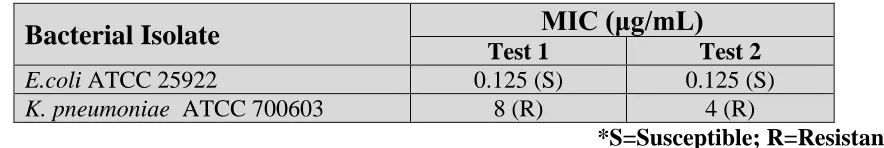
[image:24.612.94.536.351.425.2]ceftriaxone and K. pneumoniae ATCC 700603 should be resistant.
Table 4.1: Ceftriaxone MIC for Negative control (E. coli ATCC 25922) and Positive control (K. pneumoniae ATCC 700603) bacterial isolates using CLSI CAMHB macrodilution.
Bacterial Isolate
MIC (μg/mL)
Test 1 Test 2
E.coli ATCC 25922 0.125 (S) 0.125 (S)
K. pneumoniae ATCC700603 8 (R) 4 (R)
*S=Susceptible; R=Resistant
Table 4.1 shows the MIC determined for these two isolates E. coli ATCC 25922 and K.
pneumoniae ATCC 700603. The MIC of ceftriaxone for E. coli ATCC 25922 was at the lowest
concentration tested (0.125μg/mL), showing susceptibility to ceftriaxone and for K. pneumoniae
17
Table 4.2: Ceftriaxone MIC for Wild-Type and Control isolates
TEST 1
Bacterial Isolate
MIC (μg/mL)
CLSI Broth Macrodilution Bio-Rad Agar DilutionE.coli ATCC 25922 0.125 (S) 0.125 (S)
K. pneumoniae ATCC700603 ND 8 (R)
K. pneumoniae F9092593 64 (R) 32(R)
K. pneumoniae G0165470 >256 (R) >512 (R)
K. pneumoniae G2082851 256 (R) 16 (R)
TEST 2
Bacterial Isolate
MIC (μg/mL)
CLSI Broth Macrodilution Bio-Rad Agar DilutionE.coli ATCC 25922 0.125 (S) 0.125 (S)
K. pneumoniae ATCC700603 8 (R) 4 (R)
K. pneumoniae F9092593 16 (R) 16 (R)
K. pneumoniae G0165470 >256 (R) >512 (R)
K. pneumoniae G2082851 256 (R) 32 (R)
TEST 3
Bacterial Isolate
MIC (μg/mL)
CLSI Broth Macrodilution Bio-Rad Agar DilutionE.coli ATCC 25922 0.125 (S) 0.125 (S)
K. pneumoniae ATCC700603 4 (R) 4 (R)
K. pneumoniae F9092593 256 (R) 64 (R)
K. pneumoniae G0165470 >256 (R) >512 (R)
K. pneumoniae G2082851 256 (R) 64 (R)
*ND=Not determined
Table 4.2 presents results from triplicate tests performed to determine MIC of ceftriaxone
for all bacterial isolates using the CAMHB macrodilution and Bio-Rad Agar dilution methods. For
both methods, on all three tests, the MIC of ceftriaxone for the non-ESBL producing control
organism (E. coli ATCC 25922) was below 1μg/mL. Based on the CLSI breakpoint for
18
ATCC 25922. Almost identical results are observed for the ESBL-producing control organism (K.
pneumoniae ATCC 700603), for both methods. MIC for ceftriaxone for this organism was
≥4μg/mL on the Bio-Rad Agar with ceftriaxone added. Based on the CLSI breakpoint for
Enterobacteriaceae, the Bio-Rad Agar with ceftriaxone added detected the resistance of K.
pneumoniae ATCC 700603. The MIC results observed for the three Wild-Type Strains -K.
pneumoniae F9092593, K. pneumoniae G0165470, and K. pneumoniae G2082851- confirmed
their resistance to ceftriaxone. The MIC for ceftriaxone was above 4μg/mL for the three
Wild-Type Strains on all three susceptibility tests, for both methods. The Bio-Rad Agar with ceftriaxone
19
Chapter V
DISCUSSION
5.1 Discussion
Antibiotic resistance is a major public health issue (WHO, 2014). Resistance to antibiotics
leads to therapy failure which results in severe outcomes from common treatable infections.
Extended-spectrum β-lactamase (ESBL) enzymes, produced majorly by K. pneumoniae and E.coli,
facilitate resistance to ceftriaxone, cefotaxime, and aztreonam considered to be among the most
advanced antibiotics (Pitout & Laupland, 2008). Infection with K. pneumoniae strains and other
bacteria synthesizing ESBLs lead to higher mortality (Lautenbach et al., 2001; Tumbarello et al.,
2006). The presence of ESBL producing bacteria in natural environments, such as soil and water,
have been confirmed (Ben Said et al., 2015; Jouini et al., 2007; Zurfluh et al., 2013). Currently, no
standard methods exist to monitor these bacteria in natural environments. This research was
undertaken to evaluate an agar dilution method, using a growth medium –Bio-Rad Rapid E. coli 2
agar– that is designed to isolate K. pneumoniae and E. coli from the environment.
The results of this research revealed that compared to the CLSI broth macrodilution
method, the Bio-Rad agar dilution method performed well in identifying the non-ESBL producing
strain of E.coli (susceptible to ceftriaxone) and the ESBL producing strains of K. pneumoniae
(resistant to ceftriaxone). Based on the CLSI interpretative criteria, E.coli ATCC 25922 is
identified on the Bio-Rad agar with ceftriaxone added as susceptible to ceftriaxone because growth
inhibition was observed at concentrations below 1μg/mL. Growth inhibition for K. pneumoniae
ATCC 700603, K. pneumoniae F9092593, K. pneumoniae G0165470, and K. pneumoniae
20
resistant to ceftriaxone. The minimum inhibitory concentration (MIC) of two isolates K.
pneumoniae F9092593 and K. pneumoniae G2022851 did differ between repeat tests, with the
MIC of K. pneumoniae G2022851 being up to 3 dilutions higher ( 256 on Broth macrodilution; 16
on Bio-Rad agar dilution) on test one (Table 4.2). However, the variations in MIC is typical when
multiple susceptibility tests are performed.
The Bio-Rad Rapid E. coli 2 agar is a useful tool in environmental water quality studies
for direct enumeration and identification of E.coli and coliforms, which includes K. pneumoniae,
in environmental samples. The substitution of the standard Mueller-Hinton media with Bio-Rad
Rapid E. coli 2 agar media on the agar dilution susceptibility test demonstrated a high precision
level, identifying the ESBL producing and non-ESBL producing bacteria on all susceptibility tests
performed. The results of this research also showed that the Bio-Rad agar had a high accuracy in
identifying the three ESBL producing K. pneumoniae strains when compared to the Broth
macrodilution results.
The use of a standard method is critical to monitor and compare outcomes from
environmental studies demonstrating the dissemination of ESBL producing bacteria in the
environment. Monitoring these bacteria is important to determine the relevance of their presence
in natural environments where human exposure may occur. This research demonstrates the
accuracy of an agar dilution method using a differential media designed for use on environmental
samples. This method, similar to the CLSI standard susceptibility method for clinical samples,
21
5.2 Limitations
For this research, Bio-Rad Rapid E. coli 2 agar was substituted for the standard Muller
Hinton agar. MHA is recommended as the standard growth medium because it presents little or no
interference to the activity of antibiotics being tested. From the results of this research, Bio-Rad
Rapid E. coli 2 agar exhibited no interference to ceftriaxone antibiotic; this suggests that it may
work well with other β-lactam antibiotics. However, further research is necessary to confirm that
the Bio-Rad Rapid E. coli 2 agar indeed presents no interference to the activity of other antibiotics
and is suitable as an antibiotic susceptibility testing growth medium. Additionally, the sample size
for this research was restricted to three K. pneumoniae strains. Further research using a larger
sample size as well as other strains of ESBL producing bacteria such as E.coli, Enterobacter,
Pseudomonas aeruginosa, Acinetobacter baumannii is critical to validate the results from this
research. Furthermore, due to the small sample size this research was unable to determine the
sensitivity and specificity of the Bio-Rad agar dilution method.
5.3 Future Research
The results from this research are promising. However, further evaluation of the Bio-Rad
agar dilution method is necessary. Further research using β-lactam antibiotics, other than
ceftriaxone, is necessary to confirm that the Bio-Rad Rapid E. coli 2 agar presents no interference
to the activity of antibiotics and is suitable as an antibiotic susceptibility testing growth medium.
Also, a larger sample size using various strains of ESBL producing bacteria such as E.coli,
Enterobacter, Pseudomonasaeruginosa, Acinetobacter baumannii is critical to validate the results
22
REFERENCES
Ben Said, L., Jouini, A., Klibi, N., Dziri, R., Alonso, C., Boudabous, A., … Torres, C. (2015). Detection of extended-spectrum beta-lactamase (ESBL)-producing Enterobacteriaceae in
vegetables, soil and water of the farm environment in Tunisia. International Journal of
Food Microbiology, 203, 86–92. http://doi.org/10.1016/j.ijfoodmicro.2015.02.023.
Berkner, S., Konradi, S., & Schönfeld, J. (2014). Antibiotic resistance and the
environment--there and back again: Science & Society series on Science and Drugs. EMBO Reports,
15(7), 740–744. http://doi.org/10.15252/embr.201438978.
Bradford, P. (2001). Extended-Spectrum β-Lactamases in the 21st Century: Characterization,
Epidemiology, and Detection of This Important Resistance Threat. Clinical Microbiology
Reviews, 14(4), 933–951. http://doi.org/10.1128/CMR.14.4.933-951.2001.
Brooklyn Antibiotic Resistance Task Force. (2002). The Cost of Antibiotic Resistance: Effect of
Resistance among Staphylococcus aureus, Klebsiella pneumoniae, Acinetobacter
baumannii, and Pseudomonas aeruginosa on Length of Hospital Stay. Infection Control and Hospital Epidemiology, 23(2), 106–108.
Bréchet, C., Plantin, J., Sauget, M., Thouverez, M., Talon, D., Cholley, P., … Bertrand, X. (2014). Wastewater Treatment Plants Release Large Amounts of Extended-Spectrum
β-Lactamase–Producing Escherichia coli Into the Environment. Clinical Infectious
Diseases, 58(12), 1658–1665.
Bush, K., Jacoby, G., & Medeiros, A. (1995). A functional classification scheme for
beta-lactamases and its correlation with molecular structure. Antimicrobial Agents and
Chemotherapy, 39(6), 1211–1233. http://doi.org/10.1128/AAC.39.6.1211.
Casey, G. (2012). Antibiotics and the Rise of Superbugs. Kai Tiaki Nursing New Zealand,
18(10), 20-24 5p.
Clinical and Laboratory Standards Institute (CLSI). (2014). Performance standards for antimicrobial susceptibility testing; sixteenth informational supplement. CLSI document M100–S24. CLSI, Wayne, Pa.
D’Andrea, M., Arena, F., Pallecchi, L., & Rossolini, G. (2013). CTX-M-type β-lactamases: A
successful story of antibiotic resistance. International Journal of Medical Microbiology,
303(6–7), 305–317. http://doi.org/10.1016/j.ijmm.2013.02.008.
Doi, Y., Park, Y., Rivera, J., Adams-Haduch, J., Hingwe, A., Sordillo, E. M., … Paterson, D. (2013). Community-associated extended-spectrum β-lactamase-producing Escherichia
coli infection in the United States. Clinical Infectious Diseases: An Official Publication
23
George H. Talbot, Bradley, J., John E. Edwards, J., Gilbert, D., Michael Scheld, & Bartlett, J. (2006). Bad Bugs Need Drugs: An Update on the Development Pipeline from the Antimicrobial Availability Task Force of the Infectious Diseases Society of America.
Clinical Infectious Diseases, (5), 657.
Hocquet, D., Muller, A., & Bertrand, X. (2016). What happens in hospitals does not stay in
hospitals: antibiotic-resistant bacteria in hospital wastewater systems. Journal of Hospital
Infection. http://doi.org/10.1016/j.jhin.2016.01.010.
Holten, K., & Onusko, E. (2000). Appropriate prescribing of oral beta-lactam antibiotics.
American Family Physician, 62(3), 611–620.
Jorgensen, J., & Ferraro, M. (2009). Antimicrobial Susceptibility Testing: A Review of General
Principles and Contemporary Practices. Clinical Infectious Diseases, (11), 1749.
Jouini, A., Vinue, L., Ben Slama, K., Saenz, Y., Klibi, N., Hammami, S., … Torres, C. (2007). Characterization of CTX-M and SHV extended-spectrum beta-lactamases and associated
resistance genes in Escherichia coli strains of food samples in Tunisia. JOURNAL OF
ANTIMICROBIAL CHEMOTHERAPY, 60(5), 1137–1141.
Kwak, Y, Colque, P., Byfors, S., Giske, C., Möllby, R., & Kühn, I. (2015). Surveillance of antimicrobial resistance among Escherichia coli in wastewater in Stockholm during 1
year: does it reflect the resistance trends in the society? International Journal of
Antimicrobial Agents, 45(1), 25–32. http://doi.org/10.1016/j.ijantimicag.2014.09.016.
Lautenbach, E., Patel, J., Bilker, W., Edelstein, P., & Fishman, N. (2001). Extended-spectrum beta-lactamase-producing Escherichia coli and Klebsiella pneumoniae: risk factors for
infection and impact of resistance on outcomes. Clinical Infectious Diseases: An Official
Publication Of The Infectious Diseases Society Of America, 32(8), 1162–1171.
Leggiadro, R. (1997). Extended-spectrum beta-lactamases and other enzymes providing
resistance to oxyimino-beta-lactams. INFECTIOUS DISEASE CLINICS OF NORTH
AMERICA, 11(4), 875–&.
Mainous, A., Diaz, V., Matheson, E., Gregorie, S., & Hueston, W. (2011). Trends in
Hospitalizations with Antibiotic-Resistant Infections: US, 1997-2006. PUBLIC HEALTH
REPORTS, 126(3), 354–360.
Neu, H. C. (1992). The Crisis in Antibiotic Resistance. Science, 257(5073), 1064–1073.
Paterson, D., & Bonomo, R. (2005). Extended-Spectrum β-Lactamases: a Clinical Update.
Clinical Microbiology Reviews, 18(4), 657–686. http://doi.org/10.1128/CMR.18.4.657-686.2005.
Pitout, J., Hanson, N., Church, D., & Laupland, K. (2004). Population-Based Laboratory Surveillance for Escherichia coli–Producing Extended-Spectrum β-Lactamases:
Importance of Community Isolates with blaCTX-M Genes. Clinical Infectious Diseases,
24
Pitout, J., & Laupland, K. (2008). Extended-spectrum [beta]-lactamase-producing
Enterobacteriaceae: an emerging public-health concern. The Lancet Infectious Diseases,
8(3), 159–66.
Pitout, J., Nordmann, P., Laupland, K., & Poirel, L. (2005). Emergence of Enterobacteriaceae
producing extended-spectrum beta-lactamases (ESBLs) in the community. JOURNAL OF
ANTIMICROBIAL CHEMOTHERAPY, 56(1), 52–59.
Prado, T., Pereira, W., Silva, D., Seki, L., Carvalho, A., & Asensi, M. (2008). Detection of extended-spectrum β-lactamase-producing Klebsiella pneumoniae in effluents and sludge
of a hospital sewage treatment plant. Letters in Applied Microbiology, 46(1), 136–141.
http://doi.org/10.1111/j.1472-765X.2007.02275.x.
Smet, A., Martel, A., Persoons, D., Dewulf, J., Heyndrickx, M., Claeys, G., … Butaye, P. (2010). Characterization of Extended-Spectrum [beta]-Lactamases Produced by Escherichia coli Isolated from Hospitalized and Nonhospitalized Patients: Emergence of
CTX-M-15-Producing Strains Causing Urinary Tract Infections. Microbial Drug Resistance, 16(2),
129–34. http://doi.org/http://dx.doi.org.ezproxy.gsu.edu/10.1089/mdr.2009.0132.
Thomson, K., & Moland, E. (2001). Cefepime, Piperacillin-Tazobactam, and the Inoculum Effect in Tests with Extended-Spectrum β-Lactamase-Producing Enterobacteriaceae.
Antimicrobial Agents and Chemotherapy, 45(12), 3548–3554. http://doi.org/10.1128/AAC.45.12.3548-3554.2001.
Thomson, K., & Moland, E. (2000). Version 2000: the new β-lactamases of Gram-negative
bacteria at the dawn of the new millennium. Microbes and Infection, 2(10), 1225–1235.
http://doi.org/10.1016/S1286-4579(00)01276-4.
Tumbarello, M., Spanu, T., Sanguinetti, M., Citton, R., Montuori, E., Leone, F., … Cauda, R. (2006). Bloodstream Infections Caused by Extended-Spectrum-β-Lactamase-Producing Klebsiella pneumoniae: Risk Factors, Molecular Epidemiology, and Clinical Outcome.
Antimicrobial Agents and Chemotherapy, 50(2), 498–504. http://doi.org/10.1128/AAC.50.2.498-504.2006.
US Centers for Disease Control and Prevention (CDC). (2013). Antibiotic resistance threats in
the United States. Retrieved from http://www.cdc.gov/drugresistance/threat-report-2013/. Wellington, E., Boxall, A., Cross, P., Feil, E., Gaze, W., Hawkey, P..… Williams, A. (2013). The
role of the natural environment in the emergence of antibiotic resistance in
Gram-negative bacteria. The Lancet Infectious Diseases, 13(2), 155–65.
http://doi.org/http://dx.doi.org.ezproxy.gsu.edu/10.1016/S1473-3099(12)70317-1.
World Health Organization (WHO). (2014) Antimicrobial Resistant Global Surveillance Report. Retrieved from
25
Wiegand, I., Geiss, H., Mack, D., Stürenburg, E., & Seifert, H. (2007). Detection of Extended-Spectrum Beta-Lactamases among Enterobacteriaceae by Use of Semiautomated
Microbiology Systems and Manual Detection Procedures. Journal of Clinical
Microbiology, 45(4), 1167–1174. http://doi.org/10.1128/JCM.01988-06.
Wiegand, I., Hilpert, K., & Hancock, R. (2008). Agar and broth dilution methods to determine
the minimal inhibitory concentration (MIC) of antimicrobial substances. Nature
Protocols, 3(2), 163–75.
http://doi.org/http://dx.doi.org.ezproxy.gsu.edu/10.1038/nprot.2007.521.
Zurfluh, K., Hächler, H., Nüesch-Inderbinen, M., & Stephan, R. (2013). Characteristics of Extended-Spectrum β-Lactamase- and Carbapenemase-Producing Enterobacteriaceae
Isolates from Rivers and Lakes in Switzerland. Applied and Environmental Microbiology,