http://dx.doi.org/10.4236/jsea.2014.71007
Generalized
α-Entropy Based Medical Image Segmentation
Samy Sadek1, Sayed Abdel-Khalek2
1
Department of Mathematics, Faculty of Science, Sohag University, Sohag, Egypt; 2Mathematics Department, Faculty of Science, Taif Univesity, Taif, KSA.
Email: [email protected], [email protected]
Received October 19th, 2013; revised December 18th, 2013; accepted December 26th, 2013
Copyright © 2014 Samy Sadek, Sayed Abdel-Khalek. This is an open access article distributed under the Creative Commons Attri-bution License, which permits unrestricted use, distriAttri-bution, and reproduction in any medium, provided the original work is properly cited. In accordance of the Creative Commons Attribution License all Copyrights © 2014 are reserved for SCIRP and the owner of the intellectual property Samy Sadek, Sayed Abdel-Khalek. All Copyright © 2014 are guarded by law and by SCIRP as a guardian.
ABSTRACT
In 1953, Rènyi introduced his pioneering work (known as α-entropies) to generalize the traditional notion of entropy. The functionalities of α-entropies share the major properties of Shannon’s entropy. Moreover, these entropies can be easily estimated using a kernel estimate. This makes their use by many researchers in computer vision community greatly appealing. In this paper, an efficient and fast entropic method for noisy cell image segmentation is presented. The method utilizes generalized α-entropy to measure the maximum structural infor- mation of image and to locate the optimal threshold desired by segmentation. To speed up the proposed method, computations are carried out on 1D histograms of image. Experimental results show that the proposed method is efficient and much more tolerant to noise than other state-of-the-art segmentation techniques.
KEYWORDS
α-Entropy; Cell Image; Entropic Image Segmentation
1. Introduction
Instinctively, image segmentation is the process of divi- ding an image into different regions such that each re- gion is homogeneous while not the union of any two ad- jacent regions. An additional requirement would be that these regions have a correspondence to real homogene- ous regions belonging to objects in the scene [1]. Image segmentation is an elementary and significant component in many applications such as image analysis, pattern re- cognition, medical diagnosis and currently in robotic vi- sion. However, it is one of the most difficult and challen- ging tasks in image processing, and it determines the qua- lity of the final results of the image analysis. The recent developments in Digital Mammography (DM), Magnetic Resonance Imaging (MRI), Computed Tomography (CT), and other diagnostic imaging techniques provide physi- cians with high resolution images which have significant- ly assisted the clinical diagnosis. These up-to-date tech- nologies not only have a recognizably increased knowle- dge of normal and diseased anatomy for medical research but also become a significant part in diagnosis and treat- ment planning [2].
There is currently no single segmentation technique that gives satisfactory results for each medical image.
Since the pioneering work by Shannon [16,17] in 1948, entropy appears as an attention-grabbing tool in many areas of data processing. In 1953, Rènyi [8] introduced a wider class of entropies known as α -entropies. The func- tionalities of α -entropies share the major properties of Shannon’s entropy. Moreover, the α -entropies can be easily estimated using a kernel estimate. This makes their use attractive in many areas of image processing [18-20]. In this paper, we propose an efficient entropic technique for segmenting cell images which utilizes generalized Rènyi entropy. Our work for cell image segmentation has a relatively good performance in comparison to other re- lated state-of-the-art techniques [21,22].
The outline of this paper is as follows. The next sec- tion discusses the generalized form of α-entropies espe- cially generalized Rényi entropy. The proposed entropic segmentation method is explained in Section 3. Section 4 is to present the experimental results that validate the use of the proposed method. Advantages of our method and concluding remarks are outlined in Section 5.
2. Entropy of Generalized Distributions
Entropy has first appeared in thermodynamics as an in- formation theoretical concept which is intimately related to the internal energy of the system. Then it has applied across physics, information theory, mathematics and other branches of science and engineering [9]. When given a system whose exact description is not precisely known, the entropy is defined as the expected amount of infor- mation needed to exactly specify the state of the system, given what we know about the system.
Suppose P=
{
p p1, 2,,pn}
be a finite discrete pro-bability distribution that satisfies these conditions 0, 1, 2, ,
k
p ≥ k= n and
∑
nk=1pk =1. The amount of uncertainty of the distribution P, is called the entropy of the distribution, P. The Shannon entropy of the distri- bution, P, a measure of uncertainty and denoted by( )
H P ), is defined as
( )
21 log
n
k k
k
H P p p
=
= −
∑
(1)It should be noted that the Shannon entropy given by Equation (1) is additive, i.e. it satisfies the following re- lation:
(
)
( )
( )
H A+B =H A +H B (2)
for any two distributions A and B. Equation (2) states one of the most important properties of entropy, namely, its additivity: the entropy of a combined experiment con- sisting of the performance of two independent experi-
ments is equal to the sum of the entropies of these two experiments. The formalism defined by Equation (1) has been shown to be restricted to the Boltzmann-Gibbs- Shannon (BGS) statistics. However, for nonextensive sys- tems, some kind of extension appears to become neces- sary. Rènyi entropy, which is useful for describing the non- extensive systems, is defined as
Entropic segmentation for noisy mammography image.
( )
21 1 log 1 n k k
Hα P α pα
=
=
−
∑
(3)where α ≥0 and α ≠1. The real number α is called an entropic order that characterizes the degree of non- extensivity. This expression reduces to Shannon entropy in the limit α →1. We shall see that in order to get the fine characterization of Rànyi entropy, it is advantageous to extend the notion of a probability distribution, and define entropy for the generalized distributions. The cha- racterization of measures of entropy (and information) becomes much simpler if we consider these quantities as defined on the set of generalized probability distribu- tions.
Suppose
[
Ω,P]
be a probability space that is, Ω an arbitrary nonempty set, called the set of elementary events, and P a probability measure, that is, a non-ne- gative and additive set function for which P( )
Ω . Let us call a function ξ ξ ω=( )
which is defined for ω ∈ Ω1,where Ω ⊂ Ω1 . If P
( )
Ω =1 1 we call ξ an ordinary (or complete) random variable, while if( )
10 <P Ω ≤1 we call ξ an incomplete random vari- able. Evidently, an incomplete random variable can be interpreted as a quantity describing the result of an ex- periment depending on chance which is not always ob- servable, only with probability P
( )
Ω1 < 1. The distribu- tion of a generalized random variable is called a genera- lized probability distribution. Thus a finite discrete gene- ralized probability distribution is simply a sequence1, 2, , n
p p p of nonnegative numbers such that setting
{ }
1n k k
P= p = and taking
( )
1 n k k P p ϖ ==
∑
(4)where ϖ
( )
P is the weight of the distribution and( )
0 <ϖ P ≤1. A distribution that has a weight less than 1 will be called an incomplete distribution. Now, using Equation (3) and Equation (4), the Rànyi entropy for the generalized distribution can be written as
( )
12 1 1 log 1 n k k n k k p H P p α
α α =
= = −
∑
∑
(5)for statistical independent systems, defined by the fol- lowing pseudo additivity entropic formula
(
)
( )
( )
(
1)
( )
( )
H A B H A H B
H A H B
α α α
α α
α + = +
+ − ⋅ ⋅ (6)
3. Suggested Methodology
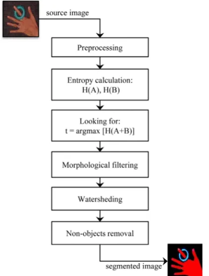
[image:3.595.70.281.428.706.2]Image segmentation problem is considered to be one of the most holy grail challenges of computer vision field especially when done for noisy images. Consequently it has received considerable attention by many researchers in computer vision community. There are many approach for image segmentation, however, these approach are still inadequate. In this work, we propose an entropic method that achieves the task of segmentation in a novel way. This method not only overcomes image noise, but also utilizes time and memory optimally. This wisely happens by the advantage of using the Rànyi entropy of genera- lized distributions to measure the structural information of image and then locate the optimal threshold depending on the postulation that the optimal threshold corresponds to the segmentation with maximum structure (i.e., max- imum information content of the distribution). The im- plementation steps of the proposed segmentation method are shown in the block diagram of Figure 1. The follow- ing sections outline in detail the process behind each step.
Figure 1. Block diagram of the proposed segmentation me- thod.
3.1. Preprocessing
Preprocessing ultimately aims at improving the image in ways that increase the opportunity for success of the other ulterior processes [17,23]. In this step, we apply a Gaussian filter to the input image prior to any process in order to reduce the amount of noise in an image.
3.2. Entropies Calculation
Suppose
{ }
pi in=1 be the probability distribution for the image. At the threshold, t this distribution is divided into two sub distributions; one for the foreground (class f) and the other for the background (class b) given by{ }
1t f
i i
P = p = and Pb=
{ }
pi ni t= +1 respectively. Thus, thegeneralized Rànyi entropies for the two distributions as functions of t are given as
( )
12 1
1 log 1
t k
f k
t k k
p H t
p
α
α α =
=
=
−
∑
∑
(7)( )
12
1
1 log 1
n k
b k t
n k k t
p H t
p
α
α α = +
= +
=
−
∑
∑
(8)3.3. Image Thresholding
Thresholding is the most often used technique to dis- tinguish objects from background. In this step an input image is converted by threshed into a binary image so that the objects in the input image can be easily separated from the background. To get the desired optimum thre- shold value t*, we have to maximize the total entropy,
( )
f b
Hα+ t . When the function f b
( )
Hα+ t is maximized, the value of parameter t that maximizes the function is believed to be the optimum threshold value [24]. Mathe- matically, the problem can be formulated as
( )
( )
( ) (
)
( )
( )
arg max
arg max 1
f b
f b f b
t H t
H t H t H t H t
α
α α α α α
∗ = +
= + + − ⋅ ⋅
(9)
3.4. Morphology-Based Operations
3.5. Overlapping Cancelation
In this step we attempt to remove the overlapping be- tween objects that perhaps happened through extensively applying the previous morphological operations. To per- form this, we first get the Euclidean Distance Transform (EDT) of the binary image. Then we apply the well- known watershed algorithm [27,28] on the resulting EDT image. The EDT ultimately converts the binary image into one where each pixel has a value equal to its dis- tance to the nearest foreground pixel. The distances are measured in Euclidean distance metric. The peaks of the distance transform are assumed to be in the centers of the objects. Then the overlapping objects can be yet easily separated.
3.6. Non-Objects Removal
This step helps in removing incorrect objects according to the object size. Sizes of objects are measured in com- parison to the total size of image. Each tiny noise object of size less than a predefined minimum threshold can be discarded. Also each object whose size is greater than the maximum threshold size can be removed as well. Note that thresholds of size used herein are often dependent on the application, and so they are considered as user-de- fined data.
4. Experimental Results
In this section, the results of the proposed approach are presented. First to investigate the proposed approach for image segmentation we began by different image histo- grams. Each of these histograms describes the “objects” and the “background”. Additionally, to verify the benefit of using the generalized Rènyi entropy, we have tried us- ing another formula of entropy (e.g. Tsallis entropy) which is given by
1
1 1
n k k p
H
α
α α =
− =
−
∑
(10)
The results of segmentation have testified to the higher efficiency of our entropic segmentation approach espe- cially when generalized Rènyi entropy is used.
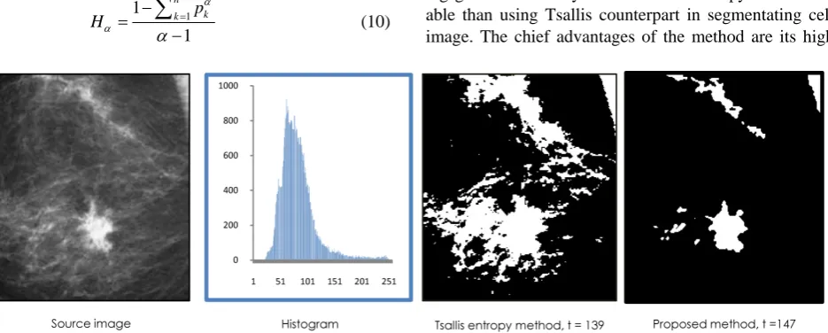
In Figure 2, an image of a mammogram showing breast cancer with a bright region (tumefaction) sur- rounded by a noisy region. The histogram roughly exem- plifies an unimodal distribution of the graylevel values. The proposed entropic method will look for regions with uniform distribution in order to find the maximum en- tropy. This will regularly take place at the peak limit. It is well-known that segmenting this type of images is typi- cally a challenging task. However the proposed method could performed well when applied on this type of im- ages. Additionally, segmentation results in the figure show that using generalized Rènyi entropy is better than using Tsallis entropy.
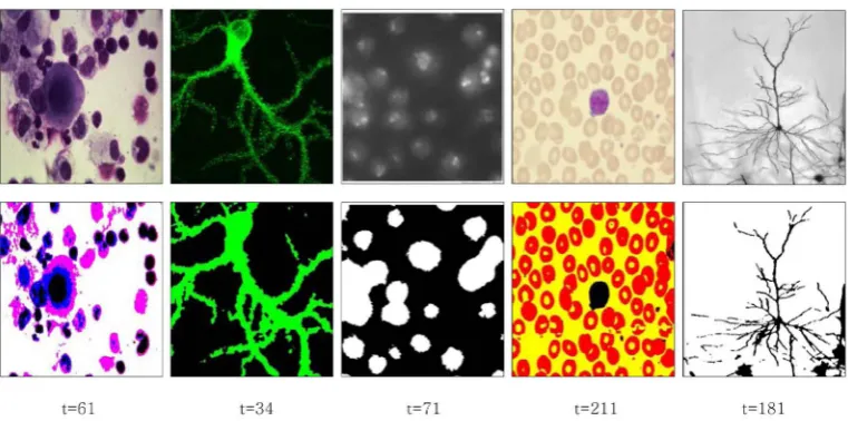
Figure 3 shows another example of our segmentation method. We present an image of a medical domain with a spatial background scattering noise; a stained brain cell that shows branching of cell dendrites-fibers that receive input from other brain cells. Several values of α are experimented. But the superior segmentation results has been obtained at α =0.9.
In Figure 4, we show the segmentation results of the proposed method on a sample of color medical images. In this example the images are segmented with α equal to 0.8.
5. Conclusion
[image:4.595.67.535.532.720.2]In this paper, we introduced a new method for cell image segmentation based on generalized α -entropy. The pro- posed method has achieved the task of segmentation in a novel way. This method has been shown to provide good results in most cases and perform well when applied to noisy cell images. The experimental results show that us- ing generalized Rènyi formalism of entropy is more vi- able than using Tsallis counterpart in segmentating cell image. The chief advantages of the method are its high
Figure 3. Entropic segmentation for a brain cell image with a spatial noise around.
Figure 4. Results of the proposed segmentation method for a sample of test images.
rapidity and its tolerance to image noise.
REFERENCES
[1] M. Albuquerque, I. A. Esquef and A. R. Gesualdi, “Image Thresholding Using Tsallis Entropy,” Pattern Recogni- tion Letters, Vol. 25, No. 9, 2004, pp. 1059-1065. http://dx.doi.org/10.1016/j.patrec.2004.03.003
[2] P.-L. Bazin and D. L. Pham, “Homeomorphic Brain Ima- ge Segmentation with Topological and Statistical Atlases,”
Medical Image Analysis, Vol. 12, No. 5, 2008, pp. 616- 625. http://dx.doi.org/10.1016/j.media.2008.06.008
[3] P.-L. Bazin and D. L. Pham, “Topology Correction of Seg- mented Medical Images Using a Fast Marching Algori- thm,” Programs in Biomedicine, Vol. 88, No. 2, 2007, pp. 182-290. http://dx.doi.org/10.1016/j.cmpb.2007.08.006
[4] J. C. Carter, D. C. Lanham, G. Bibat, S. Naidu and W. E. Kaufmann, “Selective Cerebral Volume Reduction in Rett Syndrome: A Multiple Approach MRI Study,” American Journal of Neuroradiology, Vol. 29, No. 3, 2008, pp. 436-441. http://dx.doi.org/10.3174/ajnr.A0857
[5] R. C. Gonzalez and R. E. Woods, “Digital Image Process- ing Using Matlab,” 2nd Edition, Prentice Hall, Inc., Up-
per Saddle River, 2003.
[6] W. E. L. Grimson, G. J. Ettinger, T. Kapur, M. E. Leven- ton and W. M. Wells, “Utilizing Segmented MRI Data in Image-Guided Surgery,” International Journal of Pattern Recognition and Artificial Intelligence, Vol. 11, No. 8, 1997, pp. 1367-1397.
http://dx.doi.org/10.1142/S0218001497000639
[7] V. S. Khoo, D. P. Dearnaley, D. J. Finnigan, A. Padhani, S. F. Tanner and M. O. Leach, “Magnetic Resonance Im- aging (MRI): Considerations and Applications in Radio-theraphy Treatment Planning,” Radiotherapy Oncology, Vol. 42, No. 1, 1997, pp. 1-15.
http://dx.doi.org/10.1016/S0167-8140(96)01866-X
[8] S. M. Larie and S. S. Abukmeil, “Brain Abnormality in Schizophrenia: A Systematic and Quantitative Review of Volumetric Magnetic Resonance Imaging Studies,” Jour- nal of Psychiatry, Vol. 172, 1998, pp. 110-120.
[9] I. Levner and H. Zhang, “Classification-Driven Water- shed Segmentation,” EEE Transactions on Image Pro- cessing, Vol. 16, No. 5, 2007, pp. 1437-1445.
http://dx.doi.org/10.1109/TIP.2007.894239
[image:5.595.108.489.263.452.2]Modeling,” IEEE International Symposium on Signal Pro- cessing and Information Technology (ISSPIT’10), Luxor, 2010, pp. 366-370.
http://dx.doi.org/10.1109/ISSPIT.2010.5711812
[11] A. Rényi, “On a Theorem of P. Erdǒs and Its Application in Information Theory,” Mathematica, Vol. 1, 1959, pp. 341-344.
[12] S. M. Resnick, D. L. Pham, M. A. Kraut, A. B. Zonderman and C. Davatzikos, “Longitudinal MRI Studies of Older Adults: A Shrinking Brain,” Journal of Neuroscience, Vol. 23, No. 8, 2003, pp. 3295-3301.
[13] S. Sadek, A. Al-Hamadi, M. Elmezain, B. Michaelis and U. Sayed, “Human Activity Recognition Using Temporal Shape Moments,” IEEE International Symposium on Sig- nal Processing and Information Technology (ISSPIT’10), Luxor, 2010, pp. 79-84.
http://dx.doi.org/10.1109/ISSPIT.2010.5711729
[14] S. Sadek, A. Al-Hamadi, B. Michaelis and U. Sayed, “A Fast Statistical Approach for Human Activity Recogni- tion,” International Journal of Intelligence Science (IJIS), Vol. 2, No. 1, 2012, pp. 9-15.
[15] S. Sadek, A. Al-Hamadi, B. Michaelis and U. Sayed, “An Efficient Method for Real-Time Activity Recognition,”
Proceedings of the International Conference on Soft Com- puting and Pattern Recognition (SoCPaR’10), Paris, 2010, pp. 7-10.
[16] S. Sadek, A. Al-Hamadi, B. Michaelis and U. Sayed, “An Image Classification Approach Using Multilevel Neural Networks,” Proceedings of IEEE International Confe- rence on Intelligent Computing and Intelligent Systems
(ICIS’09), Shanghai, 2009, pp. 180-183.
[17] S. Sadek, A. Al-Hamadi, B. Michaelis and U. Sayed, “Face Detection and Localization in Color Images: An Efficient Neural Approach,” Journal of Software Engineering and Applications (JSEA), Vol. 4, No. 12, 2011, pp. 682-687. http://dx.doi.org/10.4236/jsea.2011.412080
[18] S. Sadek, A. Al-Hamadi, B. Michaelis and U. Sayed, “Hu- man Action Recognition via Affine Moment Invariants,” 21st International Conference on Pattern Recognition
(ICPR’12), Tsukuba Science City, 2012, pp. 218-221. [19] S. Sadek, A. Al-Hamadi, B. Michaelis and U. Sayed, “Hu-
man Action Recognition: A Novel Scheme Using Fuzzy Log-Polar Histogram and Temporal Self-Similarity,” EU-
RASIP Journal on Advances in Signal Processing, 2011. http://dx.doi.org/10.1155/2011/540375
[20] S. Sadek, A. Al-Hamadi, A. Wannig, B. Michaelis and U. Sayed, “A New Approach to Image Segmentation via Fu- zzification of Rènyi Entropy of Generalized Distributions.
Proceedings of International Conference on Image, Sig- nal and Vision Computing (ICISVC’09), Singapore, 2009, pp. 598-603.
[21] S. Sadek, A. Al-Hamadi, B. Michaelis and U. Sayed, “A Robust Neural System for Objectionable Image Recogni- tion,” IEEE International Conference on Machine Vision
(ICMV’09), 2009, pp. 32-36.
[22] S. Sadek, M. A. Mofaddel and B. Michaelis, “Multicolor Skin Modeling with Application to Skin Detection,” Jour- nal of Computations & Modelling, Vol. 3, No. 1, 2013, pp. 153-167.
[23] C. E. Shannon and W. Weaver, “The Mathematical The- ory of Communication,” University of Illinois Press, Ur- bana, 1949.
[24] W. Tatsuaki and S. Takeshi, “When Nonextensive Entro- py Becomes Extensive,” Physica A, Vol. 301, No. 1-4, 2001, pp. 284-290.
http://dx.doi.org/10.1016/S0378-4371(01)00400-9
[25] P. Taylor, “Invited Review: Computer Aids for Decision- Making in Diagnostic Radiology—A Literature Review,”
British Journal of Radiology, Vol. 68, No. 813, 1995, pp. 945-957.
http://dx.doi.org/10.1259/0007-1285-68-813-945
[26] D. Tosun, M. E. Rettmann, X. Han, X. Tao, C. Xu, S. M. Resnick and J. L. Prince, “Cortical Surface Segmentation and Mapping,” NeuroImage, Vol. 23, No. 1, 2004, pp. S108-S118.
http://dx.doi.org/10.1016/j.neuroimage.2004.07.042
[27] C. Tsallis, S. Abe and Y. Okamoto, “Nonextensive Statis- tical Mechanics and Its Applications,” Series Lecture Notes in Physics,Springer, Berlin, 2001.
[28] A. J. Worth, N. Makris, V. S. Caviness and D. N. Kenne- dy, “Neuroanatomical Segmentation in MRI: Technolo- gical Objectives,” International Journal of Pattern Rec-ognition and Artificial Intelligence, Vol. 11, No. 8, 1997, pp. 1161-1187.