The potent anti-inflammatory agents, glucocorticoids (GCs) are frequently used for the treatment of inflammatory diseases such as arthritis, pulmonary diseases, and skin diseases. It is well known that high-dose or long-term use of GCs results in several adverse side effects including osteoporosis.1Epidemiological studies have demonstrated the high incidence of fractures at the spine (the skeletal site rich in trabecular bone), followed by fractures at the hip (the skeletal site rich in both trabecular and cortical bone).2This is probably because GC-induced osteoporosis is more evident in trabecular bone than in cortical bone.3Thus, the primary strategy for GC-induced osteoporosis seems to be the prevention of vertebral fractures.
Both vitamin K2and risedronate are widely used in the treatment of GC-induced osteoporosis in Japan,4because both drugs have been reported to be effective in reducing incidence of vertebral fractures.5-7Risedronate decreases bone turnover, increases bone
Comparison of the Effect of Vitamin K
2
and Risedronate
on Trabecular Bone in Glucocorticoid-Treated Rats:
A Bone Histomorphometry Study
Jun Iwamoto,
1Hideo Matsumoto,
1Tsuyoshi Tadeda,
1Yoshihiro Sato,
2and James K. Yeh
3 1 Institute for Integrated Sports Medicine, Keio University School of Medicine, Tokyo; 2 Department of Neurology, Mitate Hospital, Fukuoka, Japan;3 Metabolism Laboratory, Department of Medicine, Winthrop-University Hospital, New York, USA.
Purpose:To compare the effect of vitamin K2 and risedronate on trabecular bone in glucocorticoid (GC)-treated rats.
Materials and Methods: Forty-eight Sprague-Dawley female rats, 3 months of age, were randomized by the stratified weight method into 5 groups according to the following treatment schedule: age-matched control, GC administration, and GC administration with concomitant administration of vitamin K2, risedronate, or vitamin K2+ risedronate. GC
(methylprednisolone sodium succinate, 5.0 mg/kg) and risedronate (10 µg/kg) were administered subcutaneously three and five times a week, respectively. Vitamin K2(menatetrenone, 30 mg/kg) was administered orally three times a week. At the end of the 8-week experiment, bone histomorphometric analysis was performed on trabecular bone of the tibial proximal metaphysis. Results:GC administration decreased trabecular bone mass compared with age-matched controls because of decreased bone formation (mineralizing surface, mineral apposition rate, and bone formation rate) and increased bone erosion. Vitamin K2attenuated GC-induced trabecular bone loss by preventing GC-induced decrease in bone formation
(mineralizing surface) and subsequently reducing GC-induced increase in bone erosion. Risedronate prevented GC-induced trabecular bone loss by preventing GC-induced increase in bone erosion although it also suppressed bone formation (mineralizing surface, mineral apposition rate, and bone formation rate). Vitamin K2mildly attenuated suppression of bone
formation (mineralizing surface) and bone erosion caused by risedronate without affecting trabecular bone mass when administered in combination. Conclusion:The present study showed differential effect of vitamin K2and risedronate on
trabecular bone in GC-treated rats.
Key Words :Glucocorticoid, trabecular bone, bone histomorphometry, risedronate, vitamin K2
Received: February 26, 2008 Revised: May 19, 2008 Accepted: June 4, 2008
Corresponding author: Dr. Jun Iwamoto, Institute for Integrated Sports Medicine, Keio University School of Medicine, 35 Shinanomachi, Shinjuku-ku, Tokyo 160-8582, Japan.
Tel: 81-3-3353-1211, Fax: 81-3-3352-9467 E-mail: jiwamoto@sc.itc.keio.ac.jp
© Copyright:
Yonsei University College of Medicine 2009
mineral density, and prevents vertebral fractures in patients treated with GCs.5,6An iliac biopsy study clearly demonstrated
that risedronate prevented the deterioration of trabecular bone architecture, reduced the degree of mineralization, and preserved elastic modulus within the trabeculae in patients treated with GCs.8However, there are no data to show the effect of vitamin
K2on bone formation and resorption and trabecular bone in
patients treated with GCs.
Several preclinical studies using GC-treated rats have demon-strated that risedronate suppresses bone turnover and increases trabecular bone mass,9while vitamin K
2attenuates reductions
in periosteal bone formation and cortical bone mass as well as trabecular bone mass evaluated by micro computed tomogra-phic analysis.10 However, the effects of vitamin K
2on bone
mass and bone formation and resorption in trabecular bone remain uncertain. The purpose of the present study was to compare the effect of vitamin K2and risedronate on trabecular
bone in GC-treated rats.
Treatment of animals
Fifty Sprague-Dawley female rats, 3 months of age, were purchased from Hilltop Lab. Animals, Inc. (Scottdale, PA, USA). The animals were housed under local vivarium conditions (temperature 23.8˚Cand 12-h on/off light cycle), and were fed a pelleted standard chow diet containing 1.36% calcium and 2,400 IU/kg vitamin D (Rodent Diet 8604, Harlan Teklad, Madison, WI, USA), with free access to water. Following a 1-week adaptation to the new environment, the rats were randomized by the stratified weight method into 5 groups of 10 rats each according to the following treatment schedule: age-matched control, CG administration, and GC administration with concomitant administration of vitamin K2, risedronate, or vitamin
K2+ risedronate. Five-hundred mg of methylprednisolone
sodium succinate (Pharmacia & Upjohn Company, Kalamazoo, MI, USA) was reconstituted with 15 mL of bacteriostatic water and then subcutaneously administered as the GC at a dose of 5.0 mg/kg body weight three times a week. Vitamin K2
(menate-trenone, Eisai Co., Ltd., Tokyo, Japan) was suspended in 0.1 mL of 1,2-propanediol and glycerol solution at a dosage of 30 mg/kg body weight and administered by gavage into deep mouth three times a week. Risedronate (Eisai Co., Ltd., Tokyo, Japan) was dissolved in 0.1 mL of PBS solution at a dosage of 10 µg/kg body weight, and then subcutaneously administered 5 times a week. The doses of vitamin K2and risedronate were considered to
be effective in rats, in accordance with previously published data.9,11,12Two rats in the GC + vitamin K
2+ risedronate group
were deleted from the study because of a failure of GC injections during the experiment in 1 rat and a failure of making bone sections for bone histomorphometry in the other. The body weight of the rats was monitored weekly, and the total experimental
period was 8 weeks. The study was carried out at Winthrop-University Hospital, and the animals were maintained according to the National Institutes of Health (NIH) Guidelines for Care and Use of Laboratory Animals. All animal experimental protocols were approved by the Laboratory Animal Care Committee of Winthrop-University Hospital.
Preparation of specimens
All the rats were labeled with 10 mg/kg of calcein (Sigma Chemical, St. Louis, MO, USA) injected intramuscularly 10 days and 3 days before they were sacrificed. The animals were anesthetized with intraperitoneally injected ketamine at 80 mg/kg, together with xylazine at 12 mg/kg, and sacrificed by exsanguination. The left femur and right tibia were collected from every animal. The femurs were stored in a freezer (-70˚C) and processed later for the measurements of the femoral length and bone mineral density (BMD) as described below. The right tibiae were used for bone histomorphometric analysis of the tibial proximal metaphysis; the bones were fixed overnight in 40% cold ethanol, and then cut into three parts using an Isomet saw (Buehler, Lake Bluff, IL, USA). The proximal tibial metaphyses were stained with Villanueva Osteochrome Bone Stain (Polyscience, Warrington, PA, USA) for 5 days. The specimens were then dehydrated sequentially in ascending concentrations of ethanol (70%, 95%, and 100%) and xylene and then embedded in methyl methacrylate (EM Science, Gibbstown, NJ, USA) at 4˚C, in accordance with the method of Erben.13Frontal sections of the proximal tibial metaphysis were
cut at 5-µm thickness using a microtome (Leica RM2155; Leica Inc., Nussloch, Germany), transferred onto chromium-gelatin-coated slides, dried overnight under pressure at 42˚C, and coverslipped with Eukitt mounting medium (Calibrated Instruments, Hawthorne, NY, USA) for static and dynamic histomorphometric analyses.
Bone histomorphometric analysis of the tibial proximal metaphysis
A digitizing morphometric system was used to measure bone histomorphometric parameters. The system consisted of an epifluorescence microscope (Nikon E-400, OsteoMetrics, Atlanta, GA, USA), an Osteomeasure High Resolution Color Subsystem (OsteoMetrics, Atlanta, GA, USA) coupled to an IBM computer, and a morphometry program (OsteoMetrics, Atlanta, GA, USA). The parameters measured for trabecular bone included total tissue volume (TV), bone volume (BV), bone surface (BS), eroded surface (ES), single- and double-labeled surfaces (sLS and dLS, respectively), and interlabel width. These data were used to calculate percent trabecular bone volume (BV/TV), trabecular number (Tb N), trabecular thickness (Tb Th), trabecular separation (Tb Sp), ES/BS, mineralizing surface (MS)/BS [(sLS/2+dLS)/BS], mineral apposition rate (MAR), bone formation rate (BFR)/BS, and BFR/BV, in accordance with the standard nomenclature
proposed by Parfitt et al.14In the present study, the region of
trabecular bone measured was 1-4 mm distal to the lower margin of the growth plate in the proximal tibial metaphysis, which consists of secondary spongiosa. In addition to measurement of the above parameters, interlabel width beneath the growth plate was used to calculate longitudinal growth rate (LGR).
Measurements of femoral length and BMD
The length of the whole left femur was measured with a dial caliper. The BMD of the whole left femur was determined by dual energy X-ray absorptiometry (DXA) using a Hologic QDR-4500 Plus (Hologic Inc., Bedford, MA, USA). The instrument was adapted for an ultra-resolution mode, with line spacing of 0.0254 cm, resolution of 0.0127 cm, and collimation of 0.9 cm diameter. The bone was placed in a Petri dish, and to simulate soft-tissue density, tap water was poured around the bone to a depth of 1 cm. The bone mineral content and bone area were measured, and the BMD of that area was calculated by dividing bone mineral content by bone area. The coefficient of variation of
these measurements at our laboratory was less than 1.0%.15
Statistical analysis
All the data were expressed as means and standard deviation (SD). Mann-Whitney U-test was used to compare the data between the age-matched control group and other groups. Two-way factorial analysis of variance (ANOVA) was used to examine the effect of vitamin K2and risedronate and their
combination effect, using the data from the GC, GC + vitamin K2, GC + risedronate, and GC + vitamin K2+ risedronate groups.
All statistical analyses were performed using the Stat View J-5.0 program on a Macintosh computer. A significance level of
p< 0.05 was used for all the comparisons.
Effect of GC administration
[image:3.595.98.515.328.468.2]GC induced reductions in body weight and femoral BMD
Table 1.Initial and Final Body Weight and Femoral Length and Bone Mineral Density
Initial body weight Final body weight Femoral length Femoral BMD
(g) (g) (mm) (mg/cm2)
Age-matched control 287
±
13 307±
15 35.7±
0.6 277±
9GC 281
±
8 273±
11* 35.1±
0.8 263±
9�GC + vitamin K2 290
±
6 291±
16� 35.3±
0.5 265±
8�GC + risedronate 291
±
8 294±
10�35.5
±
0.6 274±
9 GC + vitamin K2+ risedronate 290±
9 289±
11� 35.4±
0.6 275±
8Effect of vitamin K2 NS NS NS
Effect of risedronate < 0.05 NS < 0.001
Effect of vitamin K2+ risedronate NS NS NS
GC, glucocorticoid; BMD, bone mineral density; NS, not significant. *p< 0.0001, �p < 0.01, �p < 0.05 vs. the age-matched control group.
Data are expressed as mean ± SD. Mann Whitney U-test was used to compare the data between the age-matched control group and other groups. Two-way factorial analysis of variance (ANOVA) was used to determine the effect of vitamin K2and risedronate and their
combination effect.
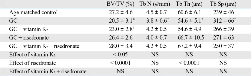
Table 2.Bone Histomorphometric Analysis of Cancellous Bone of the Proximal Tibial Metaphysis-Structural Parameters-
BV/TV (%) Tb N (#/mm) Tb Th (µm) Tb Sp (µm)
Age-matched control 27.2
±
4.6 4.5±
0.7 60.6±
6.1 239±
46GC 20.5
±
3.1* 3.8±
0.6�54.6
±
5.1�312
±
66�GC + vitamin K2 23.0
±
2.8� 4.2±
0.5 54.6±
4.9 266±
39GC + risedronate 26.4
±
2.6 4.0±
0.7 66.7±
10.5 271±
63GC + vitamin K2+ risedronate 28.0
±
3.4 4.2±
0.5 67.2±
9.4 250±
37Effect of vitamin K2 < 0.05 NS NS NS
Effect of risedronate < 0.0001 NS < 0.0001 NS
Effect of vitamin K2+ risedronate NS NS NS NS
GC, glucocorticoid; BV, bone volume; TV, total tissue volume; Tb N, trabecular number; Tb Th, trabecular thickness; Tb Sp, trabecular separation; NS, not significant.
*p < 0.01, �p < 0.05 vs. the age-matched control group.
Data are expressed as mean ±SD. mann Whitney U-test was used to compare the data between the age-matched control group and other groups. Two-way factorial analysis of variance (ANOVA) was used to determine the effect of vitamin K2and risedronate and their
combination effect.
[image:3.595.98.514.551.677.2](Table 1). GC also induced reductions in trabecular BV/TV, Tb N and Tb Th and increase in Tb Sp (Table 2), as a result of decreased bone formation (MS/BS, MAR, BFR/BS, BFR/BV) and increased bone erosion (ES/BS) (Table 3). GC reduced LGR/day (Table 3), but did not affect femoral length (Table 1).
Effect of vitamin K2administration on GC-treated rats
Vitamin K2did not affect body weight and femoral BMD
(Table 1). However, vitamin K2attenuated GC-induced
reduction in trabecular BV/TV (Table 2) by preventing GC-induced decrease in bone formation (MS/BS) and subsequently reducing GC-induced increase in bone erosion (ES/BS) (Table 3). Vitamin K2did not affect either LGR/day (Table 3) or
femoral length (Table 1).
Effect of risedronate administration on GC-treated rats
Risedronate attenuated GC-induced reduction in body weight and prevented GC-induced reduction in femoral BMD (Table 1). Risedronate also prevented GC-induced reductions in trabecular BV/TV and Tb Th (Table 2) by preventing GC-induced increase in bone erosion (ES/BS) although it also suppressed bone formation (MS/BS, MAR, BFR/BS, BFR/BV)
(Table 3). Risedronate did not affect either LGR/day (Table 3) or femoral length (Table 1).
Effect of combined administration of vitamin K2and
risedronate on GC-treated rats
There was no combination effect of vitamin K2and risedronate
on body weight and femoral length and BMD (Table 1) as well as trabecular bone structural parameters (Table 2). However, vitamin K2mildly attenuated the suppression of bone formation
(MS/BS) and bone erosion (ES/BS) caused by risedronate when administered in combination (Table 3).
Images of the proximal tibial metaphysis
Fig. 1 shows the images of the proximal tibial metaphysis, confirming the results of bone histomorphometric analysis regarding trabecular BV/TV.
The present study found the differential effect of vitamin K2
[image:4.595.40.539.373.512.2]and risedronate on trabecular bone in GC-treated rats. Vitamin
Table 3.Bone Histomorphometric Analysis of Cancellous Bone of the Proximal Tibial Metaphysis - Formative and Resorptive Parameters
LGR/day MS/BS MAR BFR/BS BFR/BV ES/BS
(µm/day) (%) (µm/day) (µm3/µm2/day) (%/yr) (%)
Age-matched control 9.6
±
0.8 16.4±
5.3 1.11±
0.14 18.6±
7.8 189±
83 2.78±
1.23GC 8.5
±
1.1�8.7
±
4.7�0.86
±
0.16�7.7
±
4.9�87
±
55�6.73
±
2.60�GC + vitamin K2 9.1
±
1.3 12.6±
3.8 0.86±
0.15� 10.9±
3.8� 120±
37� 4.18±
2.13�GC + risedronate 8.2
±
1.3�2.2
±
0.5* 0.67±
0.15* 1.4±
0.4* 13±
4* 1.83±
0.87 GC + vitamin K2+ risedronate 8.7±
1.0� 2.8±
0.9* 0.70±
0.17* 2.0±
1.0* 18±
8* 2.11±
0.73Effect of vitamin K2 NS < 0.05 NS NS NS < 0.05
Effect of risedronate NS < 0.0001 < 0.01 < 0.0001 < 0.0001 < 0.0001
Effect of vitamin K2+ risedronate NS < 0.05 NS NS NS < 0.05
GC, glucocorticoid; LGR, longitudinal growth rate; MS, mineralizing surface; BS, bone surface; MAR, mineral apposition rate; BFR, bone formation rate; BV, bone volume; ES, eroded surface; NS, not significant.
* p < 0.001, �p < 0.01, �p < 0.05 vs. the age-matched control group.
Data are expressed as mean ±SD. Mann Whitney U-test was used to compare the data between the age-matched control group and other groups. Two-way factorial analysis of variance (ANOVA) was used to determine the effect of vitamin K2and risedronate and their combination effect.
[image:4.595.42.537.573.717.2]DISCUSSION
Fig. 1.Images of the proximal tibial metaphysis. The images of the proximal tibial metaphysis confirm the results of bone histomorphometric analysis regarding trabecular BV/TV. GC, glucocorticoid; BV, bone volume; TV, total tissue volume.
K2 attenuated GC-induced trabecular bone loss by preventing
GC-induced decrease in bone formation (MS/BS) and subsequently reducing GC-induced increase in bone erosion (ES/BS). Risedronate prevented GC-induced trabecular bone loss by preventing GC-induced increase in bone erosion (ES/BS) although it also suppressed bone formation (MS/BS,
MAR, BFR/BS, BFR/BV). Vitamin K2 mildly attenuated
suppression of bone formation (MS/BS) and bone erosion (ES/BS) caused by risedronate without affecting trabecular bone mass when administered in combination.
It has been argued that rat is a poor model of GC-induced osteoporosis, because GC inhibits bone resorption by osteoclasts, resulting in a protective effect on the skeleton in mature rats16. However, GC administration has been shown to
induce losses of trabecular BV/TV, Tb N, and Tb Th in young rats10,17-20. Therefore, the clearly observed trabecular bone loss in
GC-treated young rats in the previous studies could be due, at least in part, to growth inhibition. However, GC treatment is required even in children with nephritis, systemic lupus erythematosus, asthma, juvenile idiopathic or rheumatic arthritis, lymphoblastic leukemia, and other diseases. Therefore, it is important to study the effects of drugs on GC-induced bone loss in young rats. In the present study, GC administration induced trabecular bone loss in young rats compared with age-matched controls.
GC-induced trabecular bone loss has been associated with decreased bone formation and increased bone resorption, however the key histological feature of GC-induced trabecular bone loss has been shown to be reduction in the thickness of trabecular bone, reflecting suppressed bone formation21. In the
present study, GC-induced trabecular BV/TV loss compared with age-matched controls was associated with decreases in Tb Th and Tb N and increase in Tb Sp because bone formation was decreased and bone erosion was increased by GC administration. Because bone erosion reflects the coupling of bone formation to bone resorption, both decreased bone formation and increased bone resorption could account for increased bone erosion.
Vitamin K2is known to have an anabolic action on bone;
regulation of bone formation by vitamin K2may involve
γ-carboxylation of osteocalcin and/or may be mediated by the steroid and xenobiotic receptor.22-26In the present study, vitamin K2attenuated GC-induced trabecular bone loss by preventing GC-induced decrease in bone formation (MS/BS) and subse-quently reducing GC-induced increase in bone erosion. These results suggest at least the mild anabolic effect of vitamin K2, because bone erosion reflects the coupling of bone formation to bone resorption. However, the effects of vitamin K2on trabecu-lar bone mass and bone metabolism seem to be modest.
The bisphosphonates inhibit osteoclast-mediated bone resorption, and loss of osteoclast function and apoptosis are the consequence of loss of function of one or more important signaling proteins. In particular, nitrogen-containing
bisphos-phonates, such as alendronate and risedronate, are not metabolized but can inhibit enzymes of the mevalonate pathway, thereby preventing the biosynthesis of isoprenoid compounds, which are essential for the post-translational modification of small GTPases.27 In the present study, risedronate prevented GC-induced trabecular bone loss by preventing GC-induced increase in bone erosion although it also suppressed bone formation (mineralizing surface, mineral apposition rate, and bone formation rate). Thus, risedronate ameliorated the imbalance of bone formation and resorption. These results are consistent with those of previous studies.9 However, oversuppression of bone formation as well as bone resorption is concerned.
Because vitamin K2and risedronate have different actions on bone formation and resorption, the beneficial effects of combi-nation of the 2 agents on trabecular bone could be expected. A few studies showed the effects of combined treatment with bisphosphonates and vitamin K2in other models of osteo-porosis.28-31Ito et al.29demonstrated additive effect of risedronate and vitamin K2in preventing deterioration of the trabecular bone architecture in ovariectomized rats, whereas Otomo et al.30 showed no significant additive effect of risedronate and vitamin K2 on bone turnover, bone mass/density, bone structure, mineral properties (mineral-to-matrix ratio), and bone strength evidenced by no significant effect of vitamin K2on bone parameters in ovariectomized rats. Furthermore, Iwasaki et al.31 reported that concomitant use of vitamin K2with incadronate ameliorated suppression of bone formation and more effecti-vely prevented cancellous bone loss in tail-suspended rats. In the present study, risedronate strongly suppressed bone formation and erosion, and vitamin K2mildly attenuated the suppression of bone formation (MS/BS) and bone erosion caused by risedronate without affecting trabecular bone mass when administered in combination. Bone strength reflects both bone mass and bone quality, and bone quality is determined by the bone architecture, turnover, damage accumulation, minera-lization, and matrix.32Thus, strong suppression of bone formation as well as bone resorption caused by risedronate might possibly lead to deterioration of bone quality. Vitamin K2 might have a mild anabolic effect, possibly playing a role in attenuating deterioration of bone quality in rats treated with GC and risedronate.
1. Longui CA. Glucocorticoid therapy: minimizing side effects. J Pediatr (Rio J) 2007;83(5 suppl):S163-77.
2. Van Staa TP, Leufkens HG, Cooper C. The epidemiology of corticosteroid-induced osteoporosis: a meta-analysis. Osteoporos Int 2002;13:777-87.
3. Van Staa TP, Leufkens HG, Abenhaim L, Zhang B, Cooper C. Use of oral corticosteroids and risk of fractures. J Bone Miner Res 2000; 15:993-1000.
4. Nawata H, Soen S, Takayanagi R, Tanaka I, Takaoka K, Fukunaga M, et al. Guidelines on the management and treatment of glucocorticoid-induced osteoporosis of the Japanese Society for Bone and Mineral Research (2004). J Bone Miner Metab 2005;23:105-9.
5. Wallach S, Cohen S, Reid DM, Hughes RA, Hosking DJ, Laan RF, et al. Effects of risedronate treatment on bone density and vertebral fracture in patients on corticosteroid therapy. Calcif Tissue Int 2000;67:277-85.
6. Reid DM, Hughes RA, Laan RF, Sacco-Gibson NA, Wenderoth DH, Adami S, et al. Efficacy and safety of daily risedronate in the treatment of corticosteroid-induced osteoporosis in men and women: a rando-mized trial. European Corticosteroid-Induced Osteoporosis Treatment Study. Bone Miner Res 2000;15:1006-13.
7. Tanaka I, Oshima H. Vitamin K2as a potential therapeutic agent for
glucocorticoid-induced osteoporosis. Clin Calcium 2007;17:1738-44 (in Japanese).
8. Balooch G, Yao W, Ager JW, Balooch M, Nalla RK, Porter AE, et al. The aminobisphosphonate risedronate preserves localized mineral and material properties of bone in the presence of glucocorticoids. Arthritis Rheum 2007;56:3726-37.
9. Iwamoto J, Seki A, Takeda T, Sato Y, Yamada H, Shen CL, et al. Preventive effects of risedronate and calcitriol on cancellous osteopenia in rats treated with high-dose glucocorticoid. Exp Anim 2006;55:349-55.
10. Hara K, Kobayashi M, Akiyama Y. Vitamin K2 (menatetrenone) inhibits bone loss induced by prednisolone partly through enhancement of bone formation in rats. Bone 2002;31:575-81.
11. Iwamoto J, Yeh JK, Takeda T. Effect of vitamin K2 on cortical and cancellous bones in orchidectomized and/or sciatic neurectomized rats. J Bone Miner Res 2003;18:776-83.
12. Iwamoto J, Seki A, Takeda T, Sato Y, Yamada H, Shen CL, et al. Comparative effects of risedronate and calcitriol on cancellous bone in rats with glucocorticoid-induced osteopenia. J Nutr Sci Vitaminol (Tokyo) 2006;52:21-7.
13. Erben RG. Embedding of bone samples in methylmethacrylate: an improved method suitable for bone histomorphometry, histochemistry, and immunohistochemistry. J Histochem Cytochem 1997;45:307-13. 14. Parfitt AM, Drezner MK, Glorieux FH, Kanis JA, Malluche H,
Meunier PJ, et al. Bone histomorphometry: standardization of nomenclature, symbols, and units. Report of the ASBMR Histomor-phometry Nomenclature Committee. J Bone Miner Res 1987;2:595-610.
15. Prakasam G, Yeh JK, Chen MM, Castro-Magana M, Liang CT, Aloia JF. Effects of growth hormone and testosterone on cortical bone formation and bone density in aged orchiectomized rats. Bone 1999;
24:491-7.
16. Shen V, Birchman R, Liang XG, Wu DD, Lindsay R, Dempster DW. Prednisolone alone, or in combination with estrogen or dietary calcium deficiency or immobilization, inhibits bone formation but does not induce bone loss in mature rats. Biol 1997;21:345-51.
17. Furuichi H, Fukuyama R, Izumo N, Fujita T, Kohno T, Nakamuta H, et al. Bone-anabolic effect of salmon calcitonin on glucocorticoid-induced osteopenia in rats. Bio Pharm Bull 2000;23:946-51.
18. Nitta T, Fukushima T, Nakamuta H, Koida M. Glucocorticoid-induced secondary osteopenia in female rats: a time course study as compared with ovariectomy-induced osteopenia and response to salmon calcitonin. Jpn J Pharmacol 1999;79:379-86.
19. Noa M, Mendoza S, Más R, Mendoza N, León F. Effect of D-003, a mixture of very high molecular weight aliphatic acids, on predniso-lone-induced osteoporosis in Sprague-Dawley rats. Drugs R D 2004;5:281-90.
20. Tanaka Y, Nakamura T, Nishida S, Suzuki K, Takeda S, Sato K, et al. Effects of a synthetic vitamin D analog, ED-71, on bone dynamics and strength in cancellous and cortical bone in prednisolone-treated rats. J Bone Miner Res 1996;11:325-36.
21. Manolagas SC, Weinstein RS. New developments in the pathogenesis and treatment of steroid-induced osteoporosis. J Bone Miner Res 1999;14:1061-6.
22. Hauschka PV, Lian JB, Cole DE, Gundberg CM. Osteocalcin and matrix Gla protein: vitamin K-dependent proteins in bone. Physiol Rev 1989;69:990-1047.
23. Koshihara Y, Hoshi K. Vitamin K2 enhances osteocalcin accumulation in the extracellular matrix of human osteoblasts in vitro. J Bone Miner Res 1997;12:431-8.
24. Shearer MJ. Vitamin K. Lancet 1995;345:229-34.
25. Vermeer C, Jie KS, Knapen MH. Role of vitamin K in bone metabo-lism. Annu Rev Nutr 1995;15:1-22.
26. Tabb MM, Sun A, Zhou C, Grün F, Errandi J, Romero K, et al. Vitamin K2 regulation of bone homeostasis is mediated by the steroid and xenobiotic receptor SXR. J Biol Chem 2003;278:43919-27.
27. Rogers MJ, Frith JC, Luckman SP, Coxon FR, Benford HL, Mönkkönen J, et al. Molecular mechanisms of action of bisphosphonate. Bone 1999;24 (5 Suppl):S73-9.
28. Iwamoto J, Takeda T, Sato Y. Effects of vitamin K2 on the development of osteopenia in rats as the models of osteoporosis. Yonsei Med J 2006;47:157-66.
29. Ito M. Bone mass, microstructure, and quality with bone strength - The effects of antiresorptive agents - . J Jpn Soc Bone Morphom 2002; 12:51-4 (in Japanese).
30. Otomo H, Sakai A, Ikeda S, Tanaka S, Ito M, Phipps RJ, et al. Regulation of mineral-to-matrix ratio of lumbar trabecular bone in ovariectomized rats treated with risedronate in combination with or without vitamin K2. J Bone Miner Metab 2004;22:404-14.
31. Iwasaki Y, Yamato H, Murayama H, Sato M, Takahashi T, Ezawa I, et al. Combination use of vitamin K(2) further increases bone volume and ameliorates extremely low turnover bone induced by bisphosphonate therapy in tail-suspension rats. J Bone Miner Metab 2003;21:154-60. 32. NIH Consensus Development Panel on Osteoporosis Prevention,
Diagnosis, and Therapy. Osteoporosis prevention, diagnosis, and therapy. JAMA 2001;285:785-95.