Journal of Chemical and Pharmaceutical Research, 2015, 7(8):291-299
Research Article
ISSN : 0975-7384
CODEN(USA) : JCPRC5
Performance &analysis of automated removal of head movement artifacts in
EEG using brain computer interface
M. Anto Bennet*, R. Suresh and S. Mohamed Sulaiman
Department of Electronics and Communication Engineering, VELTECH, Avadi-Chennai, Tamil Nadu, India
_____________________________________________________________________________________________
ABSTRACT
Removal of Head Movement Artifacts in EEG comes under the Domain of Bio-Medical Engineering. In Electroencephalogram (EEG), the measurements are based on the Human Brain Activities, so the output should be very accurate. But, Because of the Artifacts present in the EEG signal the Accuracy of the signal is reduced. So, improvements are needed to obtain the accurate output. Different types of artifacts occur during the EEG recording such as Head Movement Artifacts, Eye Blink Artifacts, Respiration Artifacts, Muscle Movement Artifacts, and Improper Electrode Placement Artifacts and Environmental factors. This Paper is about the removal of Head Movement Artifacts. It can be implemented by using the Independent Component Analysis (ICA) Method, which separates the multivariate components into subcomponents. EEG signal is a multivariate component signal which consists of Pure EEG signal associated with Artifacts. It can be recorded by using EEG Electrodes connected to the PC with the Necessary Amplification section. Accelerometer is used to measure the Head movement in the tri-axis. Head Movement signals are recorded separately using the Accelerometer and correlated with the EEG signal by passing the both signal to the ICA. The signals which are correlated with each other are considered as Artifacts and flagged for the removal.
Keywords: Brain–Computer Interface (BCI), Mind-Machine Interface (MMI),Brain–Machine Interface (BMI), Independent Component Analysis(ICA)
_____________________________________________________________________________________________
INTRODUCTION
Brain-Computer Interface (BCI) can help people with neuromuscular diseases (e.g. amyotrophic lateral sclera-sis (ALS), brainstem stroke, etc.) communicate. Being one of the popular approaches, event-related potential (ERP) based BCIs exploit the fact that brain potential can be modulated by human attention. For example, user attention to a rare event in an oddball paradigm can enhance the P300 component of ERP time-locked to this event. This would make the computer to under the actions of the Brain. Then, user intention (attend or not) can be detected by decoding the modulated potential. Although significant progress has been made in researching brain–computer interface technologies in recent years, the applications controlled by these interfaces have largely been designed for training or demonstration purposes. The Georgia State University (GSU) Brain Lab is devoted to researching and developing interaction techniques that will allow BCIs to be effective in real-world applications. Hence, the BCI applications will be developed because of the GSU Brain lab around the world.
Brain–computer interfaces (BCIs) are devices that translate brain signals into operational commands for technical devices. While multiple methods have been developed to extract and classify the electrical activity of the brain, the application of BCIs to the target group, for example, patients with severe physical impairment and brain damage, has rarely been considered. The electroencephalogram (EEG) is modified by motor imagery and can be used by patients with severe motor impairments (e.g., late stage of amyotrophic lateral sclerosis) to communicate with their environment. Such a direct connection between the brain and the computer is known as an EEG-based brain– computer interface (BCI).It involves the reading of EEG signal from the Human Brain to the computer with the use of EEG electrodes with appropriate Amplifiers. The idea of Brain-Computer Interfaces (BCIs) which allow the control of devices using brain signals evolved from the realm of science fiction to simple devices that currently exist. BCIs naturally present themselves to many extremely useful applications including prosthetic devices, restoring or aiding in communication and hearing, military applications, video gaming and virtual reality, and robotic control, and have the possibility of significantly improving the quality of life of many disabled individuals.
The ultimate goal of BCI research is to create a system that not only an “open loop” system that responds to users thoughts but a “closed loop” system that also gives feedback to the user. Researchers initially focused on the motor-cortex of the brain, the area which controls muscle movements, and testing on animals quickly showed that the natural learning behaviors of the brain could easily adapt to new stimuli as well as control the firing of specific areas of the brain. Though the idea of using EEG waves as input to BCIs has existed since the initial conception of BCIs, actual working BCIs based on EEG input have only recently appeared. Most EEG-BCI systems follow the paradigm of reading in and analyzing EEG data, translating that data into device output, and giving some sort of feedback to the user. The primary difficulty in creating an EEG-based BCI is the feature extraction and classification of EEG data that must be done in real-time if it is to have any use. Feature extraction deals with separating useful EEG data from noise and simplifying that data so that classification, the problem of trying to decide what the extracted data represents, can occur. There is no best way of extracting features from EEG data and modern BCIs often use several types of feature extraction including Horthy parameters (a way of describing the normalized slope of the data),wavelet transforms, Fourier transforms, and various other types of filters.
The P300 component of an event related potential is widely used in conjunction with brain–computer interfaces (BCIs) to translate the subject’s intent by mere thoughts into commands to control artificial devices [1]. In addition, they propose an online method that optimizes information transfer rates and/or accuracies. This is achieved by an algorithm which dynamically limits the number of sub trial presentations, according to the subject’s current online performance in real-time. In the first, study peak information transfer rates up to 92 bits/min with an accuracy of 100% were achieved by one subject with a mean of 32 bits/min at about 80% accuracy.
neuromuscular disabilities. With proper training, individuals can learn to modulate the amplitude of specific EEG electroencephalographic components (e.g., the 8–12 Hz rhythm and 18-26 Hz rhythm) over the sensor motor cortex and use them to control a cursor on a computer screen. Conventional spectral techniques for monitoring the continuous amplitude fluctuations fail to capture essential amplitude/phase relationships of the rhythms in a compact fashion and, therefore, are suboptimal. By extracting the characteristic rhythm for a user, the exact morphology can be characterized and exploited as a matched filter
An electroencephalographic (EEG) brain–computer interface (BCI) internet browser was designed and evaluated with 10 healthy volunteers and three individuals with advanced amyotrophic lateral sclerosis (ALS), all of whom were given tasks to execute on the internet using the browser [4]. Participants with ALS achieved an average accuracy of 73% and a subsequent information transfer rate (ITR) of 8.6 bits/min and healthy participants with no prior BCI experience over 90% accuracy and an ITR of 14.4 bits/min. FuRIA, a trainable feature extraction algorithm for noninvasive brain–computer interfaces (BCI) is based on inverse solutions and on the new concepts of fuzzy region of interest (ROI) and fuzzy frequency band [5]. FuRIA can automatically identify the relevant ROI and frequency bands for the discrimination of mental states, even for multiclass BCI. Once identified, the activity in these ROI and frequency bands can be used as features for any classifier. The evaluations of FuRIA showed that the extracted features were interpretable and can lead to high classification accuracies.
Mental state estimation is potentially useful for the development of asynchronous brain–computer interfaces [8]. In this study, four mental states have been identified and decoded from the electrocorticograms (ECoGs) of six epileptic patients, engaged in a memory reach task. A novel signal analysis technique has been applied to high-dimensional, statistically sparse ECoGs recorded by a large number of electrodes. The strength of the proposed technique lies in its ability to jointly extract spatial and temporal patterns, responsible for encoding mental state differences. Brain Computer interface (BCI) is a system which allows direct translation of brain states into actions, bypassing the usual muscular pathways [9]. A BCI system works by extracting user brain signals, applying machine learning algorithms to classify the user’s brain state, and performing a computer-controlled action. They developed an automated approach which systematically analyzes the relative contributions of different preprocessing and meta-classification approaches. They applied this procedure to three data sets drawn from BCI Competition 2003 and BCI Competition III, each of which exhibit very different characteristics. The final classification results compare favorably with those from past BCI competitions. Additionally, it analyze the relative contributions of individual preprocessing and meta-classification choices and discuss which types of BCI data benefit most from specific algorithms, the only source of control in any BCI system. Artifacts are undesirable signals that can interfere with neurological phenomena. An important determinant of the value of quantitative neuro imaging studies is the reliability of the derived information, which is a function of the data collection conditions [10]. Near infrared spectroscopy (NIRS) and electro encelphalography are independent sensing domains that are well suited to explore principal elements of the brain’s response to neuro-activation, and whose integration supports development of compact, even wearable, systems suitable for use in open environments.
Brain–machine interfaces (BMIs) provide a versatile tool for rehabilitation of severely disabled people [11]. Current BMI systems focus on the control of kinematic variables. However, this approach limits the application space of BMI technology to simulated environments. BMI systems that are aimed toward prostheses must, then, control interaction forces with their environments. They designed a BMI-driven architecture that provides a critical link between neuronal ensemble activity and real-world dynamics. P300 spellers are mainly composed of an interface, by which alphanumerical characters are presented to users, and a classification system, which identifies the target character by using acquired EEG data [12]. They proposed modifications both to the interface and to the classification system, in order to reduce the number of required stimulus repetitions and consequently boost the information transfer rate.
such changes are very similar between subjects, and thus can be reliably estimated using data from other users and utilized to construct an invariant feature space.
EXPERIMENTAL SECTION
INDEPENDENT COMPONENT ANALYSIS
[image:4.595.226.476.226.358.2]Independent component analysis (ICA) is a method for finding underlying factors or Components from multivariate (multidimensional) statistical data. What distinguishes ICA from other methods is that it looks for components that are both statistically independent, and non-gaussian. ICA is a technique to separate linearly mixed sources. For instance, define the time courses of 2 independent sources A (top) and B(bottom) as shown in Fig 1.
Fig 1 Source Signal and Artifact Signal
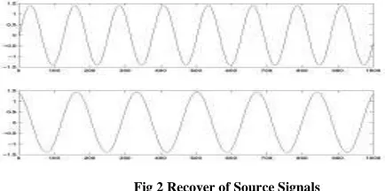
Let the two sources are mixed. The top curve is equal to A minus twice B and the bottom the linear combination is 1.73*A +3.41*B. Input these two signals into the ICA algorithm which is able to uncover the original activation of A and B.
Fig 2 Recover of Source Signals
The algorithm cannot recover the exact amplitude of the source activities. By trying this with we have to see different degree of noise that it's quite robust. ICA can only extract sources that are combined linearly shown in fig 2.
Whitening the data
[image:4.595.170.387.435.543.2]
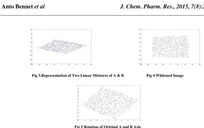
Fig 3.Representation of Two Linear Mixtures of A & B Fig 4 Whitened Image
Fig 5 Rotation of Original A and B Axis
Whitening the two linear mixtures (shown in fig 4), the variance on both axis is now equal and the correlation of the projection of the data on both axis is 0 (meaning that the covariance matrix is diagonal and that all the diagonal elements are equal). Then applying ICA only mean to "rotate" this representation back to the original A and B axis space. The whitening process is simply a linear change of coordinate of the mixed data. Once the ICA solution is found in this "whitened" coordinate frame, the ICA solution can be re projected back into the original coordinate frame shown in fig 5.
ICA algorithm
Intuitively let the ICA rotates the whitened matrix back to the original (A,B) space. It performs the rotation by minimizing the Gaussianity of the data projected on both axes (fixed point ICA).
Fig 6 Projection of 2 Arms Fig 7 Contrast of Projection of Original A,B far from Guassian
The projection on both axis is quite Gaussian (i.e., it looks like a bell shape curve shown in fig 6). By contrast the projection in the original A, B space far from gaussian.
[image:5.595.111.491.460.561.2]Fig 8 Mixing and Unmixing Matrix of ICA
Here the EEG Signals are referred as the Source Signals. The Mixing matrix is composed of both EEG signals and the Head Movement Artifact Signals. Since the Head Movement Artifact signals are generated along the EEG signal for the movement of user’s head. In the Un mixing Matrix, the Head Movement signals are alone passed along the EEG signals with the Head Movement Artifacts. Head Movement Artifacts are obtained separately with the help of Accelerometer, it is a sensor which measures the acceleration in the x,y and z axis and produces the three equivalent output voltage for the three different axis. The ICA compares the both signal, the signals correlated with each other are referred as Artifacts and are removed. So, the obtained EEG output is free from the Head Movement Artifacts shown in fig 8.
ICA IMPLEMENTATION FOR THE HEAD MOVEMENT ARTIFACTS REDUCTION
Fig 9 Removal of Head Movement Artifacts
The EEG Electrodes are attached to the user’s head along with the Accelerometer ADXL335.
The user is asked to perform a mind task along with the Head movement; the EEG signal obtained for this activity is recorded separately. Then at the same time, the Accelerometer (ADXL335) output for the above activity is recorded separately. The Accelerometer signal obtained for the three different axes x, y and z are analog signal. Hence it should be converted to the Digital format to make it readable by the computer.
RESULTS AND DISCUSSION
ACCELEROMETER READING IN PC
[image:7.595.142.436.502.656.2]
Fig 10 Accelerometer Connection to PC Fig 11 Accelerometer Interface to PC Kit
The Tri-Axial Output from the ADXL335 Accelerometer (X,Y and Z axis) is passed to the AN5,AN6 and AN7 pins of the PIC165877A.The Accelerometer is provided with the constant supply of 3V.The PIC165877A is coded in Embedded C to convert the obtained Analog Signals from the AN5,AN6 and AN7 pins into Digital Signal and transmits as a serial signal to the TX Pin and at the same time it is received at the RX pin of the Serial to USB Converter, which passes the received Digital signals to the USB Port of the PC. It is shown in Fig 10. The Accelerometer is attached to the wearable cap which is shown as in the Fig 11.
EEG AND ACCELEROMETER SIGNAL ACQUISTION
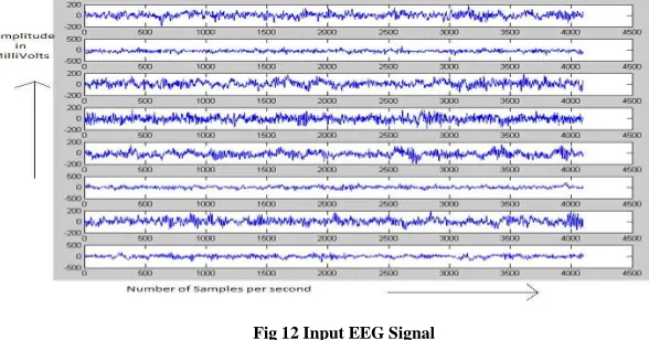
The EEG Electrodes are connected to the different lobes of the user such as in the points F3,F4,Fp1,Fp7,Pz,Cz,C3,C4
respectively Since, the EEG electrodes receive the different types of the EEG signals from the different points.These Signals are received at the range of Micro-Volts.So, the necessary amplification is provided in order to read these signal in the PC. The BRAINLAB is a software, which is used to read these obtained EEG from the different lobes in the PC and are recorded imultaneously.These signals are converted into coordinate value and are plotted into the MS-Excel sheet by using the function Export to Excel Sheet. on plotting these coordinate values in the Matlab R2012.
Fig 12 Input EEG Signal
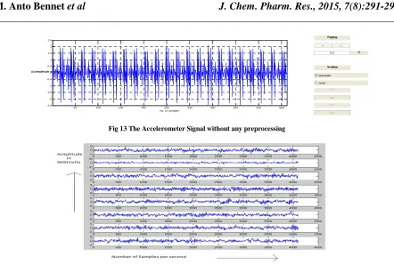
Fig 13 The Accelerometer Signal without any preprocessing
Fig 14 ICA Output for the Accelerometer Data Input
[image:8.595.142.479.411.507.2]The Fig 14 Shows the ICALAB output for the workspace data of Accelerometer Input at the sample rate of 512 Hz.
Fig 15 Reconstructed Accelerometer signal – Deflation output
[image:8.595.150.471.576.666.2]The Fig 15 shows the reconstructed accelerometer signal output, which are passed along with the EEG signal as the ICA input.
Fig 16 EEG Signals without Artifacts
Fig 16 shows the ICA Output, such that the Artifacts present in the EEG signal of the Different Lobes are removed successfully. It is the EEG signal which does not contain Head Movement Artifacts.
50 100 150 200 250 300 350 400 450 500
-2.5 -2 -1.5 -1 -0.5 0 0.5 1 1.5 2 2.5
No. of Samples
y1amplitude in mv
50 100 150 200 250 300 350 400 450 500
-60 -40 -20 0 20 40 60
xr1
50 100 150 200 250 300 350 400 450 500
0 20 40 60 80 100 120 140
No. of samples ( 512)
CONCLUSION
The Head Movement Artifacts present along the Input EEG Signal is removed successfully using the Independent Component Analysis(ICA).The EEG signal without the Head Movement Artifacts looks like a Pure EEG signal, since Head Movement Artifacts are the most interrupted portion of Artifacts in the EEG signals. The Eye Blinks Artifacts, Respiratory Artifacts can be removed easily by applying Adaptive Filters, but the Head Movement Artifacts cannot be removed by applying filters. Thus, the accelerometer is used to record the Head Movement Artifacts and applied with the EEG signal to the ICA. From the Unmixing Matrix of ICA, the EEG signal without the Head Movement Artifacts are obtained.
REFERENCES
[1] Alexander Lenhardt; MatthiasKaper; Helge J. Ritter, IEEE Transactions on Neural Systems And Rehabilitation
Engineering, 2008, 16(2), 121-129.
[2] Bernardo Dal Seno;MatteoMatteucci; Luca T. Mainardi, IEEE Transactions on Neural Systems And
Rehabilitation Engineering, 2010, 18(1), 20-27.
[3] ChuanJia; XiaorongGao; Bo HongandShangkaiGao, IEEE Transactions on Biomedical Engineering, 2011, 58(1), 200-206.
[4] Emily M. Mugle;, Carolin A.Ruf; Sebastian Halder; Michael Bensch; Andrea Kübler, IEEE Transactions on
Neural Systems And Rehabilitation Engineering,2010, 18(6), 599-601.
[5] Fabien Lotte; Anatole Lécuyer; Bruno Arnaldi,IEEE Transactions On Signal Processing,2009, 57(8), 3252-3255.
[6] Haihong Zhang;Cuntai Guan;Chuanchu Wang,IEEE Transactions on Biomedical Engineering,2008,55(6), 1754-1756.
[7] Jack DiGiovanna;BabakMahmoudi; Jose Fortes;Jose C. Principe;Justin, C. Sanchez, IEEE Transactions on
Biomedical Engineering,2009, 56(1), 54-56.
[8] Koel Das; Daniel S. Rizzuto; ZoranNenadic, IEEE Transactions on Biomedical Engineering,2009, 56(8), 2114-2116.
[9] Paul S.Hammon; Virginia R. de Sa, IEEE Transactions on Biomedical Engineering,2007, 54(3), 518-520. [10] Randall L. Barbour; Harry L. Graber; Yong Xu; Yaling Pei; Christoph H. Schmitz; Douglas S. Pfeil;AnanditaTyagi; Randy Andronica; Daniel C. Lee; San-Lian S. Barbour;J. David Nichols; Mark E. Pflieger,
IEEE Transactions on Neural Systems and Rehabilitation Engineering,2012, 20(2), 803-806.
[11] RodolpheHeliot; Amy L. Orsborn;KaruneshGanguly, IEEE Transactions on Systems, Man, And Cybernetics—
Part A: Systems And Humans, 2010, 40(4), 732-734.
[12] SercanTahaAhi; Hiroyuki Kambara; Yasuharu Koike, IEEE Transactions on Neural System Rehabilation
Engineering, 2011, 59(1), 6-8.
[13] Walter G. Besio; Hongbao Cao; Peng Zhou, IEEE Transactions on Neural Systems and Rehabilitation
Engineering, 2008, Vol. 16(2), 191-194.
[14] Weiguo Song;ArunRamakrishnan; Ubong I. Udoekwere; Simon F. Giszter, IEEE Transactions on Biomedical
Engineering, 2009, 56(11),1022-1026.