International Journal of Emerging Technology and Advanced Engineering
Website: www.ijetae.com (ISSN 2250-2459,ISO 9001:2008 Certified Journal, Volume 2, Issue 12, December 2012)
398
Thyroid Segmentation on US Medical Images: An Overview
Sheeja Agustin A
1, S. Suresh Babu
21Research Scholar, Department of Computer Science, Noorul Islam Centre for Higher Education, Noorul Islam University,
Kumaracoil, Tamil Nadu
2
Principal, T K M College of Engineering, Kollam, Kerala
Abstract- Thyroid is a small butterfly shaped gland located in the front of the neck just below the Adams apple. Thyroid is one of the endocrine gland , which produces hormones that help the body to control metabolism. Different thyroid disorders [10] includes Hyperthyroidism, Hypothyroidism, goiter, and thyroid nodules (benign/malignant).Ultrasound imaging is most commonly used to detect and classify abnormalities of the thyroid gland. Other modalities (CT/MRI) are also used. Computer aided diagnosis (CAD) help radiologists and doctors to increase the diagnosis accuracy, reduce biopsy ratio and save their time and effort. Numerous researches have been carried out in thyroid medical images and that are utilized for the diagnosis process [11]. In this paper some methods are tested to detect and segment thyroid US images.
Keyterms- Medical imaging, Thyroid, segmentation, FCM, Histogram clustering, Quad tree, Region growing etc.
I. INTRODUCTION
Image processing [5] is any form of signal processing for which the input is an image such as photograph or video frame, the output of image processing may be either an image or parameters related to the image. Image processing usually refers to digital image processing. Digital image processing is the use of computer algorithms to perform image processing on digital images. Medical imaging is the technique and process used to create images of the human body for clinical purpose or medical science including the study of normal anatomy and physiology. Different Imaging technologies are:-Radiology, Magnetic resonance imaging (MRI), Nuclear medicine, Photo acoustic imaging, Breast thermograph, Tomography and ultrasound imaging. In image processing segmentation algorithms constitute one of the main focuses of research.
Image segmentation is the process of partitioning an image into multiple segment or set of pixels used to locate object and boundaries. Each of the pixels in a region is similar with respect to some characteristics such as color, intensity or texture. Different applications of image segmentation are Medical Imaging, Locate objects in satellite images, Face recognition, Fingerprint recognition, Traffic control systems etc.
In the field of Image analysis segmentation of medical
imagesis a challenging problem due to poor resolution and
weak contrast of medical images.. Medical image segmentation [12] means Segmentation of known anatomic structures from medical images. Structures of interest include organs such as cardiac ventricles or kidneys, abnormalities such as tumors and cysts as well as other structures such as bones, vessels, brain structures etc. Now a day’s Medical image analysis has played more and more important role in many clinical procedures and in detecting different types of human diseases. Now a day’s most of the peoples have thyroid diseases.
For diagnosing thyroid diseases, Ultrasound (US) and Computerized Tomography (CT) are two of the most popular imaging modalities. US imaging is inexpensive, non-invasive and easy to use. US images are often adopted due to their cost-effectiveness and portability in smaller hospitals. The thyroid is well suited to ultrasound study because of its superficial location, size and echogenicity [12]. Computer-Aided Diagnosis (CAD) of Thyroid Ultrasound is necessary in order to delineating nodules, classifying benign/malignant and estimating the volumes of thyroid tissues to increase reliability and reduce invasive operations such as biopsy and Fine Needle Aspiration (FNA).
Thyroid produces thyroid hormones T4( thyroxin) and T3(triiodothyronine). These thyroid hormones tell the cells in the body how fast to use energy and create proteins. The thyroid gland also makes calcitonin, a hormone that helps to regulate calcium levels in the blood by inhibiting the breakdown (reabsorption) of bone and increasing calcium
excretion from the kidneys. The Hypothalamus
releases(thyrotopin-releasing hormone), which in turn causes the pituitary gland to release TSH( thyroid-stimulating hormone), TSH stimulates the thyroid gland to produce and secrete thyroid hormones. When there is sufficient thyroid hormone in the blood, the amount of TSH decreases to maintain constant amounts of thyroid
International Journal of Emerging Technology and Advanced Engineering
Website: www.ijetae.com (ISSN 2250-2459,ISO 9001:2008 Certified Journal, Volume 2, Issue 12, December 2012)
[image:2.612.68.270.132.285.2]399
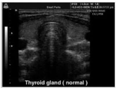
Fig I: Normal Thyroid US image
The rest of this paper is organized as follows. Section II describes about preprocessing, section III describes about
segmentation, section
IV
describes about experimentalresults .Finally, the conclusions as well as future directions are summarized in Section V.
II. PREPROCESSING
The aim of pre-processing[5] is an improvement of the image data that suppresses undesired distortions or enhances some image features relevant for further processing and analysis task. Neighboring pixels corresponding to one real object have the same or similar brightness value. Image pre-processing includes Geometric correction – adjusts locations of pixels, and pixel values and Radiometric correction – adjusts pixel values, analyst judgment Image Preprocessing.
A.Noise Reduction
Noise is an important factor that influences image quality. Noise reduction[5] is necessary to do image processing and image interpretation so as to acquire useful information that we want. In this paper the median filter is used to reduce noise in an image.
B. EDGE Detection
Edges characterize object boundaries and are therefore useful for segmentation, registration, and identification of objects in a scene. The sobel operator is used in image processing, particularly within edge detection algorithms. The Canny edge detector is an edge detection operator that uses a multi-stage algorithm to detect a wide range of edges in images.
C. Enhancement
Contrast enhancement is a technique that able to suppress speckle in thyroid ultrasound image. One of the popular methods in contrast enhancement is histogram equalization. Contrast enhancement is complete by suppressing speckles – the modulation of image brightness by random dark and bright region. The procedures start with computation of calibrated radio frequency (RF) spectra.
D. Histogram Equalization
The histogram equalization[5] is appropriate to enhance a given image. The approach is to design a transformation T (.) such that the gray values in the output is uniformly distributed in [0, 1].
III. SEGMENTATION
Segmentation [5] is a tool that used widely in many applications including image processing.One of the common applications of segmentation is in medical image analysis for clinical diagnosis that has an important role in terms of quality and quantity. Medical image segmentation methods generally have restrictions because medical images have very similar gray level and texture among the interested objects. Therefore, significant segmentation error may occur.
A.Fuzzy c-means Algorithm
Fuzzy c-means (FCM)[7] is a data clustering technique in which a dataset is grouped into n clusters with every data point in the dataset belonging to every cluster to a certain degree. For example, a certain data point that lies close to the center of a cluster[1] will have a high degree of belonging or membership to that cluster and another data point that lies far away from the center of a cluster will have a low degree of belonging or membership to that cluster.
B.Histogram Lustering
International Journal of Emerging Technology and Advanced Engineering
Website: www.ijetae.com (ISSN 2250-2459,ISO 9001:2008 Certified Journal, Volume 2, Issue 12, December 2012)
400
C. QUAD Tree
The quad tree [6] datastructure is widely used in digital
image processing for modeling spatial segmentation of
images and surfaces. A quad tree is a tree in which each
node has four descendants. Since most algorithms based on
quad trees require complex navigation between nodes, efficient traversal methods as well as efficient storage techniques are of great interest. The quad tree data structure is a tree in which each node has at most four children. In digital image processing, quad trees are used to efficiently store image segmentations.
D. Region Growing
Region growing [7] is a procedure that groups pixels or sub regions into larger regions based on predefined criteria for growth. The basic approach is to start with a set of seed points and from these grow regions by appending to each seed those neighboring pixels have predefined properties similar to the seed.
E. Random Walker
The random walker[4] algorithm is an algorithm for
image segmentation. In the first description of the algorithm,user interactively labels a small number of pixels with known labels .
The unlabeled pixels are each imagined to release a random walker, and the probability is computed that each pixel's random walker first arrives at a seed bearing each label, i.e., if a user places K seeds, each with a different label, then it is necessary to compute, for each pixel, the probability that a random walker leaving the pixel will first arrive at each seed. This computation may be determined analytically by solving a system of linear equations. After computing these probabilities for each pixel, the pixel is assigned to the label for which it is most likely to send a random walker. The image is modeled as a graph, in which each pixel corresponds to a node which is connected to neighboring pixels by edges, and the edges are weighted to reflect the similarity between the pixels. for an introduction to random walks on graphs[2]).
IV. EXPERIMENTALRESULTS
International Journal of Emerging Technology and Advanced Engineering
Website: www.ijetae.com (ISSN 2250-2459,ISO 9001:2008 Certified Journal, Volume 2, Issue 12, December 2012)
[image:4.612.89.251.121.656.2]401
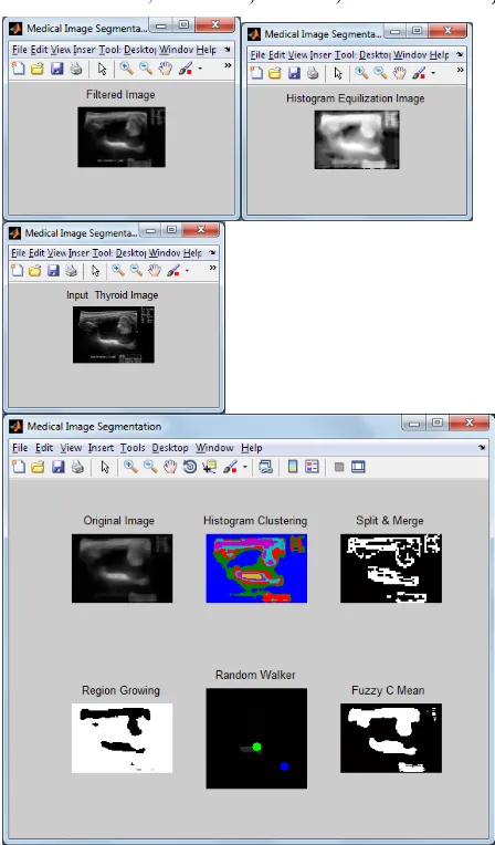
[image:4.612.323.547.128.510.2]Fig II: segmentation of thyroid UD image
International Journal of Emerging Technology and Advanced Engineering
Website: www.ijetae.com (ISSN 2250-2459,ISO 9001:2008 Certified Journal, Volume 2, Issue 12, December 2012)
[image:5.612.325.589.110.489.2]402
International Journal of Emerging Technology and Advanced Engineering
Website: www.ijetae.com (ISSN 2250-2459,ISO 9001:2008 Certified Journal, Volume 2, Issue 12, December 2012)
[image:6.612.310.560.111.536.2]403
Fig V: segmentation of thyroid with benign nodule
Fig VI: segmentation of thyroid with benign nodule
V.CONCLUSION AND FUTURE WORK
In this work, segment the thyroid images using FCM, Histogram clustering, Quad tree, Region growing and
Random Walker
methods.
The experimental results [image:6.612.48.293.141.697.2]International Journal of Emerging Technology and Advanced Engineering
Website: www.ijetae.com (ISSN 2250-2459,ISO 9001:2008 Certified Journal, Volume 2, Issue 12, December 2012)
404
REFERENCES[1 ] Francesco Masulli Andrea Schenone, A ]fuzzy clustering based segmentation system as support to diagnosis in medical imaging Artificial Intelligence in Medicine 16 (1999) 129–147
[2 ] Songül Albayrak, Fatih AmasyalıFuzzy C-means clustering on Medical Diagnostic systems.
[3 ] L. Grady: Random Walks for Image Segmentation (http:/ / www. cns. bu. edu/ ~lgrady/ grady2006random. pdf), IEEE Trans. on Pattern Analysis and Machine Intelligence, Vol. 28, No. 11, pp. 1768–1783, Nov., 2006.
[4 ] P. Doyle, J. L. Snell: Random Walks and Electric Networks, Mathematical Association of America, 1984.
[5 ] R. C. Gonzalez and R. E. Woods, Digital Image processing,Prentice-Hall, Inc., New Jersey, 2002.
[6 ] M.H. Gross, O.G. Staadt, and R.Gatti. Efficient triangular surface approximation using wavelets and quadtree data structure. IEEE Transaction On Visualization And Computer Graphics, 2(2):1–13, June 1996.
[7 ] Digital Image Processing Using MATLAB 2nd editionby Gonzalez, Woods, and Eddins © 2009
[8 ] Ali Kermani Ahmad Ayatollahi Mohammad Talebi Segmentation of Medical Ultrasound Image Based on Local Histogram Range Image 3rd International Conference on Biomedical Engineering and Informatics , 2010.
[9 ] Changming Zhu, Jun Ni, Yanbo Li1 and Guochang Gu General Tendencies in Segmentation of Medical Ultrasound Images Fourth International Conference on Internet Computing for Science and Engineering, 2009.
[10 ]Deepika Koundal1, Savita Gupta1 and Sukhwinder Singh1 “Computer-Aided Diagnosis of Thyroid Nodule: A Review”, International Journal of Computer Science & Engineering Survey (IJCSES) Vol.3, No.4, August 2012.
[11 ]Nasrul Humaimi Mahmood and Akmal Hayati Rusli “Segmentation and Area Measurement for Thyroid Ultrasound Image” International Journal of Scientific & Engineering Research Volume 2, Issue 12, December-2011
[12 ]Chuan-Yu Chang, Yue-Fong Lei, Chin-Hsiao Tseng, and Shyang-Rong Shih Thyroid Segmentation and Volume Estimation in
Ultrasound Images, IEEE Transactionson
biomedicine,2010,vol.57.no.6


