Introduction
Epithelia serve several fundamental purposes. They are barriers to the external environment, frontiers between tissues that can regulate the patterning of the embryo by limiting diffusion of signals, or they can be attachment points during morphogenesis (Shook and Keller, 2003). Epithelia are characterised by cellular junctions and an apicobasal polarity that maintain their overall architecture and coherence. During epithelial-mesenchymal transitions (EMT), contacts between cells are lost, allowing them to acquire motile behaviour, either as single migrating cells or integrated in a moving sheet of cells. This phenotypic conversion occurs at precise times and locations during embryogenesis, and in developmental processes as diverse as gastrulation movements, migration of neural crest cells, or the formation of cardiac valves (Shook and Keller, 2003; Wallingford et al., 2002). This process is reactivated in a variety of pathological conditions, including the invasive and metastatic phases of carcinomas (Thiery, 2003). A molecular understanding of the signalling pathways implicated in cell movements is only starting to emerge. In most of the cases that have been analysed, EMT are initiated by stimuli external to the epithelia. TGF, HGF/SF, FGF, EGF and Wnt family members are some of the factors that have been shown in cell cultures or in vivo to regulate the mobilisation of epithelial
cells. Downstream effectors of these signals include the Src, the Ras/MAPK as well as the slug/snailfamily of transcription factors (Locascio and Nieto, 2001; Savagner, 2001; Thiery, 2003). Depending on the cellular context, many of these signals and their effectors are also known to modulate cell specification, and to trigger or regulate cell proliferation. How these cellular decisions are made during embryogenesis is largely unknown.
The chick somite represents an attractive experimental model where EMT processes can be studied in vivo. The possibility of expressing molecules that can either activate or inhibit specific signalling pathways in the somites, by means of the electroporation technique, together with the analysis of gene expression and of cell morphology, represent powerful tools with which to analyse the regulation of EMT in a three-dimensional system.
Newly formed somites comprise an outer epithelium, the cortex, surrounding a central cavity, the somitocoel. Cortex cells are bottle-shaped and polarised (apical side facing the somitocoel), and they express, at the adherens junctions located at their apical end, the epithelial markers N-cadherin, -catenin and F-actin (Gros et al., 2005). As somites differentiate, cortex cells undergo multiple EMT processes to give rise to their derivatives: the ventral portion of the somite first dissociates The regulation of cell adhesion in epithelia is a fundamental
process governing morphogenesis in embryos and a key step in the progression of invasive cancers. Here, we have analysed the molecular pathways controlling the epithelial organisation of somites. Somites are mesodermal epithelial structures of vertebrate embryos that undergo several changes in cell adhesion during early embryonic life. We show that Wnt6 in the ectoderm overlaying the somites, but not Wnt1 in the neighbouring neural tube, is the most likely candidate molecule responsible for the maintenance of the epithelial structure of the dorsal compartment of the somite: the dermomyotome. We characterised the signalling pathway that mediates Wnt6 activity. Our
experiments suggest that the Wnt receptor molecule Frizzled7 probably transduces the Wnt6 signal. Intracellularly, this leads to the activation of the  -catenin/LEF1-dependent pathway. Finally, we demonstrate that the bHLH transcription factor paraxis, which was previously shown to be a major player in the epithelial organisation of somites, is a target of the -catenin signal. We conclude that -catenin activity, initiated by Wnt6 and mediated by paraxis, is required for the maintenance of the epithelial structure of somites.
Key words: Chick, Somite, Wnt, Paraxis, -Catenin, Epithelial-mesenchymal transition
Summary

-Catenin-dependent Wnt signalling controls the epithelial
organisation of somites through the activation of paraxis
Claudia Linker1,*,†,‡, Cynthia Lesbros1,*, Jérôme Gros1, Laura W. Burrus2, Alan Rawls3and Christophe Marcelle1,‡
1Laboratoire de Génétique et de Physiologie du Développement (LGPD). Developmental Biology Institute of Marseille (IBDM),
CNRS UMR 6545. Université de la Méditerranée, Campus de Luminy, case 907, 13288 Marseille, Cedex 09, France
2Department of Biology, San Francisco State University, 1600 Holloway Ave., San Francisco, CA 94132, USA 3School of Life Sciences, Arizona State University, Tempe, AZ 85287, USA
*These authors contributed equally to this work
†Present address: University College London, Department of Anatomy and Developmental Biology, Gower Street, London WC1E 6BT, UK ‡Authors for correspondence (e-mail: c.linker@ucl.ac.uk; marcelle@ibdm.univ-mrs.fr)
Accepted 30 June 2005
Development 132, 3895-3905
Published by The Company of Biologists 2005 doi:10.1242/dev.01961
De
into a mesenchyme, the sclerotome, which becomes organised around the notochord and neural tube and differentiates into axial cartilage and bones. The remaining dorsal epithelium, the dermomyotome, is the source of all skeletal muscles of the body and the limbs, as well as precursors for the dermis of the back (Christ and Ordahl, 1995). Skeletal muscles form in two stages: first, cells located at the epithelial borders of the dermomyotome delaminate into the nascent primary myotome to generate postmitotic myocytes (Gros et al., 2004); the second stage is characterised by the massive entry of embryonic muscle progenitors within the myotome, triggered by the EMT of the dermomyotome (Gros et al., 2005; Relaix et al., 2005; Kassar-Duchossay et al., 2005). Concomitantly, cells located in the medial domain of the dermomyotome dissociate and migrate as single cells towards the ectoderm, where they condense to form the dermis of the back (Brill et al., 1995; Olivera-Martinez et al., 2002; Gros et al., 2005; Ben Yair et al., 2005). Finally, limb, tongue and diaphragm muscle precursors dissociate from the epithelial border of the lateral dermomyotome and migrate as single cells to later differentiate into their cognate muscles (Bladt et al., 1995; Brand-Saberi et al., 1996; Dietrich et al., 1999; Heymann et al., 1996).
The analysis of paraxis (Tcf15) mutant mice has shown that the epithelial organisation of somites is crucial for the later morphogenesis of trunk muscles. These mutants display somites that are unable to organise themselves into an epithelium, and although the specification of skeletal muscles takes place correctly, the spatial organisation of their muscles is grossly altered (Burgess et al., 1996; Wilson-Rawls et al., 1999). These data indicate that muscle progenitors of the body use the epithelial sheet of the dermomyotome as a scaffold on which they organise. Thus, an important step towards a comprehension of the organisation of the trunk skeletal muscles lies in an understanding of the tissue interactions and the molecular networks that regulate the epithelial organisation of somites.
Embryonic manipulations have shown that both the ectoderm and the dorsal neural tube can promote the induction or the maintenance of dermomyotome structure (Borycki and Emerson, 2000; Burgess et al., 1995; Correia and Conlon, 2000; Dietrich et al., 1998; Dietrich et al., 1997; Fan and Tessier-Lavigne, 1994; Goulding et al., 1991; Maroto et al., 1997; Reshef et al., 1998; Spence et al., 1996). In vivo and in vitro experiments support a model where Wnt proteins mediate this dorsalising activity of the neural tube and ectoderm (Capdevila et al., 1998; Fan et al., 1997; Maroto et al., 1997; Wagner et al., 2000; Münsterberg et al., 1995; Stern et al., 1995; Galli et al., 2004). Whether or not these tissues and molecules regulate the epithelial organisation of somites has not been systematically addressed in these experiments. Importantly, Pax3, which has been generally used as a dorsal somitic marker, cannot be considered as an adequate epithelium marker, because it is expressed in paraxis-null mice, where the epithelial structure is not present (Burgess et al., 1996; Wilson-Rawls et al., 1999). Moreover in the Pax3
knockout mice, the epithelial structure of the somite is initially normal, indicating that Pax3 does not play an active role in this process (Daston et al., 1996). This is not due to a functional redundancy with Pax7, which is expressed in somites in a similar fashion as Pax3, because in Pax3/Pax7double knockout mice, epithelial somites initially form (Frédéric Relaix and Margaret Buckingham, personal communication).
While the phenotype of paraxis-null mice clearly implicates this molecule in the epithelialisation of the somite, the mechanisms that regulate its expression are poorly understood. Studies in chick and mouse indicate that paraxis expression is initiated independently of surrounding structures in a mesoderm autonomous way (Correia and Conlon, 2000; Linker et al., 2003; Palmeirim et al., 1998). Sosic and colleagues (Sosic et al., 1997) have shown that ectoderm provides signals that maintains paraxis expression and the epithelial organisation of somites, but they have not addressed the role of Wnt in this process. Wnt6 is the only Wnt known to be expressed in the ectoderm overlying the anterior segmental plate and early somites of the chick embryo (Cauthen et al., 2001; Marcelle et al., 2002; Schubert et al., 2002; Rodriguez-Niedenführ et al., 2003), which makes it a likely candidate to play a role in this process. Schmidt and collaborators (Schmidt et al., 2004) have recently shown that Wnt6 promotes the epithelial organisation of the segmental plate mesenchyme. However, the effect of this molecule in later events of somite differentiation, such as dermomyotome formation and dermis specification, was not addressed.
In this study, we have analysed the tissue and molecular interactions that regulate the epithelial organisation of somites. Through a systematic removal of tissues surrounding the somites, followed by a time-course analysis, we show that the ectoderm is not responsible for the formation, but for the maintenance of the epithelial structure of the somite. Our experiments confirm that Wnt6 is the molecule from the ectoderm responsible for this effect. In addition, we show evidence that this action is specific to Wnt6, while Wnt1 exerts a distinct function in the medial compartment of the dermomyotome. We then characterised the intracellular signalling pathway activated by Wnt6 in the somites. Three Frizzled receptors, Fz1, Fz2and Fz7are expressed in epithelial somites and in the dermomyotome (Linker et al., 2003). Although the expression of all three is lost upon removal of the ectoderm, Wnt6 maintains Fz7, but not Fz1or Fz2expression, suggesting a specific role for Fz7 in mediating Wnt6 activity. Blocking the activity of -catenin or Lef1 in the dermomyotome leads to a loss of somite cell polarisation, and a de-epithelialisation of the dermomyotome, indicating that Wnt6 activity is mediated by a -catenin-dependent pathway. Finally, we demonstrate that paraxisis a target of the -catenin signal and that its prolonged expression is sufficient to counter the de-epithelialisation of the dermomyotome that normally takes place during late embryonic development. These results establish that a -catenin activity, initiated by Wnt6 through Fz7 and mediated by paraxis, is required for the maintenance of the epithelial organisation of somites.
Materials and methods
Embryonic manipulations and in situ hybridisation
Fertilised chick eggs were obtained from a local commercial source. They were incubated at 39°C in a humid atmosphere for the indicated time. The procedures to perform in vivo surgeries, separation of ectoderm from somites with an impermeable membrane and injection of cells have been described (Linker et al., 2003). All the experiments were performed at the rostral region of the PSM, in order not to interfere with either segmentation or border formation processes, which can in turn influence mesoderm differentiation processes. Whole-mount in situ hybridisation was performed as described
De
previously (Henrique et al., 1995). The following probes were used this study: probes for chick Frizzled 1, Frizzled 2 and Frizzled 7 (see Linker et al., 2003); a 400 bp PCR fragment of the chick Wnt6 was used [kindly provided by A. McMahon (Hollyday et al., 1995)]; a 1380 bp long chick paraxisprobe [a kind gift from E. Olson (Sosic et al., 1997)]; and a quail 900 bp Pax3 probe (Stark et al., 1997).
Antibodies, immunohistochemistry, confocal analysis on cryosections
Phalloidin Alexa 456 (Molecular Probes) was used to detect F-actin. The following antibodies were used: a mouse monoclonal against N-cadherin (Sigma C-8538) and a polyclonal antibody against GFP (Torrey Pines Biolabs). Sections were examined with a confocal microscope Zeiss LSM 510 Meta.
Injection of Wnt6-expressing cells and electroporation Electroporation was performed as described (Scaal et al., 2004). For the observation of electroporated somites, embryos were fixed in 4% formaldehyde, cleared in 90% glycerol/H2O and examined with a confocal microscope. Image stacks (50-100 m thick) were treated with Metamorph (Universal Imaging) and Imaris (Bitplane) image analysis software for image visualisation and 3D reconstruction. The full-size mouse paraxis cDNA clone (provided by A. Rawls), a dominant-negative form of Xenopus-catenin (kindly provided by P. McCrea) (Montross et al., 2000), and a dominant-negative form of chick Lef1 [kind gift of J. C. Ispizua-Belmonte (Kengaku et al., 1998)] were cloned into the pCLGFPA electroporation vector (Scaal et al., 2004), which drives the gene of interest under the control of a CMV/chick -actin enhancer/promoter. The pCLGFPA vector also contains the coding sequence for the reporter gene e-GFP under the control of a SV40 promoter/enhancer. Cell counting was statistically analysed. Results were different from each other with a 95% confidence; P<0.005 were evaluated by a Student’s t-test. COS cells were transfected using the Lipofectamine agent (Gibco-BRL) with 1 g of the pHYK vector containing the full-length mouse Wnt6 cDNA (obtained from A. McMahon and L. Burrus) (Burrus and McMahon, 1995). Forty-eight hours after transfection, cells were collected by trypsinisation, labelled with DiI (DM-DiI, Molecular Probes) and pressure-injected into somites of stage 12HH developing embryos using a Picospritzer (General Valve Corporation), as described previously (Linker et al., 2003).
Results
Previous experiments have shown that the ectoderm alone (Kardon et al., 2002; Kuratani et al., 1994; Sosic et al., 1997; Spence et al., 1996) or in combination with the neural tube (Dietrich et al., 1997) is required for the specification of the dorsal compartment of somites, recognised by the expression of Pax3. Apparent discrepancies between authors might be due to experiments performed at different levels of the segmental plate, where it is now recognised that two regions exist. Caudally a non-determined region, where Wnt and Notch signalling regulate segmentation, and rostrally a determined region, where segments are positioned and differentiation is allowed by the lack of FGF8 and the presence of a retinoic acid signal (Diez del Corral and Storey, 2004; Dubrulle et al., 2001; Moreno and Kintner, 2004; Palmeirim et al., 1997). Thus, we performed the following experiments at the rostral level of the segmental plate, where cells are competent to respond to environmental signals.
The ectoderm maintains the epithelial structure of the dermomyotome
We have investigated the role of the tissues surrounding the
paraxial mesoderm on somite epithelialisation. This was carried out over an extended period of time to follow the progression of the phenotypes obtained after these manipulations. We have analysed the expression of paraxisand of the Wnt receptors Frizzled 1, Frizzled 2 and Frizzled 7.
In these experimental conditions, our results demonstrate that the surgical separation of the rostral segmental plate mesoderm from axial (neural tube and notochord, Fig. 1A) or lateral (lateral plate mesoderm, Fig. 1B) structures had no visible effects on the formation of epithelial somites (Fig. 1C,D) or on the expression of paraxis (10/11, Fig. 1F), Fz1 (8/8, not shown), Fz2 (11/11, not shown) and Fz7 (9/9, Fig. 1C-E,G).
[image:3.612.312.561.338.600.2]We then tested whether the ectoderm overlying the segmental plate and somites plays a role in the formation, and/or the maintenance, of an epithelial dermomyotome. Ectoderm was separated from the paraxial mesoderm by the positioning of a tantalum foil between these tissues (Fig. 2A). Embryos were then allowed to develop for varying times, and their morphology and gene expression were examined. After 3 hours incubation, two somites had formed under the membrane (Fig. 2B), their epithelial structure was normal in
Fig. 1.Axial or lateral structures do not affect the epithelial structure of the dermomyotome. (A) Schematic of the experiment: separation of the anterior PSM from axial structures (neural tube and
notochord) by making a slit through the three germ layers of the embryo. Embryos were then cultured for 18-20 hours. (B) Scheme of the separation of PSM from lateral structures. (C,D) Transverse sections of embryos in E and G. (E,F) Expression of Fz7and paraxis is maintained after separation of the PSM from axial structures. The residual expression of paraxisand Fz7along the neural tube is due to remnants of somitic tissue and ectoderm that have not been
completely separated from the neural tube (and confirmed on sections, not shown). (G) Expression of Fz7is not affected after separation of the PSM from lateral structures.
De
section (not shown) and they expressed the three Fz proteins normally (Fz19/9, Fz26/6 not shown and Fz77/7, Fig. 2B). The same result was obtained if embryos were re-incubated for 6 or 8 hours (not shown, 13/13). Twelve hours of separation from the ectoderm did not affect the morphology of somites (Fig. 2I). However, the dorsal epithelium, did not express paraxis(9/9, Fig. 2G), pax3(8/8, Fig. 2E) or any of the three Frizzled (Fz110/10, Fz28/8 not shown, Fz710/10; Fig. 2C,I). Finally, when embryos were allowed to develop for 24 hours after the manipulation, the dermomyotome was no longer recognisable. Instead, the entire somite presented a mesenchyme-like structure (Fig. 2J) and the expression of all the markers was completely abolished (Fz118/18, Fz216/20 not shown, and Fz717/17, Fig. 2D,J; paraxis 13/13, Fig. 2H;
pax38/8, Fig. 2F).
Altogether, these data indicate that the rostral segmental plate grown separated from the ectoderm is capable of forming epithelial somites, of differentiating into an epithelial dermomyotome and a sclerotome, and of initiating its normal genetic program of differentiation. However, the ectoderm is
required to maintain the expression of these genes and the epithelial organisation of the dermomyotome.
Overlapping and unique properties of Wnt6 and Wnt1 on somites
Wnt6 is the only member of the Wnt family known to be expressed in the ectoderm overlying the segmental plate and newly formed somites, in the chick embryo (Marcelle et al., 2002; Schubert et al., 2002; Rodriguez-Niedenfüihr, 2003). For this reason, Wnt6 was the best candidate to be playing a role in the maintenance of the epithelial structure of the dermomyotome.
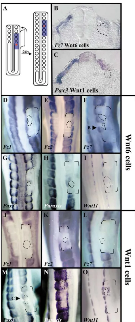
To test this hypothesis, Wnt6-expressing cells (DiI-labelled) were injected in the anterior segmental plate, the ectoderm above the injected region was separated from segmental plate by an impermeable membrane as described before, and embryos were allowed to develop for 24 hours (Fig. 3A). Under these conditions, paraxis (26/31, Fig. 3H) and pax3 (11/18, Fig. 3G) expression was rescued in dermomyotomal cells close to the injection site. Interestingly, Wnt-6 injected cells were able to rescue Fz7expression (9/12, Fig. 3B,F), but failed to rescue Fz1(15/15, Fig. 3D) or Fz2(14/21, Fig. 3E) expression. In transverse sections, we could also clearly observe an epithelial dermomyotme-like structure around the Wnt6-expressing cells, at the place where paraxisand Fz7expression was rescued (Fig. 3B). Control, untransfected cells did not rescue the expression of any of these genes (Fz79/11, paraxis
11/12; pax38/8, not shown), or the epithelial structure of the dermomyotome.
A number of studies have implicated Wnt molecules from the neural tube in the patterning of the somites, as well as the control of their epithelialisation. To test whether the observed effects were a specific response to Wnt6, we performed the same set of experiments using Wnt1-expressing cells. Under these conditions, we observed that Wnt1 is not able to rescue
Fz1 (11/13, Fig. 3J) or Fz7 expression (17/18, Fig. 3L). By contrast, paraxis(12/13, Fig. 3N) and pax3(18/19, Fig. 3M) expression was rescued around the injected cells in most embryos, whereas Fz2 expression was distinctively, albeit faintly, activated in 60% of the analyzed embryos (8/13, Fig. 3K). On sections, we could observe that an epithelial, dermomyotome-like structure was present around the Wnt1 cells (Fig. 3C). Further confirmation that Wnt1 and Wnt6 display distinct function in somite differentiation came from the comparison of their role in the patterning of the dermomyotome. As Wnt1 regulates Wnt11 expression in the dorsomedial lip of the dermomyotome (Marcelle et al., 1997), we compared the induction of Wnt11 by Wnt1 with that of Wnt6. Whereas Wnt1-expressing cells were able to rescue, and ectopically induce, Wnt11 expression (5/5, Fig. 3O), Wnt6-expressing cells were never able to do so under the same experimental conditions (6/6, Fig. 3I).
Together, these data indicate that Wnt1 and Wnt6 activate overlapping and distinct cellular responses in the paraxial mesoderm. Although it is tempting to postulate from our data that Wnt6 and Wnt1 mediate their activity through Fz7 and Fz2, respectively, further analysis of this process will be needed to determine whether this is indeed the case. We observed that Wnt1 and Wnt6 are both able to rescue the epithelial organisation of somites. However, as the epithelial organisation of somites was never perturbed in the absence of Fig. 2. Ectoderm maintains the epithelial structure of somites.
(A) Schematic of the procedure: separation of the anterior PSM from overlying ectoderm by the introduction of an impermeable
membrane (blue), followed by 3-24 hours of culture. The position of the membrane at the end of the experiment matches perfectly the region where no staining is observed (see C-H). (B-D) Expression of Fz7after 3 (B), 12 (C) or 24 (D) hours of culture. The position of the membrane is indicated in B. (E,F) Expression of Pax3after 12 (E) or 24 (F) hours of culture. (G,H) Expression of paraxisafter 12 (G) or 24 (H) hours of culture. (I,J) Transverse sections of C (I) and D (J) at the positions indicated.
De
[image:4.612.42.296.75.380.2]neural tube (the source of Wnt1), this indicates that Wnt6 is the endogenous molecule regulating the epithelial organisation of somites in the embryo. Thus, it is likely that under our experimental conditions, Wnt1 mimics the activity of Wnt6 on the epithelial organisation of somites. Alternatively, Wnt1 (or a Wnt1-like molecule in the neural tube) and Wnt6 cooperate for the epithelialisation of the somite: Wnt from the neural tube might provide additional
epithelialisation function, resulting in a ‘hyper-epithelialisation’ in the medial dermomyotome. Indeed, the medial border of the dermomyotome (the DML) displays a stronger expression of adherens junction molecules ( -catenin, N-cadherin and actin) than the rest of the dermomyotome (Gros et al., 2005). This should result in an increased cell adhesion in the DML, which might be functionally relevant during myotome formation. At present, we cannot exclude any of the two possibilities.
Wnt6 mediates its activity through the -catenin pathway
The results described above indicate that Wnt6 signalling maintains the epithelial structure of the dermomyotome. The binding of Wnt to Frizzled receptors can initiate a variety of cellular responses (Huelsken and Birchmeier, 2001; Veeman et al., 2003). As -catenin and its co-effector molecule Lef1 are expressed in the dermomyotomal compartment (Marcelle et al., 2002; Schmidt et al., 2000), we analysed their possible function as mediators of the Wnt6 signal.
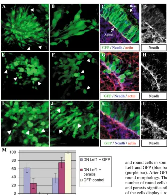
This was performed by the electroporation of dominant-negative forms of -catenin or Lef1 molecules in newly formed somites (Kengaku et al., 1998; Montross et al., 2000). Constructs containing either of the factors, together with the GFP reporter, were electroporated in the dorsal region of newly formed epithelial somites (somites I to IV). An expression plasmid containing only GFP served as control. After 24 hours of incubation, embryos were analysed by confocal microscopy. Control dermomyotomal cells exhibited a typical pseudo-epithelial morphology, with bottle-shaped cells extending long cytoplasmic filopodia towards the ectoderm, and expressing at their apical end the adherens junction markers N-cadherin and actin (Fig. 4A-D). By contrast, somites electroporated with DN -catenin (Fig. 4E) or DN Lef1 constructs (Fig. 4F-H) displayed a disorganised dermomyotome composed of round cells, which have lost the majority or their cytoplasmic extensions. In those cells, N-cadherin was redistributed evenly along the cell membrane (Fig. 4G,H). In embryos electroporated with DN Lef1, we did not observe any significant increase in activated caspase 3 staining, indicating that round cells were not dying (not shown).
[image:5.612.52.287.71.630.2]These data show that blockage of the Wnt -catenin canonical pathway promotes the de-epithelialisation of dermomyotomal cells, implying that Wnt6, which is secreted by the ectoderm, initiates a -catenin-dependent cascade that is required for the maintenance of the epithelial structure of the dermomyotome.
Fig. 3. Specific functions of Wnt6 and Wnt1 in the epithelialisation and patterning of somites. (A) Schematic of the procedure: separation of the PSM from overlying ectoderm with a membrane (blue) after the injection of Wnt-expressing cells (in red) and followed by 24 hours in culture. (B) Transverse section through F. (C) Transverse section through M. (D-I) Expression of markers after the injection of Wnt6- (D-I) and Wnt1-expressing (J-O) cells: (D) Fz1; (E) Fz2; (F) Fz7; (G) pax3; (H) paraxis; (I) Wnt11. Wnt-expressing cells were stained with fixable CM Tracker DiI; their exact position, defined under UV examination, was recorded and is indicated in each panel by broken lines. The corners of the impermeable membranes are indicated in each embryo.
De
paraxis acts downstream of -catenin to maintain the epithelialisation of the dermomyotome
The phenotype of paraxis-null mice clearly implicates this molecule in somite epithelialisation (Burgess et al., 1996). Thus, an attractive possibility was that Wnt6 secreted by the ectoderm interacts with Fz7 activating the -catenin-dependent cascade responsible for regulation of the expression of paraxis, which controls the epithelial morphology of the dermomyotome.
The first requirement for such hypothesis is that Fz7
expression should precede paraxisexpression in the presomitic mesoderm during development. To test this, we have analysed the expression of Fz7 and paraxisin two halves of the same embryo. By doing this, we were able to directly compare the expression of each gene in embryonic tissues at the exact same age and axial level (Fig. 5A). This analysis clearly showed that
Fz7expression precedes paraxisexpression in the presomitic mesoderm (Fig. 5D, 5/5). A second requirement is that the expression of Fz7 should be downregulated before the expression of paraxiswhen the ectoderm is removed. To test how the expression of these two genes is lost in the absence of ectoderm, we placed two membranes, one at each side of the embryo. Embryos were then reincubated and one half was tested for paraxisand the other for Fz7expression (Fig. 5B).
As expression of both genes is lost after 12 hours (Fig. 2), we decided to decrease the time of incubation. After 10 hours of incubation, Fz7 expression was greatly reduced, or absent, while in the same embryos exhibits a robust expression of
paraxis(Fig. 5E, 11/11). These results support the hypothesis that paraxiscould be a downstream target of Fz7 activation. To test this directly, we have analysed paraxis expression in dermomyotomal cells where either -catenin or Lef1 activity was inhibited. Following the electroporation of somites with DN--catenin (Fig. 5C, black arrowheads) or DN-Lef1 (Fig.
5F-G), we observed that paraxis expression was
downregulated in the electroporated cells, whereas in control cells (Fig. 5C, white arrowheads) or in GFP electroporated cells, paraxis was expressed normally (not shown). These results strongly suggest that Wnt signalling mediated by  -catenin is required for maintenance of paraxisexpression in the dermomyotome.
[image:6.612.43.380.345.738.2]Finally, we tested whether paraxisis sufficient to rescue the de-epithelialisation observed after an arrest of the Wnt canonical signalling. Dorsal somitic cells were electroporated with both constructs DN Lef1 and paraxis, and analysed as described. Control embryos were electroporated with a mix of GFP and DN Lef1 constructs (this allow to have the same concentration of DN Lef1 plasmid in both conditions).
Fig. 4. Wnt6 activates the -catenin pathway. Projections of confocal dorsal views (A,B,E,F,I,J) and confocal transverse sections (C,D,G,H,K,L) of somites electroporated with GFP (A-D), DN  -catenin (E), DN Lef1 (F-H), DN Lef1 and GFP (1:1, J), DN Lef1 and paraxis (1:1, J-L). Sections are stained with a combination of phalloidin red, recognising F-actin (in red) and an antibody directed against N-cadherin (in blue), together with the GFP (in green). Sections in D,H,L show the N-cadherin staining only. (A-D) Control, GFP electroporated somites with typical long bottle-shaped cells (arrowheads in A) that display the adherens junction-specific markers F-actin and N-cadherin at their apical end (C,D). (B) Enlargement of cells in A. (E-H) Dermomyotome cells
electroporated with DN -catenin (E) or DN Lef1 (F-H) display a round morphology with a clear redistribution of the adherens junction markers at the plasma membrane (G,H). (J-L) Dermomyotome cells expressing DN Lef1 together with paraxis display a normal epithelial-like morphology (J) with adherens-junction marker at the apical end (K,L). (M) Quantification of experiments shown in A-L, where coloured bars represent the percentage of epithelial and round cells in somites electroporated with GFP only (yellow bar), DN Lef1 and GFP (blue bar), or a combination of DN Lef1 and paraxis (purple bar). After GFP electroporation, only 1.5% of the cells displayed a round morphology. The electroporation of DN Lef1+GFP increases the number of round cells to 62.5%, while the co-electroporation of DN Lef1 and paraxis significantly rescues the epithelial morphology, as only 24.8% of the cells display a round morphology. Standard deviations are
indicated.
De
Whereas co-electroporation of DN-Lef1 and GFP led to the de-epithelialisation of dermomyotomal cells (Fig. 4I), which adopted a round shape as observed previously (62.5% of GFP-positive cells were round; Fig. 4M), co-electroporation of DN-LEF1 with paraxis rescued the pseudo-stratified epithelium phenotype (in this case, only 24.8% of GFP-positive cells were round Fig. 4M), with bottle-shape cells extending filopodia towards the ectoderm and displaying the typical adherens junction markers at their apical end (Fig. 4J-L).
These results demonstrate that paraxisacts downstream of the Wnt/-catenin pathway to maintain the epithelial organisation of somites.
Wnt6 or paraxis expression are sufficient to counter the de-epithelialisation of the dermomyotome
Between day 3 and day 6 of development, the dermomyotome
[image:7.612.43.325.73.268.2]de-epithelialise to give rise to the dermis of the back (Brill et al., 1995; Christ and Ordahl, 1995; Couly and Le Douarin, 1988; Olivera-Martinez et al., 2000; Olivera-Martinez et al., 2002; Gros et al., 2005). The de-epithelialisation of the dermomyotome is progressive, first observed in the cervical region of an E3 embryo (stage 18 HH), in a central domain of the dermomyotome. Then it advances posteriorly along the embryonic axis and towards the borders of the dermomyotome; the medial and lateral borders of the dermomyotome are the last to dissociate between E5 and E6 (Marcelle et al., 2002; Scaal et al., 2004; Gros et al., 2005). Interestingly, Wnt6 expression, which is homogeneous in the ectoderm located above newly formed somites, is progressively downregulated in all but the ectoderm located above the somitic borders (Marcelle et al., 2002). This is followed in the dermomyotome by a similar loss in the expression of paraxis, which is initially
Fig. 5. -Catenin regulates paraxis expression. (A) Schematic of the experiment: embryos were halved with one half processed for Fz7expression and the other half processed for paraxisexpression, as seen in D. (B) Schematic of the procedure: impermeable membranes were positioned at each side of the embryo and the embryos were allowed to grow for 10 hours. Embryos were then cut in half and processed for Fz7 andparaxisas before (E). (C) Somites (dorsal view) electroporated with a DN  -catenin construct lose paraxisexpression in the
electroporated region (black arrowheads), whereas control somites express paraxis in the entire dermomyotome (white arrowheads). (F,G) Dorsal electroporation of DN Lef1 construct. Electroporated cells, which express GFP (G), do not express paraxis (F).
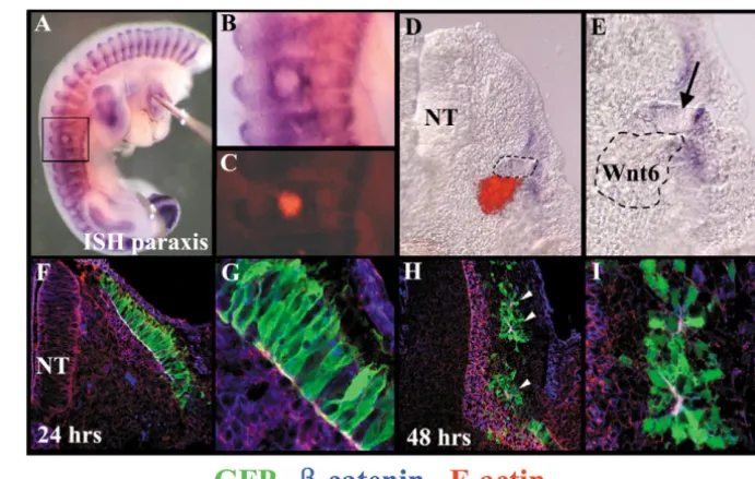
Fig. 6.Maintaining a continuous source of Wnt6 or paraxis counters the
de-epithelialisation of the dermomyotome. (A) Expression of Paraxis in an embryo injected with Wnt6-expressing cells at E2.5 and incubated for 48 hours.
(B) Enlargement of the boxed area in A, showing the maintenance of a high paraxis expression around the injected cells. (C) Fluorescence image of B, showing the position of the DiI-labelled cells.
(D) Transverse section of the same embryo. (E) Enlargement of D. Arrow indicates a dermomyotome-like epithelial structure located in the vicinity of the injected cells (outlined). (F-I) Confocal pictures of sections of embryos electroporated with paraxis CLGFPA in the dorsal region of the dermomyotome, stained with antibodies against GFP (in green), -catenin (in blue) and phalloidin red, recognising F-actin (in red), (F,G) Twenty-four hours after electroporation, the morphology (shown
with GFP staining) and polarity (recognised by F-actin and -catenin staining at the adherens junctions) of cells overexpressing paraxis is normal. (G) Enlargement of F. (H,I) Forty-eight hours after electroporation, cells over-expressing paraxis organise in clusters of cells that maintain contacts through their apical ends, expressing F-actin and -catenin (arrowheads). (I) An enlargement of H.
De
[image:7.612.213.559.483.703.2]homogeneous and becomes progressively restricted to the epithelial borders of the dermomyotome (Fig. 6A,B). Although the reason for the Wnt6 progressive spatial restriction in the ectoderm is unknown, an interesting hypothesis is that it could account for the de-epithelialisation of the dermomyotome. To analyze this possibility, we have injected Wnt6-expressing cells under the medial region of the dermomyotome at E2.5, and the embryos were left to develop for 48 hours. In this way, we presented a continuous source of Wnt6 to a central region of the dermomyotome that should otherwise have undergone de-epithelialisation. Under these conditions, high level of
paraxisexpression was maintained around the source of Wnt6 (8/8, Fig. 6A-C), while de-epithelialisation did not take place (Fig. 6D,E). These observations indicate that the dissociation of the dermomyotome observed between E3 and E6 may be due to the disappearance of the epithelium-maintaining signal, Wnt6. Evidently, this observation does not exclude the possibility that dissociation-promoting signals may also participate in this process.
While the analyses of the paraxis mouse mutant demonstrate that paraxis is a necessary player during somite epithelialisation, it is unclear whether this process requires the activity of one or a combination of molecular inputs or pathways. To test whether paraxis function is sufficient to maintain the epithelial structure of the somite, we electroporated the construct for paraxis in newly formed somites, and we analyzed the morphology of the dermomyotome 24 hours after electroporation (i.e. at a time when the dermomyotome is epithelial) and 48 hours after electroporation (when dermomyotome de-epithelialisation is well under way). After 24 hours, we did not observe any significant difference in cell morphology of electroporated cells or in the distribution and intensity of the molecular markers for epithelial junctions when compared with normal cells (n=6; Fig. 6F,G). By contrast, 48 hours after electroporation, we observed that most paraxis-expressing dermomyotome cells organised in small clusters of cells that maintain cell contacts through their apical ends, recognised by the expression of actin and -catenin (n=5; Fig. 6H,I, arrowheads). In a control embryo at a similar stage of somite differentiation, most dermomyotome cells should have migrated into the myotome (Gros et al., 2005). Although the presence of adherens junction implies that paraxis-expressing cells were polarised, they did not display the typical bottle-shape morphology of a bona fide dermomyotomal cell (compare Fig. 6G with 6I). Together, these results show that the overexpression of paraxis is sufficient to maintain the epithelial organisation of dermomyotome cells.
Discussion
Ectoderm maintains the epithelial structure of the dermomyotome
In this study, we have analysed the signals controlling the epithelial structure of the somite. These experiments were performed in the determined region, where somite differentiation can take place. By performing a precise time-course analysis of the effect of ectoderm removal on somite morphology and gene expression, we have shown that ectoderm overlying the rostral segmental plate is required for the maintenance of the expression of paraxis, pax3 and
Frizzled, and the epithelial organisation of the dermomyotome. Although these data confirm the crucial role that ectoderm plays on paraxial mesoderm patterning and morphology, as shown previously (Kardon et al., 2002; Kuratani et al., 1994; Schmidt et al., 2004; Sosic et al., 1997; Spence et al., 1996), they also complement these studies by enabling us to differentiate between an inductive or a maintenance role of the ectoderm in this process. In our study, we never observed a compensatory role of the neural tube in the epithelialisation of the somite (Dietrich et al., 1997; Schmidt et al., 2004). It is possible that the different level of surgery (determined versus non-determined region of the segmental plate) can partially explain the divergent outcomes of similar experiments (see below). Ectoderm has also been shown to be required for somite border formation (Correia and Conlon, 2000; Sosic et al., 1997). It will be interesting to determine whether similar or distinct signals from the ectoderm regulate both processes in the embryo.
Finally, our observation that genes implicated in cell epithelialisation (e.g. paraxis) and cell differentiation (e.g.
pax3) are regulated in a similar manner by the presence or absence of the ectoderm (and by Wnt6) underlines the close interconnection of both processes during mesoderm maturation. Although this link has been demonstrated in many developmental and pathological EMTs, it remains largely unexplained at a molecular level. Recent discoveries on the role of GSK-3 provide important insights into this crucial question: in addition to its role in mediating the canonical  -catenin dependent Wnt signal, GSK3 acts as a molecular sensor for a number of signalling pathways (MAPK-, PI(3)K/Akt-, SHH/Gli-dependent pathways) to regulate the activity of the zinc-finger transcription factor snail, which is a major player during epithelial-mesenchymal transitions (Zhou et al., 2004). It will be important to determine whether such molecular networks play a role in both the differentiation process and the EMT of the dermomyotome.
Wnt6-Fz7: a specific pathway regulating the epithelial structure of the dermomyotome?
Our experiments indicate that Wnt6 secreted by the ectoderm is responsible for the maintenance of the epithelial structure of the dermomyotome. Wnt6-expressing cells are able to rescue the epithelial structure of the somites when ectoderm is removed. Under these conditions, dermomyotome cells maintain the expression of Fz7, whereas the expression of Fz1
and Fz2is not maintained. As Fz1, Fz2 and Fz7 are the only Frizzled receptors identified by PCR in early somites (Linker et al., 2003), this observation suggests that Fz7is the receptor that mediates the signal transduction of Wnt6 in somites. The Wnt6 signal also maintains the expression of paraxisand pax3. These data confirm those recently obtained by Schmidt et al. (Schmidt et al., 2004). However, we significantly extend their findings by showing that the effect that we observed after Wnt6 ectopic expression is specific to this molecule, showing that Wnt1 has a distinct action on somites. Wnt1 is able to maintain
Fz2 expression (instead of Fz7 as Wnt6) and to induce the formation of a dorsomedial, Wnt11-positive compartment in the dermomyotome, whereas Wnt6 is unable to do so.
These data show that in vertebrates, Wnts can control the activity of their receptors through a modulation of their transcription. A similar regulatory mechanism has been
De
observed in the Drosophila wing imaginal disc, where Wingless inhibits the transcription of Drosophila Fz2, contributing to the generation of a gradient of its activity (Cadigan et al., 1998; Lecourtois et al., 2001). Later during wing development, Wingless is capable of activating
Drosophila Fz3transcription (Sato et al., 1999; Sivasankaran et al., 2000). The fact that different Wnts maintain the transcription of different Fz receptors argues for the specificity of the ligand-receptor interaction. Our data suggest that Wnt6 mediates its response through Fz7, whereas Wnt1 binds Fz2.
Paraxis is a downstream target of the -catenin signalling activated by Wnt6
Intracellularly, Wnt molecules can signal through multiple pathways: the canonical pathway, which leads to the stabilisation of -catenin in the cytoplasm that in turn translocates to the nucleus and binds TCF/LEF proteins to regulate transcription (Huelsken and Birchmeier, 2001); and the non-canonical pathways, the signal transduction of which is very diverse and includes Ca2+flux, JNK, and both small and heterotrimeric G proteins (Veeman et al., 2003). Although the activation of the non-canonical pathway has been generally linked to the control of cell architecture and movement in the embryo, in this work we show that this is not general to all tissues. In the somites, Wnt6 signals through the -catenin pathway, to activate paraxistranscription, that in turns control the maintenance of its epithelial structure. The molecular mechanism by which paraxis controls the architecture of the dermomyotome is unclear. Recent work from Nakaya et al. (Nakaya et al., 2004), has implicated the small GTPases Cdc42 and Rac1 in paraxial mesoderm epithelialisation. Correct levels of the activity of both proteins are necessary for PSM cells to incorporate into the epithelium (Nakaya et al., 2004). Interestingly, they also show that the epithelial morphology induced by paraxis requires the activity of Rac1, and they suggest that paraxis indirectly controls the function of Rac1 and/or Cdc42. Together with our own data, it is tempting to propose that the -catenin-dependent signal regulates the epithelialisation of somites at least in part via the activation of paraxis expression, that in turns control the expression of cytoskeletal modulators of the Rac1/Cdc42/RhoA family. It is noteworthy that some of these modulators [Pebble protein in
Drosophila(Smallhorn et al., 2004), and the Cap1 and Quattro proteins in zebrafish (Daggett et al., 2004)] have been specifically involved in the migration and delamination of mesodermic cells. Further analyses will be needed to establish the participation of these molecules during somite formation and differentiation.
Importantly,paraxisis not only expressed in epithelial cells, but also in non-epithelial cells such as the sclerotome, or muscle progenitor cells migrating into the limb bud (Delfini et al., 2000). This indicates that paraxisability to maintain the epithelial structure of the dermomyotome is dependent upon a favourable cellular context, indicating that additional factor(s) might act in synergy with paraxis to regulate somite epithelialisation.
Epithelialisation: a crucial step of mesoderm morphogenesis
As mesoderm cells mature, they progress rostrally in the PSM and become competent to respond to both intrinsic and extrinsic differentiation signals (Dubrulle et al., 2001; Fan and Tessier-Lavigne, 1994; Linker et al., 2003; Stern et al., 1995; Stern and Hauschka, 1995; Tajbakhsh et al., 1998; Zhang et al., 2001). The description of the paraxis-null mutant mouse strongly suggests that somite epithelialisation is not an essential step in the differentiation of the somite derivatives. In these mice, myogenic differentiation is initiated correctly, but throughout the entire somite rather than only at its medial border. As a consequence, myocytes form and elongate, but in a disorganised fashion (Wilson-Rawls et al., 1999). This observation suggests that the epithelial structure of the dermomyotome provides a scaffold on which myocytes orient themselves in space. Alternatively, epithelialisation might be required for the correct expression of positional signals that are required for the orientation of the myocytes.
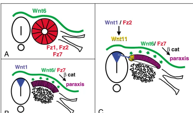
Together, our data allow us to propose an integrated model for Wnts function during somite epithelialisation and differentiation (Fig. 7). First, at the posterior segmental plate level, a signal from axial structures is required for the epithelialisation of somites. That signal can be mimicked by Wnt6-expressing cells (Schmidt et al., 2004); however, Wnt6 cannot be the endogenous Wnt molecule acting at this level, as it is only expressed in the ectoderm. Wnt3a, which is strongly expressed in the caudal PSM and neural tube (Marcelle et al., 1997), could represent this factor. Second, the Wnt6 signal, which
Wnt6/ Fz7 Wnt1
paraxis β cat
paraxis Wnt6
Fz1, Fz2 Fz7
Wnt6/Fz7 Wnt1 /Fz2
paraxis β cat
paraxis Wnt11
Wnt11
A
[image:9.612.45.371.547.738.2]B
C
Fig. 7.A model for the sequential action of Wnts on somite epithelialisation and compartimentalisation. (A) The Wnt6 signal, secreted by the ectoderm, binds to Fz7 receptor expressed by the cells of the segmental plate. (B) This interaction activates the canonical,  -catenin pathway that controls the expression of paraxisand maintains the epithelial structure of the dorsal part of the somite, while the ventral region disaggregates to form the sclerotome. (C) Cells in the medial-most region of the dermomyotome receive a Wnt1 signal from the dorsal neural tube, which activates Fz2 receptor and induces Wnt11expression in this region, thus defining the dorsomedial compartment of the dermomyotome.
De
is secreted by the ectoderm, probably binds the Fz7 receptor in segmental plate cells to control paraxisexpression through the activation of the canonical, -catenin pathway. The permanent source of Wnt6 from the ectoderm allows the maintenance of the epithelial structure and paraxisexpression in dorsal cells, while ventral cells (that receive Hedgehog signals) lose paraxis expression, undergo an EMT and differentiate into sclerotome. Finally, cells located in the medial-most region of the dermomyotome receive a Wnt1 signal from the dorsal neural tube, which, maybe through Fz2 receptors, imposes a medial character on these cells through the induction of Wnt11 expression. As somites differentiate, cells from the four borders of the dermomyotome generate myocytes. The decrease of Wnt6 and paraxis expression in the central region of the dermomyotome probably triggers its de-epithelialisation, allowing dorsally the migration of dermis precursors and ventrally that of muscle progenitors. The addition of a permanent source of Wnt6 or paraxis during this period maintains the central dermomyotomal cells in an epithelial state, impeding their migration and further differentiation.
We are indebted to Anne-Gaelle Boricky, Costis Papanayotou and Claudio Stern for useful comments on the manuscript and advice. This study was funded by grants from the Actions Concertées Incitatives (ACI), the Association Française contre les Myopathies (AFM), the Fondation pour le Recherche Médicale (FRM) and the Association pour le Recherche sur le Cancer (ARC) to C.M.; by NIH MBRS (SO6 GM52588) and AREA (1 R15 HD4204501) grants to L.W.B.; and by an NIH RIMI (P20 MD000262) grant to San Francisco State University. C. Linker was a fellow of the Boehringer Ingelheim Fonds, and C. Lesbros and J.G. were fellows of the Ministère de l’Education Nationale de la Recherche et des Technologies (MENRT).
References
Ben-Yair, R. and Kalcheim, C. (2005). Lineage analysis of the avian dermomyotome sheet reveals the existence of single cells with both dermal and muscle progenitor fates.. Development132, 689-701.
Bladt, F., Riethmacher, D., Isenmann, S., Aguzzi, A. and Birchmeier, C.
(1995). Essential role for the c-met receptor in the migration of myogenic precursor cells into the limb bud. Nature376, 768-771.
Borycki, A. G. and Emerson, C. P., Jr(2000). Multiple tissue interactions and signal transduction pathways control somite myogenesis. Curr. Top. Dev. Biol.48, 165-224.
Brand-Saberi, B., Wilting, J., Ebensperger, C. and Christ, B.(1996). The formation of somite compartments in the avian embryo. Int. J. Dev. Biol.
40, 411-420.
Brill, G., Kahane, N., Carmeli, C., von Schack, D., Barde, Y. A. and Kalcheim, C.(1995). Epithelial-mesenchymal conversion of dermatome progenitors requires neural tube-derived signals: characterization of the role of Neurotrophin-3. Development121, 2583-2594.
Burgess, R., Cserjesi, P., Ligon, K. L. and Olson, E. N.(1995). Paraxis: a basic helix-loop-helix protein expressed in paraxial mesoderm and developing somites. Dev. Biol.168, 296-306.
Burgess, R., Rawls, A., Brown, D., Bradley, A. and Olson, E. N.(1996). Requirement of the paraxis gene for somite formation and musculoskeletal patterning. Nature384, 570-573.
Burrus, L. W. and McMahon, A. P.(1995). Biochemical analysis of murine Wnt proteins reveals both shared and distinct properties. Exp. Cell Res.220, 363-373.
Cadigan, K. M., Fish, M. P., Rulifson, E. J. and Nusse, R.(1998). Wingless repression of Drosophila frizzled 2 expression shapes the Wingless morphogen gradient in the wing. Cell93, 767-777.
Capdevila, J., Tabin, C. and Johnson, R. L.(1998). Control of dorsoventral somite patterning by Wnt-1 and beta-catenin. Dev. Biol.193, 182-194.
Cauthen, C. A., Berdougo, E., Sandler, J. and Burrus, L. W.(2001).
Comparative analysis of the expression patterns of Wnts and Frizzleds during early myogenesis in chick embryos. Mech. Dev.104, 133-138.
Christ, B. and Ordahl, C. P. (1995). Early stages of chick somite development. Anat. Embryol. (Berl)191, 381-396.
Correia, K. M. and Conlon, R. A.(2000). Surface ectoderm is necessary for the morphogenesis of somites. Mech. Dev.91, 19-30.
Couly, G. and Le Douarin, N. M.(1988). The fate map of the cephalic neural primordium at the presomitic to the 3-somite stage in the avian embryo.
Development103, 101-113.
Daggett, D. F., Boyd, C. A., Gautier, P., Bryson-Richardson, R. J., Thisse, C., Thisse, B., Amacher, S. L. and Currie, P. D.(2004). Developmentally restricted actin-regulatory molecules control morphogenetic cell movements in the zebrafish gastrula. Curr. Biol.14, 1632-1638.
Daston, G., Lamar, E., Olivier, M. and Goulding, M.(1996). Pax-3 is necessary for migration but not differentiation of limb muscle precursors in the mouse. Development122, 1017-1027.
Delfini, M., Hirsinger, E., Pourquie, O. and Duprez, D.(2000). Delta 1-activated notch inhibits muscle differentiation without affecting Myf5 and Pax3 expression in chick limb myogenesis. Development127, 5213-5224.
Dietrich, S., Schubert, F. R. and Lumsden, A. (1997). Control of dorsoventral pattern in the chick paraxial mesoderm. Development 124, 3895-3908.
Dietrich, S., Schubert, F. R., Healy, C., Sharpe, P. T. and Lumsden, A.
(1998). Specification of the hypaxial musculature. Development125, 2235-2249.
Dietrich, S., Schubert, F. R., Gruss, P. and Lumsden, A.(1999). The role of the notochord for epaxial myotome formation in the mouse. Cell. Mol. Biol.45, 601-616.
Diez del Corral, R. and Storey, K. G.(2004). Opposing FGF and retinoid pathways: a signalling switch that controls differentiation and patterning onset in the extending vertebrate body axis. BioEssays 26, 857-869.
Dubrulle, J., McGrew, M. J. and Pourquie, O. (2001). FGF signaling controls somite boundary position and regulates segmentation clock control of spatiotemporal Hox gene activation. Cell106, 219-232.
Fan, C. M. and Tessier-Lavigne, M. (1994). Patterning of mammalian somites by surface ectoderm and notochord: evidence for sclerotome induction by a hedgehog homolog. Cell79, 1175-1186.
Fan, C. M., Lee, C. S. and Tessier-Lavigne, M.(1997). A role for WNT proteins in induction of dermomyotome. Dev. Biol.191, 160-165.
Galli, L. M., Willert, K., Nusse, R., Yablonka-Reuveni, Z., Nohno, T., Denetclaw, W. and Burrus, L. W.(2004). A proliferative role for Wnt-3a in chick somites. Dev. Biol.269, 489-504.
Goulding, M. D., Chalepakis, G., Deutsch, U., Erselius, J. R. and Gruss, P.(1991). Pax-3, a novel murine DNA binding protein expressed during early neurogenesis. EMBO J.10, 1135-1147.
Gros, J., Scaal, M. and Marcelle, C. (2004). A two-step mechanism for myotome formation in chick. Dev. Cell6, 875-882.
Gros, J., Manceau, M., Thomé, V. and Marcelle, C.(2005). A common somitic origin for embryonic muscle progenitors and satellite cells. Nature
435, 954-958.
Henrique, D., Adam, J., Myat, A., Chitnis, A., Lewis, J. and Ish-Horowicz, D.(1995). Expression of a Delta homologue in prospective neurons in the chick. Nature375, 787-790.
Heymann, S., Koudrova, M., Arnold, H., Koster, M. and Braun, T.(1996). Regulation and function of SF/HGF during migration of limb muscle precursor cells in chicken. Dev. Biol.180, 566-578.
Hollyday, M., McMahon, J. A. and McMahon, A. P.(1995). Wnt expression patterns in chick embryo nervous system. Mech. Dev.52, 9-25.
Huelsken, J. and Birchmeier, W. (2001). New aspects of Wnt signaling pathways in higher vertebrates. Curr. Opin. Genet. Dev. 11, 547-553.
Kardon, G., Heanue, T. A. and Tabin, C. J.(2002). Pax3 and Dach2 positive regulation in the developing somite. Dev. Dyn.224, 350-355.
Kassar-Duchossoy, L., Giacone, E., Gayraud-Morel, B., Jory, A., Gomes, D. and Tajbakhsh, S. (2005). Pax3/Pax7 mark a novel population of primitive myogenic cells during development. Genes Dev. 19, 1426-1431.
Kengaku, M., Capdevila, J., Rodriguez-Esteban, C., De La Pena, J., Johnson, R. L., Belmonte, J. C. and Tabin, C. J.(1998). Distinct WNT pathways regulating AER formation and dorsoventral polarity in the chick limb bud. Science280, 1274-1277.
Kuratani, S., Martin, J. F., Wawersik, S., Lilly, B., Eichele, G. and Olson, E. N.(1994). The expression pattern of the chick homeobox gene gMHox suggests a role in patterning of the limbs and face and in compartmentalization of somites. Dev. Biol.161, 357-369.
Lecourtois, M., Alexandre, C., Dubois, L. and Vincent, J. P. (2001).
De
Wingless capture by Frizzled and Frizzled2 in Drosophila embryos. Dev. Biol.235, 467-475.
Linker, C., Lesbros, C., Stark, M. R. and Marcelle, C.(2003). Intrinsic signals regulate the initial steps of myogenesis in vertebrates. Development
130, 4797-4807.
Locascio, A. and Nieto, M. A.(2001). Cell movements during vertebrate development: integrated tissue behaviour versus individual cell migration.
Curr. Opin. Genet. Dev. 11, 464-469.
Marcelle, C., Stark, M. R. and Bronner-Fraser, M.(1997). Coordinate actions of BMPs, Wnts, Shh and noggin mediate patterning of the dorsal somite. Development124, 3955-3963.
Marcelle, C., Lesbros, C. and Linker, C.(2002). Somite patterning: a few more pieces of the puzzle. Results and problems in cell differentiation. In
Vertebrate Myogenesis, Vol. 38 (ed. B. Brand-Saberi), pp. 81-108. Berlin-Heidelberg: Springer-Verlag.
Maroto, M., Reshef, R., Munsterberg, A. E., Koester, S., Goulding, M. and Lassar, A. B.(1997). Ectopic Pax-3 activates MyoD and Myf-5 expression in embryonic mesoderm and neural tissue. Cell89, 139-148.
Montross, W. T., Ji, H. and McCrea, P. D.(2000). A beta-catenin/engrailed chimera selectively suppresses Wnt signaling. J. Cell Sci. 113, 1759-1770.
Moreno, T. A. and Kintner, C.(2004). Regulation of segmental patterning by retinoic acid signaling during Xenopus somitogenesis. Dev. Cell6, 205-218.
Münsterberg, A. E., Kitajewski, J., Bumcrot, D. A., McMahon, A. P. and Lassar, A. B.(1995). Combinatorial signaling by Sonic hedgehog and Wnt family members induces myogenic bHLH gene expression in the somite.
Genes Dev. 9, 2911-2922.
Nakaya, Y., Kuroda, S., Katagiri, Y. T., Kaibuchi, K. and Takahashi, Y.
(2004). Mesenchymal-epithelial transition during somitic segmentation is regulated by differential roles of Cdc42 and Rac1. Dev. Cell7, 425-438.
Olivera-Martinez, I., Coltey, M., Dhouailly, D. and Pourquie, O.(2000). Mediolateral somitic origin of ribs and dermis determined by quail-chick chimeras. Development127, 4611-4617.
Olivera-Martinez, I., Missier, S., Fraboulet, S., Thelu, J. and Dhouailly, D.
(2002). Differential regulation of the chick dorsal thoracic dermal progenitors from the medial dermomyotome. Development129, 4763-4772.
Palmeirim, I., Henrique, D., Ish-Horowicz, D. and Pourquie, O.(1997). Avian hairy gene expression identifies a molecular clock linked to vertebrate segmentation and somitogenesis. Cell91, 639-648.
Palmeirim, I., Dubrulle, J., Henrique, D., Ish-Horowicz, D. and Pourquie, O. (1998). Uncoupling segmentation and somitogenesis in the chick presomitic mesoderm. Dev. Genet.23, 77-85.
Relaix, F., Rocancourt, D., Mansouri, A. and Buckingham, M. (2005). A Pax3/Pax7-dependent population of skeletal muscle progenitor cells. Nature
435, 948-953.
Reshef, R., Maroto, M. and Lassar, A. B.(1998). Regulation of dorsal somitic cell fates: BMPs and Noggin control the timing and pattern of myogenic regulator expression. Genes Dev.12, 290-303.
Rodriguez-Niedenfuhr, M., Dathe, V., Jacob, H. J., Prols, F. and Christ, B.
(2003). Spatial and temporal pattern of Wnt-6 expression during chick development. Anat. Embryol.206, 447-451.
Sato, A., Kojima, T., Ui-Tei, K., Miyata, Y. and Saigo, K. (1999). Dfrizzled-3, a new Drosophila Wnt receptor, acting as an attenuator of Wingless signaling in wingless hypomorphic mutants. Development126, 4421-4430.
Savagner, P. (2001). Leaving the neighborhood: molecular mechanisms involved during epithelial-mesenchymal transition. BioEssays 23, 912-923.
Scaal, M., Gros, J., Lesbros, C. and Marcelle, C. (2004). In ovo electroporation of avian somites. Dev. Dyn.229, 643-650.
Schmidt, C., Stoeckelhuber, M., McKinnell, I., Putz, R., Christ, B. and Patel, K. (2004). Wnt 6 regulates the epithelialisation process of the segmental plate mesoderm leading to somite formation. Dev. Biol.271, 198-209.
Schmidt, M., Tanaka, M. and Munsterberg, A.(2000). Expression of (beta)-catenin in the developing chick myotome is regulated by myogenic signals.
Development127, 4105-4113.
Schubert, F. R., Mootoosamy, R. C., Walters, E. H., Graham, A., Tumiotto, L., Munsterberg, A. E., Lumsden, A. and Dietrich, S.(2002). Wnt6 marks sites of epithelial transformations in the chick embryo. Mech. Dev.
114, 143-148.
Shook, D. and Keller, R.(2003). Mechanisms, mechanics and function of epithelial-mesenchymal transitions in early development. Mech. Dev.120, 1351-1383.
Sivasankaran, R., Calleja, M., Morata, G. and Basler, K. (2000). The
Wingless target gene Dfz3 encodes a new member of the Drosophila Frizzled family. Mech. Dev.91, 427-431.
Smallhorn, M., Murray, M. J. and Saint, R. (2004). The epithelial-mesenchymal transition of the Drosophila mesoderm requires the Rho GTP exchange factor Pebble. Development131, 2641-2651.
Sosic, D., Brand-Saberi, B., Schmidt, C., Christ, B. and Olson, E. N.
(1997). Regulation of paraxis expression and somite formation by ectoderm-and neural tube-derived signals. Dev. Biol.185, 229-243.
Spence, M. S., Yip, J. and Erickson, C. A.(1996). The dorsal neural tube organizes the dermamyotome and induces axial myocytes in the avian embryo. Development122, 231-241.
Stark, M. R., Sechrist, J., Bronner-Fraser, M. and Marcelle, C.(1997). Neural tube-ectoderm interactions are required for trigeminal placode formation. Development124, 4287-4295.
Stern, H. M. and Hauschka, S. D.(1995). Neural tube and notochord promote in vitro myogenesis in single somite explants. Dev. Biol.167, 87-103.
Stern, H. M., Brown, A. M. and Hauschka, S. D.(1995). Myogenesis in paraxial mesoderm: preferential induction by dorsal neural tube and by cells expressing Wnt-1. Development121, 3675-3686.
Tajbakhsh, S., Borello, U., Vivarelli, E., Kelly, R., Papkoff, J., Duprez, D., Buckingham, M. and Cossu, G.(1998). Differential activation of Myf5 and MyoD by different Wnts in explants of mouse paraxial mesoderm and the later activation of myogenesis in the absence of Myf5. Development125, 4155-4162.
Thiery, J. P.(2003). Epithelial-mesenchymal transitions in development and pathologies. Curr. Opin. Cell Biol.15, 740-746.
Veeman, M. T., Axelrod, J. D. and Moon, R. T.(2003). A second canon. Functions and mechanisms of beta-catenin-independent Wnt signaling. Dev. Cell5, 367-377.
Wagner, J., Schmidt, C., Nikowits, W., Jr and Christ, B. (2000). Compartmentalization of the somite and myogenesis in chick embryos are influenced by wnt expression. Dev. Biol.228, 86-94.
Wallingford, J. B., Fraser, S. E. and Harland, R. M.(2002). Convergent extension: the molecular control of polarized cell movement during embryonic development. Dev. Cell2, 695-706.
Wilson-Rawls, J., Hurt, C. R., Parsons, S. M. and Rawls, A. (1999). Differential regulation of epaxial and hypaxial muscle development by paraxis. Development126, 5217-5229.
Zhang, X. M., Ramalho-Santos, M. and McMahon, A. P. (2001). Smoothened mutants reveal redundant roles for Shh and Ihh signaling including regulation of L/R asymmetry by the mouse node. Cell105, 781-792.
Zhou, B. P., Deng, J., Xia, W., Xu, J., Li, Y. M., Gunduz, M. and Hung, M. C. (2004). Dual regulation of Snail by GSK-3beta-mediated phosphorylation in control of epithelial-mesenchymal transition. Nat. Cell. Biol. 6, 931-940.