R E S E A R C H A R T I C L E
Open Access
Gene expression profiling reveals underlying
molecular mechanism of hepatoprotective effect
of
Phyllanthus niruri
on thioacetamide-induced
hepatotoxicity in Sprague Dawley rats
Zahra A Amin
1†, Mohammed A Alshawsh
2†, Mustafa Kassim
3, Hapipah M Ali
4and Mahmood A Abdulla
5*Abstract
Background:The liver plays an essential role in the body by regulating several important metabolic functions. Liver injury is associated with the distortion of these functions causing many health problems. Pharmaceutical drugs treat liver disorders but cause further damage to it. Hence, herbal drugs are used worldwide and are becoming increasingly popular.
Methods:The hepatoprotective activity ofPhyllanthus niruri(PN) was evaluated against liver cirrhosis induced by thioacetamide (TAA) in male Sprague Dawley rats. Rats received intraperitoneal injections of thioacetamide (TAA, 200 mg/kg, b.w. three times weekly) for eight weeks. Daily treatments with plant extract (200 mg/kg) were administered orally for eight weeks. At the end of the study, hepatic damage was evaluated by monitoring transforming growth factor (TGFβ), collagenα1 (Collα1), matrix metalloproteinase-2 (MMP2) and tissue inhibitor of matrix metalloproteinase-1 (TIMP1) gene expression by real-time PCR. Moreover, different chromatographic techniques including column chromatography, thin layer chromatography, and Ultra Performance Liquid
Chromatography (UPLC) with Liquid Chromatography/Mass Spectrometry (LC/MS) were used to isolate the active constituents of the plant.
Results:The results revealed that treatment with PN significantly reduced the effect of thioacetamide toxicity and exhibited effective hepatoprotective activity. The mechanism of the hepatoprotective effect of PN is proposed to be by normalizing ROSs. Additionally, PN treatment regulated the expression of TGFβ, Collα1, MMP2, and TIMP1 genes. In the active fraction ofP. niruri, the isolated chemical constituents were 4-O-caffeoylquinic acid and quercetin 3-O-rhamnoside.
Conclusions:The results of the present study indicate that PN ethanol extracts possess hepatoprotective activity that is most likely because of the isolated chemical constituents.
Keywords:Phyllanthus niruri, Hepatoprotective, Gene expression, Active constituents
* Correspondence:[email protected]
†Equal contributors
5
Department of Biomedical Science, Faculty of Medicine, Kuala Lumpur 50603, Malaysia
Full list of author information is available at the end of the article
Background
Phyllanthus niruri has been used in folk medicine as an antipyretic, analgesic, or anti-inflammatory treatment, and treatment of other symptoms suggests antihistamine effects. Moreover, the decoction of the whole plant has been used orally against diarrhea and topically to treat jaundice. Crushed leaves together with leaves of Eupatorium odoratumand lime are applied on boils [1]. Previous studies have revealed the therapeutic potential of Phyllanthus niruri to treat genitourinary infections, venereal diseases, and kidney or bladder stones. More-over,P. niruriis reported to act as a urinary inhibitor of calcium oxalate crystallization and an effective treatment for urolithiasis by interfering in the growth and aggrega-tion of calcium oxalate crystals [2-4]. The reported anti-hyperuricemic action might be because of its uricosuric activity through an xanthine oxidase inhibitory effect [5]. Many reports in the literature have verified the pro-tective activity of Phyllanthus niruri against various drug- and toxin-induced hepatic disorders. Earlier stud-ies [6] have shown that extracts ofP. nirurihave demon-strated hepatoprotective activity against the carbon tetrachloride induced lipid peroxidation in the livers of rats, which was determined by raised serum enzyme levels. Although the effects of aqueous extracts ofP. niruri against carbon tetrachloride (CCL4)-induced liver, kidney and testes injuries have been studied [7], Manjrekaret al. concluded that the hepatoprotective and antioxidant acti-vity of this plant was associated with adverse effects on kidney and testes. In the study by Bhattacharjeeet al. [8], the hepatoprotective potential of the protein isolated from P. niruriagainst CCL4-induced liver damage was investi-gated. These results suggested that this protein protected the liver against oxidative stress and stimulated liver repair mechanisms. Additionally, Harish et al. [6] investigated the antioxidant activity of extracts of P. niruri against CCL4-induced liver damage. They demonstrated that membrane lipid peroxidation (LPO) inhibition was con-firmable by pre-treatment with the extracts.
In our previous research, we proved thatP. niruri pos-sesses hepatoprotective activity against thioacetamide-induced liver cirrhosis. Acute toxicity was studied, and the results demonstrated that P. niruriextract was non toxic when applied to SD rats. Significant differences were ob-served between thioacetamide-treated rats (200 mg/kg) and high or low dose (200 mg/kg and 100 mg/kg) P. niruri-treated rats in the body and liver weights, total antioxidant capacity, liver biochemical parameters, oxida-tive stress enzyme and lipid peroxidation levels. Moreover, P. niruritreatment effectively restored the histological and morphological observations closer to their normal appea-rances [9].
The goal of this study was to study the mechanism that induces the hepatoprotective activity ofPhyllanthus
niruri ethanol extract in protecting liver cirrhosis induced by thioacetamide in Sprague Dawley rats by monitoring the expression of transforming growth factor beta (TGFβ1), tissue inhibitors of metalloproteinases (TIMP1), matrix metalloproteinase (MMP2), and collagen alpha (Collα1) gene expression by real-time PCR. More-over, the active constituents of thePhyllanthus niruriwere isolated by separating the crude extract into several frac-tions using flash column chromatography and thin layer chromatography. Subsequently, the immunomodulatory ac-tivity for all fractions was tested to examine their abilities to proliferate human peripheral blood mononuclear cells (PBMCs). LC/MS was performed on the fraction that exhibited higher proliferation activity on the PBMCs.
Methods
Preparation of plant extract
Phyllanthus niruri plant was gained from Ethno Re-sources Sdn Bhd, identified and a voucher specimen (voucher number KLU46618) was kept. By the method of Zahraet al.[10], a crude ethanol extract was prepared by drenching 100 g of it in 1000 mL of 95% ethanol (1:10 w/v) for 72 hours at 25°C. The mixture was filtered and distilled under reduced pressure at 45°C by a rotary evaporator. The crude extract was maintained at −20°C until further experiments were done.
Chemicals and apparatus
Experimental design
The animal protocol was agreed by the Ethics Commit-tee for Animal Experimentation, Medicine Faculty, Uni-versity Malaya, Kuala Lumpur, Malaysia, under Ethic number PM/28/08/2010/MAA (R). The animals were cared for according to the “Guide for the Care and Use of Laboratory Animals”, published by the National Aca-demy of Science. The animals were provided with water and standard pelletad libitum.
Sprague Dawley rats (24 male) weighing 190–260 grams were divided into 3 groups randomly of 8 rats each. The experimental groups were as follows: healthy controls (vehicle control group), thioacetamide controls (liver cirrhosis control), and the P. niruri treatment group. The plant extract was suspended in Tween 20 (10%) and administered by oral gavage (5 mL/kg body weight). Thioacetamide was suspended in dH2O (2 mL/kg body weight) and injected intraperitoneally to the rats.
Group no.1(normal group) treated daily with Tween 20 orally and injected with dH2O three times weekly intra-peritoneally (ip) for two months. Group no. 2 (TAA group) treated daily with Tween 20 and injected with TAA (200 mg/kg) three times weekly for two months. The above mentioned procedure was following the method of Alshawshet al. [11].Group no. 3(PN-treated group) treated daily with the PN extract (200 mg/kg) orally and injected with TAA (200 mg/kg) three times weekly for two months.
Sample collection
After two months, each rat was fasted for 24 hours prior to sacrificing. A perfusion was performed under keta-mine and xylazine (1:10 v/v) anesthesia, and rats were quickly sacrificed by exsanguination of the jugular vein. A small portion of the livers were kept immediately in an RNAlatersolution (Applied Biosystems) for gene ex-pression analysis and kept at−80°C until the purification experiment was performed. The gene expression assays were performed using the following TaqMan gene ex-pression workflow: RNA isolation and purification, RNA transcription RNA to cDNA, and amplification of cDNA and target genes by real-time PCR. The gene assays used in this study were transforming growth factor beta (TGFβ1), tissue inhibitors of metalloproteinases (TIMP1), matrix metalloproteinase (MMP2), and collagen alpha (Collα1), and hypoxanthine phosphoribosyltransferase 1 (Hprt1) and peptidylprolyl isomerase A (Ppia) were used as housekeeping genes.
Gene expression profile RNA isolation and purification
RNA was extracted from the frozen liver using a QIAamp RNA Blood Mini Kit following the manufacturer’s protocol. Briefly, 30 mg of frozen RNAlater-stabilized liver tissue was
weighed immediately (without allowing the tissue to thaw), then disrupted and grinded by using a mortar and pestle. QIAshredder spin columns were used for homogenization and separation of the tissue lysates. The column digestion of DNA was performed during RNA purification using an RNase-free DNase set according to the manufacturer’s in-structions. Finally, total RNA was stored at −70°C until further use.
RNA purity was quantified using a spectrophotometer using a 10 mm quartz cuvette. With a 40× dilution fac-tor, absorbance was measured at 230, 260, 280, and 320 nm, and the following ratios were calculated: 260/ 280 and 260/320.
RNA integrity was measured by agarose gels electro-phoresis. Electrophoresis buffer tris-borate-EDTA (10× TBA) was prepared to fill the electrophoresis tank and cast the gel. A 0.5% (w/v) solution of agarose in electro-phoresis buffer was prepared. Ethidium bromide was added to the molten gel to a final concentration (0.5μg/ mL) and then mixed thoroughly by gentle swirling. A small-toothed comb (allows 1μL sample/well) was posi-tioned on the plate to form complete wells. The 60°C agarose solution was poured into the mold and allowed to set at room temperature for 30–45 minutes. The gel was mounted into the tank, and the electrophoresis buf-fer was added to cover the gel at a depth of 1 mm.
RNA samples (1μL each) were loaded after mixing with the loading dye. Subsequently, the RNAs were allowed to migrate toward the positive anode. The gel ran for 30 mi-nutes at 95 V until the migrated distance was 75% through the gel. The gels were examined under UV light to observe the discrete 18S and 28S ribosomal RNA bands.
Reverse transcription of RNA to cDNA
cDNA was synthesized using High Capacity RNA-to-cDNA Master Mix in a reaction plate according to the manufac-turer’s instructions.
Amplification of cDNA by real-time PCR
Real-time PCR was performed using a StepOnePlus Sys-tem and TaqMan Fast Advanced Master Mix (Applied Biosystems). The total reaction volume (10μL) consisted of the following: 1μL cDNA (20 ng), 5μL TaqMan Fast Advanced Master Mix, 0.5μL of each TaqMan Gene Ex-pression Assay, and 3.5 μL ultrapure DNase-free water. The cycle parameters were as follows: UNG incubation at 50°C for 2 min, polymerase activation at 95°C for 20 s, denaturation at 95°C for 1 s and then annealing and extension at 60°C for 20 s.
Real-time PCR data normalization
isomerase A (Ppia), were used for normalization. The comparative CT method was used to calculate to calcu-late the relative amount of the transcripts in all groups, and genes were normalized to the endogenous controls. The final value was normalized to the Hprt1 and Ppia genes and qualified to the normal control values of the investigated genes. The formula is as follows:
ΔΔCT¼ΔCT sampleð Þ−ΔCT normalð Þ
WhereΔCT is the difference in CT between the targeted gene and housekeeping controls by minimizing the average CT of the controls. The fold-change calculated as:2-ΔΔCT.
Chromatography profile
Flash column chromatography (FCC)
Plant fractionations were performed following the method of Fraga et al. [12]. Flash column chromatography was performed on silica gel 60 (0.063–0.200 mm, 70–230 mesh) from (Merck, Germany) using a Kontes column (2 × 30 cm) with an EYEL-4 pump (Rikakikai, Tokyo, Japan). The elution process to extract plant fractions (1 g/5 mL methanol) began with the most non-polar solvent (n-hex-ane), and then a continuous gradient elution (n-hexane – ethyl acetate – methanol – acetonitrile) was applied that concluded with the most polar solvent (dH2O), which was purified by a Milli-Q water purification system (Millipore).
Thin layer chromatography (TLC)
The obtained fractions were dissolved in methanol at 10 mg/mL to perform thin layer chromatography (TLC) with silica gel F254 (20 × 20 cm, 0.2 mm) plates. The analyses were achieved in the following: n-hexane – ethyl acetate, ethyl acetate – methanol, methanol – acetonitrile, and acetonitrile–water.
Ultra Performance Liquid Chromatography (UPLC) and Liquid Chromatography/Mass Spectrometry (LC/MS) A Waters Synapt HDMS system in TOF-mode was used to perform Ultra Performance Liquid Chromatography (UPLC)
and HDMS-mode was used to perform mass spectrometry that was equipped with an ACQUITY PDA Detector and ACQUITY UPLC BEH C18column (1.7μm, 2.1 × 50 mm).
The flow rate was 0.5 mL/min, and the injection volume was 3μL. The analyses were performed using binary gra-dients of Milli-Q water (with 0.1% formic acid) (solvent A) and HPLC grade acetonitrile (with 0.1% formic acid) (solv-ent B) with the following elution profile: from 0 min: 0% (B) in (A); 7 min: 100% (B) in (A); 10 min: 100% (B) in (A); 11 min: 0% (B) in (A).
Statistical analysis and data management
Real-time PCR data were analyzed using GenEx pro-gram. (GenEx software, www.multid.se); fold changes were calculated, andT-test was used to examine the dif-ferences between groups for all genes. “The Dictionary of Natural Products on DVD”software (CRC Press, Tay-lor and Francis Group, www.netbeans.org) was used to analyze the chromatography profiling data.
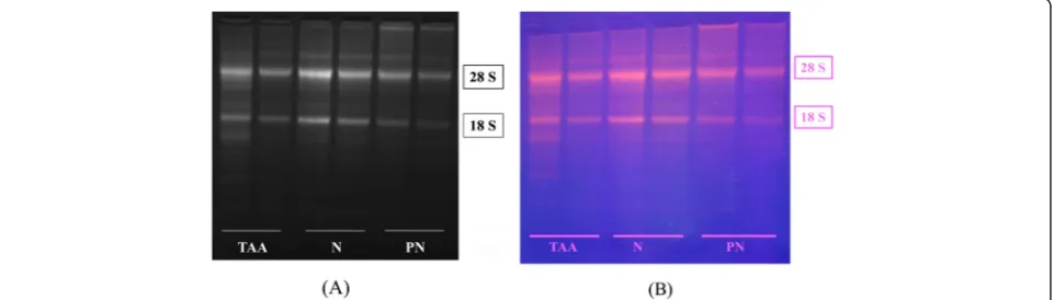
Results Integrity of RNA
[image:4.595.59.540.567.704.2]Total RNA was extracted from the liver tissues, and the quantity of RNA was determined by reading the absorb-ance at 260 nm spectrophotometrically with an ND-2000 NanoDrop Spectrophotometer (Thermo Fisher Sci-entific, Wilmington, DE, USA). The ratio of the absor-bance readings at 260 nm and 280 nm was used to indicate the quality of RNA. The 260/280 ratio for our RNA preparation ranged from 1.6–2.1; these values sug-gested good quality RNA. The integrity of RNA was checked by agarose gel electrophoresis. Discrete 28S and 18S ribosomal RNA bands were obtained in each case. The 28S rRNA band was approximately twice as large as the 18S rRNA band, indicating that the extracted RNA was intact and could be used in RT-PCR. Figure 1 shows a typical ethidium bromide-stained agarose RNA gel.
Real-time PCR analysis
Ct values of real-time PCR data were calculated using GenEX software and normalized to the reference genes HPRT1 and Ppia. The analysis showed significant diffe-rences in mRNA expression levels of the investigated genes between the controls and TAA- and PN-treated rats. In the control rats, the mRNA levels of TGFβ, collα1, MMP2 or TIMP1 were unchanged suggesting that the hepatic satellite cells (HSCs) were in their qui-escent state (Figure 2). In the TAA-treated group, hep-atic expression levels of all investigated genes were upregulated. The upregulation was significant at (P< 0.01) in the TAA-treated group compared with the PN-treated group for the genes TGFβ (1.677 ± 0.120), Collα1 (47.062 ± 7.716) and MMP2 (14.500 ± 3.528). However, the difference was non-significant for TIMP1 (1.738 ± 0.486).
Oral administration of PN before cirrhosis induction prevented and resolved the activation of HSCs, and the
remaining cells expressed decreased levels of TGFβ, Collα1, and MMP2 compared with the TAA-treated group as shown in Figure 2.
Chromatography profile
[image:5.595.55.540.329.684.2]After crude extraction of P. niruri, the ethanol extract was objected to flash column chromatography to sepa-rate the constituents of the extracts according to mo-lecular size, momo-lecular mass, and polarity. Therefore, 12 fractions were obtained, and by performing thin layer chromatography, the subsequent fractions with the same retention factor and spot colors after visualizing under UV light at 240 nm and 360 nm were combined to yield five fractions (PNF1, PNF2, PNF3, PNF4, and PNF5). The best resolutions of plates were given by acetonitrile-water. Subsequently, the immunomodulatory activity for all fractions was tested to examine their abilities to pro-liferate human peripheral mononuclear cells (PBMCs). As shown in Figure 3, PN fractions showed high activities
as a percent of viability to proliferate PMBCs; the frac-tion with the highest activity was PNF1.
LC/MS was performed on the PNF1 fraction, which exhibited higher activity to proliferate the PBMCs. Sub-sequently, by LC/MS/MS using the positive ionization
mode, four peaks were observed from PNF1 (Figures 4, 5, 6 and Table 1). However, only peak numbers 2 and 4 were identified. Peak number 2 (RT = 4.454 min, λ= 221 and 280 nm, MW = 355) (Figure 5) had [M + H]+ at m/z 356 and was identified as caffeoylquinic acid
%
P
B
M
C
s C
e
ll v
ia
b
ilit
y
CTRL 200 100 50 25
12.5 200 100 50 2512.5 200 100 50 2512.5 200 100 50 2512.5 200 100 50 25 12.5
0 200 400 600
PNF1 PNF2 PNF3 PNF4 PNF5
Phyllanthus niruri's Fractions *
*
*
*
*
*
*
[image:6.595.57.543.88.326.2]* *
Figure 3The effects ofP. nirurifractions on human peripheral blood mononuclear cell (PBMC) proliferation.Data are expressed as the mean ± SEM for triplicates; (*) indicates significance versus the control group (CTRL = dH2O) atP≤0.05.
[image:6.595.56.542.439.708.2](an isomer of chlorogenic acid) with fragments at m/z 340 (loss of CH3) and predominant fragments at m/z 191,165,151, and 147. Peak number 4 (Figure 6) (RT = 7.96 min, λ= 225 nm, MW = 448) had [M + H-H2O]+ atm/z430 and was identified as quercetin 3-O-rhamnoside with the loss of H2O, and with the loss of rhamnoside, the ion appeared at m/z 303 and was identified as
quercetin with other fragments at m/z 219, 205 and 165 [13,14].
Discussion
[image:7.595.57.540.88.319.2]The objective of this study was to determine the roles of transforming growth factorβ(TGFβ1), metalloproteinase-2 (MMP2), collagen αI (Collα1) and tissue inhibitor of
Figure 5UV max spectra and mass spectrum (TOF MS ES+) of peak no. 2 inP. niruriF1 (identified as 4-O-caffeoylquinic acid).
[image:7.595.61.537.444.715.2]metalloproteinase-1 (TIMP1) in preventing thioacetamide-induced liver cirrhosis in rats.
These results demonstrated that the mRNA expression levels of TGFβ1, Collα1, MMP2, and TIMP1 were un-changed in the control group; this supports the hypothesis that hepatic satellite cells (HSCs) were still in their quies-cent state. However, these HSCs were activated by the presence of TAA and led to the high production of ECM and consequently high expression of TGFβ, Collα1, MMP2, and TIMP1.
PN treatment successfully prevented the high synthesis of ECM and reduced the mRNA expression of TGFβ, Collα1, and MMP2 compared with the TAA-treated group.
Most studies of human liver diseases and animal models of progressive fibrosis have demonstrated that TIMP1 mRNA expression was upregulated at early stages of fibrosis and because TIMP1 functions not only reduce MMP activity but also act on the suppression of apoptosis by HSCs [15]. In our findings, hepatic reduc-tion in TIMP1 mRNA expression in the TAA-treated group can be explained as a consequence of increased HSC apoptosis [16].
Figure 7 shows the putative mechanism of the alter-ation of mRNA levels of the investigated genes in
TAA-treated rats. First, TAA bioactivates into thioacetamide-S-oxide and other ROSs [17,18], and activates the HSCs, which, in turn releases more ECM and subsequently in-creases TGFβ gene expression that affects the release of collagen α and MMP1 and then TIMP1. Therefore, scar tissue develops, and the liver losses its normal functions, anatomical shape and architecture [19,20]. Treatment with PN significantly reduced the effect of thioacetamide toxicity as follows: 1) removing the causative stimuli of TAA, neutralizing ROSs by their high antioxidant con-tent and attenuating endogenous antioxidant enzymes to their normal levels; 2) maintaining HSCs in their quies-cent state; and 3) increasing the release of TIMP1 to counter balance MMP2 and complete remodeling of the hepatocyte cellular system that preserves or sustains nor-mal liver function, shape, and appearance.
[image:8.595.59.542.102.142.2]These findings confirm the previous findings of Wills and Asha [21] who suggested that the hepatoprotective role of Lygodium flexuosum plant extract is because of the reduced mRNA levels of growth factors, proinflammatory cytokines, and other signaling mole-cules, which are involved in hepatic fibrosis including TGFβ1, procollagen-I, and TIMP1. Additionally, Chen et al. [22] demonstrated the hepatoprotective effects of Table 1 Putative identification of main components ofP. niruri’s active fraction
Plant fraction Rt.time (min) λ(nm) Molecular weight [M + H]m/z MS/MS fragmentation Tentative identification Molecular formula
PNF1 4.437 221, 280 355 356 340, 191, 165 , 151, 147 4-O-Caffeolquinic acid C16H18O9
[image:8.595.61.538.467.712.2]7.92 225 448 430 303, 219, 205, 165 Quercetin3-O-rhamnoside C21H20O11
silymarin against TAA-induced liver damage to be caused by downregulation of hepatic MMP2, TIMP1, TGFβ1, COLα1 and other genes in the mouse model of chronic liver fibrosis. Although the antifibrotic and hepatoprotective properties of the silibinin– phosphat-idylcholine-vitamin E complex in the rat model of liver fibrosis stimulated by bile duct ligation and dimethyl-nitrosamine administration have been postulated to cause reduced mRNA expression levels of procollagen type I, TGFβ1, TIMP1, and MMP2, the administration of the complex has also been reported to reduce hep-atic stellate cell activation and proliferation with colla-gen deposition [23].
The isolated chemical constituents included in P. niruri (4-O-caffeoylquinic acid and quercetin 3-O-rhamnoside) can further interpret the abovementioned hepatoprotective activity. 4-O-Caffeoylquinic acid, which is classified as a tannin, has been isolated previously from P. niruri and proven to possess antioxidant, immunomodulatory, and hepatoprotective effects in severalin vivoandin vitroassays [24-26]. However, quercetin 3-O-rhamnoside belongs to the flavonoid group of compounds that exhibits a wide range of pharmacological benefits including antimicrobial, antiviral, antioxidant, gastroprotective, hepatoprotective, anti-inflammatory and chemopreventive effects [27-30]. Moreover, potential working mechanisms of flavonoids du-ring injuries and tissue damage include the following: the interference of≥3 different free radical producing systems and an increase in function of the endogenous antioxidants CAT, SOD, and GPX [31]. Quercetin 3-O-rhamnoside has been confirmed inPhyllanthusspecies and other spe-cies of the Euphorbiaceae family [32-34]. Finally, both iso-lated chemical compounds from P. niruri (tannins and flavonoids) are classified as “polyphenols”, which are one of the most frequent and ever-present groups of plant metabolites, and have an important role in human and animal diets [35]. Recent studies have shown that plant-derived polyphenols are promising nutraceuticals for the control of various disorders, such as cardiovas-cular, neurological, and neoplastic diseases [36]. In addition, plant-derived polyphenolic compounds have hepatoprotective activity against different types of liver damage inducers, such as CCL4 [37], paracetamol [38], and thioacetamide [39], which explains the high interest and initiation of many studies to evaluate the biological activity and bioavailabilities of polyphenolic compounds.
Conclusion
This present study contributes significant knowledge to our understanding of the mechanism that underlies the hepatoprotective effect of Phyllanthus niruri, which is suggested to be through the regulation of TGFβ, Collα1, MMP2, and TIMP1 genes expression. The iso-lated chemical compounds (4-O-caffeoylquinic acid and
quercetin 3-O-rhamnoside) ofPhyllanthus nirurimight have direct consequences for hepatoprotective activity. Therefore, promising approaches from this study must focus on TGFβ, Collα1, MMP2, and TIMP1 genes ex-pression to develop new therapy for the treatment of liver cirrhosis.
Competing interests
The authors declare that they have no competing interests.
Authors’contributions
Conceived and designed the experiments: ZA MS, performed the experiments: ZA MS, analyzed the data: ZA MS MK, contributed reagents/ materials/analysis tools: MA HM ZA MS, wrote and revised the paper: ZA MA. All authors read and approved the final manuscript.
Acknowledgements
The authors express gratitude to the University of Malaya for the financial supports PV047/2011B, Um.c/625/1/HIR (151) and FL011-2012.
Author details
1Department of Pharmacognosy, College of Pharmacy, Hawler Medical
University, Erbil 44001, Iraq.2Department of Pharmacology, Faculty of
Medicine, University of Malaya, Kuala Lumpur 50603, Malaysia.3Department
of Anesthesiology, Faculty of Medicine, University of Malaya, Kuala Lumpur 50603, Malaysia.4Department of Chemistry, Faculty of Science, University of
Malaya, Kuala Lumpur 50603, Malaysia.5Department of Biomedical Science,
Faculty of Medicine, Kuala Lumpur 50603, Malaysia.
Received: 26 January 2013 Accepted: 1 July 2013 Published: 5 July 2013
References
1. Ong H, Norzalina J:Malay herbal medicine in Gemencheh, Negri Sembilan, Malaysia.Fitoterapia1999,70(1):10–14.
2. Freitas A, Schor N, Boim M:The effect of Phyllanthus niruri on urinary inhibitors of calcium oxalate crystallization and other factors associated with renal stone formation.BJU Int2002,89(9):829–834.
3. Boim MA, Heilberg IP, Schor N:Phyllanthus niruri as a promising alternative treatment for nephrolithiasis.Int Braz J Urol2010,
36(6):657–664.
4. Barros ME, Lima R, Mercuri LP, Matos JR, Schor N, Boim MA:Effect of extract of Phyllanthus niruri on crystal deposition in experimental urolithiasis.Urol Res2006,34(6):351–357.
5. Murugaiyah V, Chan KL:Mechanisms of antihyperuricemic effect of
Phyllanthus niruriand its lignan constituents.J Ethnopharmacol2009,
124(2):233–239.
6. Harish R, Shivanandappa T:Antioxidant activity and hepatoprotective potential of Phyllanthus niruri.Food Chem2006,95(2):180–185.
7. Manjrekar A, Jisha V, Bag P, Adhikary B, Pai M, Hegde A, Nandini M:Effect of Phyllanthus niruri Linn. treatment on liver, kidney and testes in CCl4 induced hepatotoxic rats.Indian J Exp Biol2008,46(7):514–520. 8. Bhattacharjee R, Sil P:Protein isolate from the herb, Phyllanthus niruri L.
(Euphorbiaceae), plays hepatoprotective role against carbon tetrachloride induced liver damage via its antioxidant properties.Food Chem Toxicol 2007,45(5):817–826.
9. Amin ZA, Bilgen M, Alshawsh MA, Ali HM, Hadi AHA, Abdulla MA:
Protective Role of Phyllanthus niruri Extract against Thioacetamide-Induced Liver Cirrhosis in Rat Model.Evidence-Based Complementary and
Alternative Medicine2012,2012.
10. Zahra AA, Kadir FA, Mahmood AA, AA A h, Suzy SM, Sabri SZ, Latif II, Ketuly K:Acute toxicity study and wound healing potential of Gynura procumbens leaf extract in rats.Journal of Medicinal Plants Research2011,
5(12):2551–2558.
12. Fraga CG, Martino VS, Ferraro GE, Coussio JD, Boveris A:Flavonoids as antioxidants evaluated by in vitro and in situ liver chemiluminescence.
Biochem Pharmacol1987,36(5):717–720.
13. Bravo L, Goya L, Lecumberri E:LC/MS characterization of phenolic constituents of mate (Ilex paraguariensis, St. Hil.) and its antioxidant activity compared to commonly consumed beverages.Food Res Int2007,
40(3):393–405.
14. Seeram NP, Lee R, Scheuller HS, Heber D:Identification of phenolic compounds in strawberries by liquid chromatography electrospray ionization mass spectroscopy.Food Chem2006,97(1):1–11.
15. Yoshiji H, Kuriyama S, Yoshii J, Ikenaka Y, Noguchi R, Nakatani T, Tsujinoue H, Yanase K, Namisaki T, Imazu H:Tissue inhibitor of metalloproteinases‐1 attenuates spontaneous liver fibrosis resolution in the transgenic mouse.
Hepatology2002,36(4):850–860.
16. Arthur MJP:Fibrogenesis II.Metalloproteinases and their inhibitors in liver fibrosis. American Journal of Physiology-Gastrointestinal and Liver Physiology 2000,279(2):G245–G249.
17. Chilakapati J, Shankar K, Korrapati MC, Hill RA, Mehendale HM:Saturation toxicokinetics of thioacetamide: role in initiation of liver injury.Drug
Metab Dispos2005,33(12):1877–1885.
18. Yogalakshmi B, Viswanathan P, Anuradha CV:Investigation of antioxidant, anti-inflammatory and DNA-protective properties of eugenol in thioacetamide-induced liver injury in rats.Toxicology2010,268(3):204–212. 19. Wang H, Zhao YP, Gao CF, Ji Q, Gressner AM, Yang ZX, Weiskirchen R:
Transforming growth factorβ1 gene variants increase transcription and are associated with liver cirrhosis in Chinese.Cytokine2008,43(1):20–25. 20. Mormone E, George J, Nieto N:Molecular pathogenesis of hepatic fibrosis
and current therapeutic approaches.Chem Biol Interact2011,193(3):225–231. 21. Wills P, Asha V:Protective mechanism of Lygodium flexuosum extract in
treating and preventing carbon tetrachloride induced hepatic fibrosis in rats.Chem Biol Interact2007,165(1):76–85.
22. Chen IS, Chen YC, Chou CH, Chuang RF, Sheen LY, Chiu CH:
Hepatoprotection of silymarin against thioacetamide‐induced chronic liver fibrosis.J Sci Food Agric2012,92(7):1441–1447.
23. Di Sario EB A, Taffetani S, Omenetti A, Candelaresi C, Marzioni M, De Minicis S, Benedetti A:Hepatoprotective and antifibrotic effect of a new silybin– phosphatidylcholine–Vitamin E complex in rats.Dig Liver Dis2005,
37:869–876.
24. Silva BM, Andrade PB, Valentão P, Ferreres F, Seabra RM, Ferreira MA:
Quince (Cydonia oblonga Miller) fruit (pulp, peel, and seed) and jam: antioxidant activity.J Agric Food Chem2004,52(15):4705–4712.
25. Adzet T, Camarasa J, Laguna JC:Hepatoprotective activity of polyphenolic compounds from Cynara scolymus against CCl4 toxicity in isolated rat hepatocytes.J Nat Prod1987,50(4):612–617.
26. Kapil A, Koul I, Suri O:Antihepatotoxic effects of chlorogenic acid from Anthocephalus cadamba.Phytother Res1995,9(3):189–193.
27. Kähkönen MP, Hopia AI, Vuorela HJ, Rauha JP, Pihlaja K, Kujala TS, Heinonen M:
Antioxidant activity of plant extracts containing phenolic compounds.
J Agric Food Chem1999,47(10):3954–3962.
28. Zayachkivska O, Konturek S, Drozdowicz D, Konturek P, Brzozowski T, Ghegotsky M:Gastroprotective effects of flavonoids in plant extracts.
J Physiol Pharmacol2005,56(Suppl 1):219–231.
29. Ozcelik B, Orhan I, Toker G:Antiviral and antimicrobial assessment of some selected flavonoids.Zeits Chrift Fur Natur Forschung C2006,
61(9/10):632.
30. Oh H, Kim DH, Cho JH, Kim YC:Hepatoprotective and free radical scavenging activities of phenolic petrosins and flavonoids isolated from < i > Equisetum arvense</i>.J Ethnopharmacol2004,95(2):421–424. 31. Nijveldt RJ, van Nood E, van Hoorn DEC, Boelens PG, van Norren K, van
Leeuwen PAM:Flavonoids: a review of probable mechanisms of action and potential applications.Am J Clin Nutr2001,74(4):418–425.
32. Zhang YJ, Abe T, Tanaka T, Yang CR, Kouno I:Two new acylated flavanone glycosides from the leaves and branches of Phyllanthus emblica.
Chem Pharm Bull2002,50(6):841–843.
33. Calixto JB, Santos A, Cechinel FV, Yunes RA:A review of the plants of the genus Phyllanthus: their chemistry, pharmacology, and therapeutic potential.Medicinal research reviews1998,18(4):225.
34. Kuti JO, Konuru HB:Antioxidant capacity and phenolic content in leaf extracts of tree spinach (Cnidoscolus spp.).J Agric Food Chem2004,
52(1):117–121.
35. Bravo L:Polyphenols: chemistry, dietary sources, metabolism, and nutritional significance.Nutr Rev1998,56(11):317–333.
36. Ullah MF, Khan MW:Food as medicine: potential therapeutic tendencies of plant derived polyphenolic compounds.Asian Pac J Cancer Prev2008,
9(2):187–196.
37. Shimoda H, Tanaka J, Kikuchi M, Fukuda T, Ito H, Hatano T, Yoshida T:
Walnut polyphenols prevent liver damage induced by carbon tetrachloride and d-galactosamine: hepatoprotective hydrolyzable tannins in the kernel pellicles of walnut.J Agric Food Chem2008,
56(12):4444–4449.
38. Chen X, Sun CK, Han GZ, Peng JY, Li Y, Liu YX, Lv YY, Liu KX, Zhou Q, Sun HJ:
Protective effect of tea polyphenols against paracetamol-induced hepatotoxicity in mice is significanly correlated with cytochrome P450 suppression.World journal of gastroenterology: WJG2009,15(15):1829. 39. Madani H, Talebolhosseini M, Asgary S, Naderi G:Hepatoprotective activity
of Silybum marianum and Cichorium intybus against thioacetamide in
rat.Pak J Nutr2008,7(1):172–176.
doi:10.1186/1472-6882-13-160
Cite this article as:Aminet al.:Gene expression profiling reveals underlying molecular mechanism of hepatoprotective effect of
Phyllanthus nirurion thioacetamide-induced hepatotoxicity in Sprague Dawley rats.BMC Complementary and Alternative Medicine201313:160.
Submit your next manuscript to BioMed Central and take full advantage of:
• Convenient online submission
• Thorough peer review
• No space constraints or color figure charges
• Immediate publication on acceptance
• Inclusion in PubMed, CAS, Scopus and Google Scholar
• Research which is freely available for redistribution