Evaluation of Oxacillin and Cefoxitin Disk Diffusion and MIC
Breakpoints Established by the Clinical and Laboratory
Standards Institute for Detection of
mecA
-Mediated Oxacillin
Resistance in
Staphylococcus schleiferi
H. K. Huse,aS. A. Miller,aS. Chandrasekaran,aJ. A. Hindler,a S. D. Lawhon,bD. A. Bemis,cL. F. Westblade,d R. M. Humphries,a*on behalf of theStaphylococcus Ad HocWorking Group of the CLSI Antimicrobial Susceptibility Testing Subcommittee
aUCLA David Geffen School of Medicine, Los Angeles, California, USA
bTexas A&M University College of Veterinary Medicine, College Station, Texas, USA
cUniversity of Tennessee College of Veterinary Medicine, Knoxville, Tennessee, USA
dWeill Cornell Medicine, New York, New York, USA
ABSTRACT Staphylococcus schleiferi is a beta-hemolytic, coagulase-variable
colo-nizer of small animals that can cause opportunistic infections in humans. In
vet-erinary isolates, the rate of mecA-mediated oxacillin resistance is significant, with
reported resistance rates of ⬎39%. The goal of this study was to evaluate
oxacil-lin and cefoxitin disk diffusion (DD) and MIC breakpoints for detection of mecA
-mediated oxacillin resistance in 52 human and 38 veterinary isolates ofS.
schleif-eri. Isolates were tested on multiple brands of commercial media and according
to Clinical and Laboratory Standards Institute (CLSI) methods. Zone diameters
and MIC values were interpreted using CLSI breakpoints (CLSI, Performance
Stan-dards for Antimicrobial Susceptibility Testing. M100-S27, 2017) for Staphylococcus aureus/Staphylococcus lugdunensis, coagulase-negative staphylococci (CoNS), and
Staphylococcus pseudintermedius. Results were compared to those of mecA PCR.
Twenty-nine of 90 (32%) isolates were mecA positive. Oxacillin inhibition zone
sizes and MICs interpreted by S. pseudintermediusbreakpoints reliably
differenti-ated mecA-positive and mecA-negative isolates, with a categorical agreement
(CA) of 100% and no very major errors (VMEs) or major errors (MEs) for all
me-dia. For cefoxitin DD results interpreted using S. aureus/S. lugdunensisand CoNS
breakpoints, CA values were 85% and 75%, respectively, and there were 72% and
64% VMEs, respectively, and 0 MEs. For cefoxitin MICs interpreted using S.
au-reus/S. lugdunensis breakpoints, CA was 81%, and there were 60% VMEs and no MEs. Our data demonstrate that oxacillin DD or MIC testing methods using the
current S. pseudintermedius breakpoints reliably identify mecA-mediated oxacillin
resistance in S. schleiferi, while cefoxitin DD and MIC testing methods perform
poorly.
KEYWORDS breakpoints, cefoxitin,mecA, oxacillin, PBP2a,Staphylococcus schleiferi
S
taphylococcus schleiferiis an emerging zoonotic pathogen that colonizes the skin and mucosal surfaces of small animals (1–3). Isolates form medium to large, nonpigmented, beta-hemolytic colonies on 5% sheep blood agar (4, 5). The species isfurther divided into S. schleiferi subsp. schleiferi and S. schleiferi subsp. coagulans.
Staphylococcus schleiferisubsp.schleiferiwas first isolated from human specimens, in
1988 (4), while S. schleiferi subsp. coagulans was first isolated from dogs with otitis
externa, in 1990 (5). The subspecies have high levels of DNA homology but differ in
Received17 October 2017 Returned for modification5 November 2017 Accepted24 November 2017
Accepted manuscript posted online29 November 2017
CitationHuse HK, Miller SA, Chandrasekaran S, Hindler JA, Lawhon SD, Bemis DA, Westblade LF, Humphries RM, on behalf of the
Staphylococcus Ad HocWorking Group of the
CLSI Antimicrobial Susceptibility Testing Subcommittee. 2018. Evaluation of oxacillin and cefoxitin disk diffusion and MIC breakpoints established by the Clinical and Laboratory Standards Institute for detection of
mecA-mediated oxacillin resistance in
Staphylococcus schleiferi. J Clin Microbiol
56:e01653-17.https://doi.org/10.1128/JCM .01653-17.
EditorSandra S. Richter, Cleveland Clinic Copyright© 2018 American Society for Microbiology.All Rights Reserved. Address correspondence to R. M. Humphries, [email protected].
*Present address: R. M. Humphries, Accelerate Diagnostics, Inc., Tucson, Arizona, USA.
crossm
on May 17, 2020 by guest
http://jcm.asm.org/
several phenotypic characteristics, including clumping factor, tube coagulase, and
urease production.S. schleiferisubsp.schleiferiis clumping factor positive, tube
coag-ulase negative, and urease negative, whileS. schleiferi subsp.coagulans is clumping
factor negative, tube coagulase positive, and urease positive (4, 5). Matrix-assisted laser desorption ionization–time of flight mass spectrometry (MALDI-TOF MS) can reliably
identifyS. schleiferito the species level, but further biochemical testing is needed to
differentiate the two subspecies (6).
In small animals, S. schleiferi most frequently colonizes the skin, nares, ears, and
rectum of dogs, where it can cause inflammatory skin disease, otitis externa, and otitis
media (2, 3, 5, 7–14). Though found at a lower prevalence than that in dogs,S. schleiferi
has also been isolated from healthy cats (1), cats with inflammatory skin disease (1), and parrots (2). Oxacillin resistance rates vary but can be high, with some studies reporting oxacillin resistance for 39 to 73% of veterinary isolates (7–9, 15, 16). Staphylococcal
cassette chromosomemectype IV (SCCmecIV) has been identified inS. schleiferisubsp.
coagulans(16).S. schleiferihas also been reported to carry SCCmectypes I and IV (17, 18). In addition to oxacillin resistance, reduced susceptibility to clindamycin,
erythro-mycin, and fluoroquinolones has been reported (13, 19, 20). In a small study,S. schleiferi
isolates were negative for beta-lactamase production (21).
Though it is primarily a veterinary pathogen,S. schleiferican also cause opportunistic
infections in humans. Cases of endophthalmitis (22), endocarditis (23, 24), bacteremia (25), osteomyelitis (26), and wound (27), surgical site (27), and pacemaker (28) infections
have been reported.S. schleiferiwas implicated in an outbreak of surgical site infections
and was originally misidentified asStaphylococcus aureus(29). Interestingly, while both
subspecies can cause infection,S. schleiferisubsp.schleiferiis more prevalent in causing
human infections (27, 29).
S. schleiferi has been reported to give false-positive results in latex agglutination
tests forS. aureusidentification, at rates of 25 to 75% (30). Use of MALDI-TOF MS will
likely increase the number of S. schleiferi isolates identified, and the high oxacillin
resistance rates in this species are concerning. Many clinical laboratories use cefoxitin disk diffusion (DD) testing for detection of oxacillin resistance in staphylococci other thanS. pseudintermedius, as outlined by the Clinical and Laboratory Standards Institute (CLSI) M100-S27 document (31, 32). Previous data supporting the use of cefoxitin as a surrogate agent for detection of oxacillin resistance in coagulase-negative
staphylo-cocci (CoNS) were largely derived fromStaphylococcus epidermidisisolates (33).
How-ever, the most accurate methods for detectingmecA-mediated oxacillin resistance in
other CoNS species have yet to be determined. Some studies have shown that cefoxitin
disk testing has low sensitivity for detecting oxacillin resistance inS. schleiferiveterinary
isolates andS. pseudintermedius(15, 33, 34).
The goal of the present study was to evaluate oxacillin and cefoxitin DD and broth
microdilution (BMD) MIC testing for detection ofmecA-mediated oxacillin resistance in
52 human and 38 veterinary isolates ofS. schleiferi. Oxacillin-resistant staphylococci are
resistant to all beta-lactam antibiotics except for new anti-methicillin-resistantS. aureus
(anti-MRSA) cephalosporins. We demonstrate that oxacillin DD results interpreted by
the CLSI M100-S27 breakpoints for S. pseudintermedius reliably detectmecA-positive
andmecA-negativeS. schleiferiisolates, while cefoxitin is an unreliable surrogate agent.
(The results of this study were presented to the CLSI Antimicrobial Susceptibility Testing Subcommittee in June 2017, leading to the addition of specific breakpoints for
oxacillin disk diffusion and MIC testing ofS. schleiferifor the forthcoming 28th edition
of the M100 document.)
MATERIALS AND METHODS
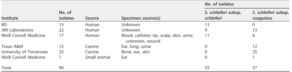
Specimens.A total of 90S. schleiferiisolates were included in this study (Table 1). Human isolates (n⫽52) were submitted by Becton Dickinson and Company (BD) (n⫽13), JMI Laboratories (n⫽22), and Weill Cornell Medicine (n⫽17). Canine and other small animal isolates (n⫽38) were obtained from the Texas A&M University College of Veterinary Medicine (n⫽12), the University of Tennessee College of Veterinary Medicine (n⫽25), and Weill Cornell Medicine (n⫽1). Isolates were identified to the species or subspecies level at each institution, using the corresponding standard operating procedures.
on May 17, 2020 by guest
http://jcm.asm.org/
species identifications were confirmed by urease testing at the Ronald Reagan UCLA Medical Center, Los Angeles, CA. Urease testing on urea agar slants (BD) was performed according to the manufacturer’s protocol, with the slants incubated at 35°C in ambient air for 24 h.
mecA PCR.Fifty-four isolates with knownmecA genotypes were submitted. For the remaining isolates, colony PCR was performed on 18- to 24-h isolates grown on 5% sheep blood agar plates (SBAP). The following primers were used to amplify a 533-bp product from themecAgene: 5=-AAAATCGATGGTAA AGGTTGGC-3=and 5=-AGTTCTGCAGTACCGGATTTGC-3=(35). A pipette tip was used to transfer a pinpoint amount of an isolated colony to a 25-l PCR mixture containing 12.5l AmpliTaq Gold 360 master mix (Applied Biosystems, Thermo Fisher Scientific, Waltham, MA), 0.5M (each)mecAprimers, and 11.25l water. Reaction mixtures were incubated at 95°C for 10 min, followed by 35 cycles of 95°C for 15 s, 55°C for 30 s, and 72°C for 30 s. The final extension was performed at 72°C for 7 min. As a positive control, colony PCR was performed onS. aureusATCC 43300, amecA-positive strain, and 1l purified genomic DNA from the same strain. PCR products were visualized on a 4% precast agarose gel (Lonza, Basel, Switzerland).
Antimicrobial susceptibility testing (AST).Isolates were stored as described previously (34) and subcultured twice on 5% SBAP before testing. DD and BMD tests were performed as described by the CLSI (31, 36). DD was evaluated on Mueller-Hinton agar (MHA) plates obtained from 3 vendors: Remel (Lenexa, KS), Hardy Diagnostics (Santa Maria, CA), and BD. Disks containing 1g oxacillin and 30g cefoxitin (BBL, BD) were used. BMD was performed by the CLSI reference method, using frozen-form panels containing cation-adjusted Mueller-Hinton broth (CA-MHB) with cefoxitin (unsupplemented) or oxacillin supplemented with 2% NaCl. BMD panels were made by Thermo Fisher. MIC tests using CA-MHB from 3 different manufacturers (Difco, BD, and Oxoid) were evaluated on a single panel. Oxacillin and cefoxitin were tested in 2-fold dilutions at concentrations ranging from 0.015g/ml to 32g/ml. Isolated colonies grown overnight on SBAP at 35 to 37°C in ambient air were resuspended in 0.85% saline to obtain a 0.5 McFarland standard. The suspensions were used to inoculate all DD and MIC plates per CLSI recommendations (31, 36). DD test plates were incubated at 35°C in ambient air, and zones of inhibition were measured at 16 to 18 h for oxacillin and 24 h for cefoxitin. During preliminary studies, zones of inhibition were read with both transmitted and reflected light, and there was no difference noted between the two methods. Therefore, for the final study, both oxacillin and cefoxitin zones of inhibition were read using reflected light. BMD test plates were incubated at 35°C in ambient air and read at 16 to 20 h for cefoxitin and 24 h for oxacillin.S. aureusATCC 25923 andS. aureusATCC 29213 were used as quality control strains for DD and BMD tests, respectively.
[image:3.585.39.548.83.210.2]Data analysis.Zone diameters and MIC values were interpreted using breakpoints for the following organisms, obtained from the CLSI M100-S27 document: (i)S. aureus/S. lugdunensis; (ii) CoNS, excluding S. lugdunensis andS. pseudintermedius; and/or (iii) S. pseudintermedius(Table 2) (32). Results were
TABLE 1Summary of study isolates submitted by various institutions
Institute
No. of
isolates Source Specimen source(s)
No. of isolates
S. schleiferisubsp. schleiferi
S. schleiferisubsp. coagulans
BD 13 Human Unknown 13 0
JMI Laboratories 22 Human Unknown 9 13
Weill Cornell Medicine 17 Human Blood, catheter tip, scalp, skin, urine, unknown, wound
11 6
Texas A&M 12 Canine Ear, lung, urine 0 12
University of Tennessee 25 Canine Bone, ear, skin 0 25
Weill Cornell Medicine 1 Small animal Ear 0 1
Total 90 33 57
TABLE 2Breakpoints used for prediction ofS. schleiferi mecA-mediated oxacillin resistance in this studya
Organism
Oxacillin breakpoint Cefoxitin breakpoint
DD inhibition
zone (mm) MIC (g/ml) DD inhibition zone (mm)
MIC (g/ml)
S R S R S R S R
S. aureus/S. lugdunensis NA NA ⱕ2 ⱖ4 ⱖ22 ⱕ21 ⱕ4 ⱖ8
Coagulase-negative staphylococci, exceptS. lugdunensisandS. pseudintermedius
NA NA ⱕ0.25 ⱖ0.5 ⱖ25 ⱕ24 NA NA
S. pseudintermediusb ⱖ18 ⱕ17 ⱕ0.25 ⱖ0.5 NA NA NA NA
aFrom the CLSI M100-S27 document (32). NA, not applicable; S, susceptible; R, resistant.
bIn the 28th edition of the CLSI M100 document, the guidance forS. pseudintermediuswill also apply toS.
schleiferi.
on May 17, 2020 by guest
http://jcm.asm.org/
[image:3.585.42.374.597.712.2]compared to the results ofmecAPCR, which was considered the gold standard for oxacillin resistance. Categorical agreement (CA), very major errors (VMEs), and major errors (MEs) were calculated as previously described (37). CA was determined usingmecAPCR results as the reference to define isolates as resistant or susceptible. VMEs were counted as identifications of isolates that weremecApositive but oxacillin or cefoxitin susceptible. MEs were defined as identifications of isolates that weremecAnegative but oxacillin or cefoxitin resistant.
Discrepancy analysis.For any isolates with oxacillin results that were discordant withmecAPCR results, testing was repeated by both AST and PCR. If the error resolved, it was excluded as an error.
PBP2a testing.Penicillin binding protein 2a (PBP2a) testing was performed on 54S. schleiferiisolates by use of Alere PBP2a SA culture colony test kits (Alere Inc., Scarborough, ME) and Oxoid PBP2=latex agglutination test kits (Thermo Fisher Scientific, Waltham, MA). Only 54 isolates were tested due to a limited number of available testing kits. Isolates were chosen based onmecAPCR results to give an almost even distribution ofmecA-positive andmecA-negative isolates (28 and 26, respectively). Forty-three isolates wereS. schleiferisubsp.coagulans, and 11 wereS. schleiferisubsp.schleiferi. Thirty-eight isolates were from animals (allS. schleiferisubsp.coagulans), and 16 were from humans (5S. schleiferi subsp.coagulansisolates and 11S. schleiferisubsp.schleiferiisolates). Colonies from SBAP used for DD and BMD inoculum preparation were tested for noninduced PBP2a expression according to the manu-facturers’ instructions forS. aureus. S. aureusATCC 43300 andS. aureus25923 were used as positive and negative controls, respectively.
RESULTS
Isolates.Ninety isolates were tested in this study (Table 1). Fifty-two (58%) were isolated from human specimens (blood, wounds, urine, ears, catheters, skin, and the scalp). Thirty-eight (42%) isolates were urine, bone, lung, pyoderma (of the ear), or skin isolates from canines or other small animals. Thirty-three (37%) isolates were identified as S. schleiferi subsp. schleiferi, all of which were isolated from human specimens.
Fifty-seven (63%) isolates were identified asS. schleiferisubsp.coagulans; 19 (33%) of
these were isolated from human specimens, and 38 (67%) were isolated from animals.
mecAPCR.Twenty-nine (32%) isolates weremecApositive. All of themecA-positive
isolates wereS. schleiferisubsp.coagulans, and four of these were isolated from human
specimens. Sixty-one (68%) isolates weremecA negative. Of these, 33 (54%) were S.
schleiferisubsp.schleiferiand 28 (46%) wereS. schleiferisubsp.coagulans.
Cefoxitin DD and BMD testing.Results from the cefoxitin DD and BMD tests are summarized in Table 3 and Fig. 1. For cefoxitin tests, neither the zones of inhibition nor
the MICs showed clear divisions betweenmecA-positive andmecA-negative isolates for
any medium (either MHA or CA-MHB) brand tested (Fig. 1; see Fig. S1 and S2 in the supplemental material). For DD testing, 17 isolates showed only faint growth on Remel MHA medium (Fig. 2), and thus zones of inhibition could not be measured. Therefore, a total of 253 data points were collected for all media (Fig. 1A).
DD and MIC results were interpreted using the cefoxitin breakpoints listed in Table
2. On applying the CLSI M100-S27S. aureus/S. lugdunensisbreakpoints for DD testing
using cefoxitin, the CA values for BD, Hardy, and Remel media were 78%, 76%, and 71%, respectively. For cefoxitin, there were 20/29 (69%), 22/29 (76%), and 21/29 (72%) VMEs for the BD, Hardy, and Remel media, respectively. There were no MEs because all
mecA-negative isolates were susceptible by DD testing on all media tested, with zone
sizes ranging from 29 to 45 mm (Fig. 1). For BMD, all brands performed similarly with cefoxitin. The CA values and numbers of VMEs for BD, Difco, and Remel CA-MHB were 81% and 12/29 (41%), 81% and 12/29 (41%), and 80% and 11/29 (38%), respectively. There were no MEs for any of the CA-MHB medium brands tested. On applying the CoNS breakpoints for DD testing using cefoxitin, the CA value and percentage of VMEs were 81% and 59% for BD medium, 77% and 72% for Hardy medium, and 75% and 62% for Remel medium (Table 3). There were no MEs with application of the CoNS break-points.
Overall, for cefoxitin DD results interpreted usingS. aureus/S. lugdunensisand CoNS
breakpoints, CA was 85% and 75%, respectively, and there were 63/87 (72%) and 56/87
(64%) VMEs, respectively. Cefoxitin MICs interpreted using S. aureus/S. lugdunensis
breakpoints yielded an overall CA of 81% and 52/87 (60%) VMEs. There were no MEs for either DD or MIC results interpreted using any breakpoints.
Oxacillin DD and BMD testing.Results from the oxacillin DD and BMD tests are summarized in Table 4 and Fig. 1. In contrast to the cefoxitin results, there was a clear
on May 17, 2020 by guest
http://jcm.asm.org/
TABLE 3 Performances of cefoxitin DD and BMD testing for detection of mecA -mediated oxacillin resistance in S. schleiferi Breakpoints Summary BD medium Hardy medium Remel medium Difco CA-MHB BD CA-MHB Oxoid CA-MHB CA (%) No. of errors/no. of isolates (%) CA (%) No. of errors/no. of isolates (%) CA (%) No. of errors/no. of isolates (%) CA (%) a No. of errors/no. of isolates (%) CA (%) No. of errors/no. of isolates (%) CA (%) No. of errors/no. of isolates (%) CA (%) No. of errors/no. of isolates (%) VMEs MEs VMEs MEs VMEs MEs VMEs MEs a VMEs MEs VMEs MEs VMEs MEs CLSI M100-S27 DD breakpoints S. aureus/S. lugdunensis 85 63/87 (72) 0/166 (0) 78 20/29 (69) 0/61 (0) 76 22/29 (76) 0/61 (0) 71 21/29 (72) 0/44 (0) CoNS staphylococci (except for S. lugdunensis and S. pseudintermedius ) 75 56/87 (64) 0/166 (0) 81 17/29 (59) 0/61 (0) 77 21/29 (72) 0/61 (0) 75 18/29 (62) 0/44 (0) CLSI M100-S27 MIC breakpoints S. aureus/S. lugdunensis 81 52/87 (60) 0/183 (0) 81 12/29 (41) 0/61 (0) 81 12/29 (41) 0/61 (0) 80 11/29 (38) 0/61 (0) aWe were unable to read zones for 17 mecA -negative isolates due to poor growth; these were not included in the denominator.
on May 17, 2020 by guest
http://jcm.asm.org/
division betweenmecA-positive andmecA-negative isolates for both zones of inhibition
and MICs for all medium brands tested (Fig. 1; Fig. S3 and S4). On applying the S.
pseudintermedius breakpoints for DD testing, CA was 100% for all medium brands. There were no VMEs or MEs. On Remel medium, 16 isolates did not grow well enough
for reading of the oxacillin DD zones, so the denominator (n) was 45.
For oxacillin BMD, CA values with theS. aureus/S. lugdunensisbreakpoints were 98%,
92%, and 89% for Difco, BD, and Oxoid CA-MHB, respectively. The numbers of VMEs for Difco, BD, and Oxoid CA-MHB were 6/29 (21%), 7/29 (24%), and 10/29 (35%), respec-tively. There were no MEs for the three medium brands tested. The oxacillin MIC
breakpoints for CoNS andS. pseudintermediusare the same. CA was 100%, and there
were no VMEs or MEs for the three medium brands.
Overall, for oxacillin DD results interpreted using theS. pseudintermediusbreakpoints,
overall CA was 100%, and there were no VMEs or MEs. Oxacillin MICs interpreted using the
FIG 1Distributions of cefoxitin and oxacillin growth inhibition zone diameters and MICs as determined by DD and BMD testing on all media tested. M100-S27 DD and MIC breakpoints are shown for S. aureus/S. lugdunensis (Sa/Sl), coagulase-negative staphylococci (excludingS. lugdunensisandS. pseudintermedius) (CoNS), andS. pseudintermedius. R, resistant; S, susceptible. (A) Cefoxitin DD testing (for BD, Hardy, and Remel media,n⫽253). (B) Oxacillin DD testing (for BD, Hardy, and Remel media,n⫽253). (C) Cefoxitin MICs (for Difco, BD, and Oxoid media,n⫽270). (D) Oxacillin MICs (for Difco, BD, and Oxoid media,n⫽270).
FIG 2Growth of a single strain ofS. schleiferion BD (left), Remel (middle), and Hardy (right) MHA plates with oxacillin (OX) and cefoxitin (FOX) disks.
on May 17, 2020 by guest
http://jcm.asm.org/
[image:6.585.41.476.74.323.2] [image:6.585.41.403.576.714.2]TABLE 4 Performances of oxacillin DD and BMD testing for detection of mecA -mediated oxacillin resistance in S. schleiferi Breakpoints Summary BD medium Hardy medium Remel medium Difco CA-MHB BD CA-MHB Oxoid CA-MHB CA (%) a No. of errors/no. of isolates (%) CA (%) No. of errors/no. of isolates (%) CA (%) No. of errors/no. of isolates (%) CA (%) a No. of errors/no. of isolates (%) CA (%) No. of errors/no. of isolates (%) CA (%) No. of errors/no. of isolates (%) CA (%) No. of errors/no. of isolates (%) VMEs MEs a VMEs MEs VMEs MEs VMEs MEs a VMEs MEs VMEs MEs VMEs MEs CLSI M100-S27 DD breakpoints S. pseudintermedius 100 0/87 (0) 0/167 (0) 100 0/29 (0) 0/61 (0) 100 0/29 (0) 0/61 (0) 100 0/29 (0) 0/45 (0) CLSI M100-S27 MIC breakpoints S. aureus/S. lugdunensis 91 23/87 (26) 0/183 (0) 98 6/29 (21) 0/61 (0) 92 7/29 (24) 0/61 (0) 89 10/29 (35) 0/61 (0) Coagulase-negative staphylococci (except for S. lugdunensis and S. pseudintermedius ) 100 0/87 (0) 0/183 (0) 100 0/29 (0) 0/61 (0) 100 0/29 (0) 0/61 (0) 100 0/29 (0) 0/61 (0) S. pseudintermedius b 100 0/87 (0) 0/183 (0) 100 0/29 (0) 0/61 (0) 100 0/29 (0) 0/61 (0) 100 0/29 (0) 0/61 (0) aWe were unable to read zones for 16 mecA -negative isolates; these were not included in the denominator. bIn the 28th edition of the CLSI M100 document, the guidance for S. pseudintermedius will also apply to S. schleiferi .
on May 17, 2020 by guest
http://jcm.asm.org/
S. aureus/S. lugdunensisbreakpoints yielded an overall CA of 91%, and there were 23/87 (26%) isolates with VMEs and no MEs. Overall CA for oxacillin MICs interpreted using CoNS andS. pseudintermediusbreakpoints was 100%, with no VMEs or MEs.
Discrepant analysis.One isolate testedmecAPCR negative but was PBP2a positive
and oxacillin resistant by DD and BMD testing. When all tests were repeated, themecA
PCR was positive, so this error was excluded from our analyses.
PBP2a testing.Compared tomecAPCR as the gold standard, PBP2a results showed
100% CA for all 54S. schleiferiisolates tested, using both Alere PBP2a SA culture colony
test kits and Oxoid PBP2=latex agglutination test kits.
DISCUSSION
The widespread adoption of MALDI-TOF MS for bacterial identification has allowed clinical laboratories to better identify staphylococci to the species level (6, 38). Rapid adoption of this technology, combined with an increasing immunocompromised pop-ulation and close contact between humans and companion animals, has increased the number and diversity of clinically significant CoNS isolates identified (39). This increase
leads to the issue of determining which methods are best for detectingmecA-mediated
oxacillin resistance in CoNS, which has not been critically evaluated for many species.
Here we present oxacillin and cefoxitin DD and BMD data for detection ofmecA
-mediated oxacillin resistance in 90 human and veterinary isolates ofS. schleiferi. PCR for
mecA was used to define oxacillin resistance. The correlation between oxacillin
resis-tance andmecAdetection has previously been reported to be 93 to 95% (12, 16). In
previous studies, the mechanism of oxacillin resistance inmecA-negative isolates was
not determined (12, 16).
While a similar study has been performed on veterinary isolates (15), our study included 52 human isolates, MIC testing, MHA manufactured by BD, Hardy, and Remel, and CA-MHB manufactured by Difco, BD, and Oxoid. The 3 brands of CA-MHB performed similarly for both oxacillin and cefoxitin MIC testing (Tables 3 and 4). Some isolates did not grow satisfactorily on Remel MHA to produce a readable zone of inhibition (Fig. 2). However, for those isolates that grew well, major differences in performance between brands were not noted (Fig. 1; Tables 3 and 4). Nonetheless, laboratories should be cognizant of medium-to-medium variability for commercial MHAs.
For DD testing of staphylococci other thanS. pseudintermedius, the 2017 guidance
for the CLSI reference method uses cefoxitin as a surrogate agent for detecting
mecA-mediated oxacillin resistance (31, 32). Our data show that cefoxitin DD testing
does not accurately predict the presence ofmecAinS. schleiferiby use of the M100-S27
breakpoints for CoNS. Although cefoxitin DD testing accurately identified mecA
-negative isolates, VME rates were unacceptably high forS. schleiferion all media. Our
findings are similar to those of previous studies that have shown species-dependent results for cefoxitin disk diffusion testing of CoNS. Compared to PBP2a testing results,
the overall sensitivity of cefoxitin DD testing was only 25% for 150 isolates of S.
intermedius andS. schleiferi (15). In a study of 170 isolates of mecA-positive CoNS,
cefoxitin DD testing failed to identify fivemecA-positive isolates ofS. simulans(33). For
S. saprophyticus, cefoxitin DD testing was 100% sensitive but only 56% specific
com-pared tomecAPCR (40). Finally, the VME rate for cefoxitin DD testing forS.
pseudin-termediuswas 29.7% compared to the mecA PCR results (34). The findings from our study may be problematic for laboratories that identify CoNS by use of phenotypic
methods alone because S. schleiferi can be misidentified as S. aureus, leading to
erroneous oxacillin susceptibility results.
As a result of this study, along with the data demonstrated previously for S.
pseudintermedius(28), the CLSI recently removed cefoxitin DD testing as an option for
confirming that non-S. epidermidisCoNS isolates from serious infections with oxacillin
MICs in the 0.5- to 2.0-g/ml range are truly oxacillin resistant. Laboratories should
confirm susceptibility for such isolates bymecA or PBP2a tests. At present, the Alere
PBP2a SA culture colony test is FDA cleared for PBP2a testing inS. aureus, while the
Oxoid PBP2=latex agglutination test kit is FDA cleared for testing PBP2a inS. aureusand
on May 17, 2020 by guest
http://jcm.asm.org/
induced CoNS. In this study, PBP2a testing was 100% sensitive and 100% specific for
identifyingmecA-positive andmecA-negative isolates when colonies were tested
with-out induction. ForS. schleiferi, the Oxoid PBP2=latex agglutination test kit has shown
85 to 100% CA between PBP2a expression and oxacillin resistance (3, 15), while the Alere PBP2a SA culture colony test has shown 100% CA (41). Additionally, the CLSI
Staphylococcus Ad Hoc Working Group will continue to systematically evaluate the
performance of current recommended phenotypic testing options for predictingmecA
-mediated oxacillin resistance in the genusStaphylococcus.
Oxacillin DD and BMD testing performed most reliably in detectingmecA-mediated
oxacillin resistance inS. schleiferi. Our findings are similar to those of Bemis et al. (15),
who tested 43 S. schleiferi canine isolates for PBP2a and correlated the results with
those of oxacillin and cefoxitin DD testing. Using breakpoints for animal isolates, they found 100% CA between PBP2a testing and oxacillin DD testing for both subspecies but
only 0% and 46% CA between PBP2a testing and cefoxitin DD testing forS. schleiferi
subsp.schleiferiandS. schleiferisubsp.coagulans, respectively (15). Additionally, Wu et
al. (34) found that oxacillin DD testing performed better than cefoxitin DD testing for
detecting mecA inS. pseudintermedius, a common colonizer of dogs and cats. One
limitation to our study is that all 29 mecA-positive isolates wereS. schleiferi subsp.
coagulansbecause we were unable to obtainmecA-positiveS. schleiferisubsp.schleiferi
isolates. There may be differences inmecA-positiveS. schleiferisubsp.schleiferiDD and
BMD testing that could not be examined in this study.
In summary, we have shown that oxacillin DD and BMD testing methods using the
current S. pseudintermedius breakpoints accurately identify mecA-mediated oxacillin
resistance inS. schleiferi. Cefoxitin DD and BMD were unreliable for identifying oxacillin
resistance due to the large number of false-susceptible results observed. The results from this study were presented to the CLSI Antimicrobial Susceptibility Testing Sub-committee in June 2017, leading to specific breakpoints for oxacillin DD and MIC
testing ofS. schleiferi.
SUPPLEMENTAL MATERIAL
Supplemental material for this article may be found athttps://doi.org/10.1128/JCM
.01653-17.
SUPPLEMENTAL FILE 1,PDF file, 2.0 MB.
ACKNOWLEDGMENTS
We acknowledge those at the Ronald Reagan UCLA Medical Center, Weill Cornell Medicine, the Texas A&M University College of Veterinary Medicine, the University of Tennessee College of Veterinary Medicine, BD, and JMI Laboratories who contributed isolates and/or participated in the testing of isolates. We also thank Thermo Fisher Scientific for making the BMD panels used in this study and Alere for providing PBP2a kits.
REFERENCES
1. Abraham JL, Morris DO, Griffeth GC, Shofer FS, Rankin SC. 2007. Surveil-lance of healthy cats and cats with inflammatory skin disease for colo-nization of the skin by methicillin-resistant coagulase-positive staphylo-cocci and Staphylococcus schleiferi ssp. schleiferi. Vet Dermatol 18: 252–259.https://doi.org/10.1111/j.1365-3164.2007.00604.x.
2. Briscoe JA, Morris DO, Rosenthal KL, Shofer FS, Rankin SC. 2009. Evalu-ation of mucosal and seborrheic sites for staphylococci in two popula-tions of captive psittacines. J Am Vet Med Assoc 234:901–905.https:// doi.org/10.2460/javma.234.7.901.
3. Griffeth GC, Morris DO, Abraham JL, Shofer FS, Rankin SC. 2008. Screening for skin carriage of methicillin-resistant coagulase-positive staphylococci andStaphylococcus schleiferiin dogs with healthy and inflamed skin. Vet Dermatol 19:142–149.https://doi.org/10.1111/j.1365-3164.2008.00663.x. 4. Freney J, Brun Y, Bes M, Meugnier H, Grimont F, Grimont PAD, Nervi C,
Fleurette J. 1988.Staphylococcus lugdunensissp. nov. andStaphylococcus schleiferisp. nov., two species from human clinical specimens. Int J Syst Evol Microbiol 38:168 –172.
5. Igimi S, Takahashi E, Mitsuoka T. 1990.Staphylococcus schleiferisubsp. coagulanssubsp. nov., isolated from the external auditory meatus of dogs with external ear otitis. Int J Syst Bacteriol 40:409 – 411.https://doi .org/10.1099/00207713-40-4-409.
6. Argemi X, Riegel P, Lavigne T, Lefebvre N, Grandpre N, Hansmann Y, Jaulhac B, Prevost G, Schramm F. 2015. Implementation of matrix-assisted laser desorption ionization–time of flight mass spectrometry in routine clinical laboratories improves identification of coagulase-negative staphylococci and reveals the pathogenic role of Staphylococ-cus lugdunensis. J Clin Microbiol 53:2030 –2036.https://doi.org/10.1128/ JCM.00177-15.
7. Beck KM, Waisglass SE, Dick HL, Weese JS. 2012. Prevalence of meticillin-resistantStaphylococcus pseudintermedius(MRSP) from skin and carriage sites of dogs after treatment of their resistant or meticillin-sensitive staphylococcal pyoderma. Vet Dermatol 23:369 –375, e66 – e67. https://doi.org/10.1111/j.1365-3164.2012.01035.x.
8. Cain CL, Morris DO, O’Shea K, Rankin SC. 2011. Genotypic relatedness
on May 17, 2020 by guest
http://jcm.asm.org/
and phenotypic characterization ofStaphylococcus schleiferisubspecies in clinical samples from dogs. Am J Vet Res 72:96 –102.https://doi.org/ 10.2460/ajvr.72.1.96.
9. Frank LA, Kania SA, Hnilica KA, Wilkes RP, Bemis DA. 2003. Isolation of Staphylococcus schleiferifrom dogs with pyoderma. J Am Vet Med Assoc 222:451– 454.https://doi.org/10.2460/javma.2003.222.451.
10. Kania SA, Williamson NL, Frank LA, Wilkes RP, Jones RD, Bemis DA. 2004. Methicillin resistance of staphylococci isolated from the skin of dogs with pyoderma. Am J Vet Res 65:1265–1268.https://doi.org/10.2460/ajvr .2004.65.1265.
11. May ER, Kinyon JM, Noxon JO. 2012. Nasal carriage ofStaphylococcus schleiferifrom healthy dogs and dogs with otitis, pyoderma or both. Vet Microbiol 160:443– 448.https://doi.org/10.1016/j.vetmic.2012.06.020. 12. Penna B, Mendes W, Rabello R, Lilenbaum W. 2013. Carriage of
methi-cillin susceptible and resistantStaphylococcus schleiferiamong dog with or without topic infections. Vet Microbiol 162:298 –299.https://doi.org/ 10.1016/j.vetmic.2012.08.022.
13. Vanni M, Tognetti R, Pretti C, Crema F, Soldani G, Meucci V, Intorre L. 2009. Antimicrobial susceptibility of Staphylococcus intermedius and Staphylococcus schleiferi isolated from dogs. Res Vet Sci 87:192–195. https://doi.org/10.1016/j.rvsc.2009.01.011.
14. Yamashita K, Shimizu A, Kawano J, Uchida E, Haruna A, Igimi S. 2005. Isolation and characterization of staphylococci from external auditory meatus of dogs with or without otitis externa with special reference to Staphylococcus schleiferisubsp. coagulansisolates. J Vet Med Sci 67: 263–268.https://doi.org/10.1292/jvms.67.263.
15. Bemis DA, Jones RD, Hiatt LE, Ofori ED, Rohrbach BW, Frank LA, Kania SA. 2006. Comparison of tests to detect oxacillin resistance in Staphylococ-cus intermedius,Staphylococcus schleiferi, andStaphylococcus aureus iso-lates from canine hosts. J Clin Microbiol 44:3374 –3376.https://doi.org/ 10.1128/JCM.01336-06.
16. Roberts S, O’Shea K, Morris D, Robb A, Morrison D, Rankin S. 2005. A real-time PCR assay to detect the Panton Valentine leukocidin toxin in staphylococci: screeningStaphylococcus schleiferisubspeciescoagulans strains from companion animals. Vet Microbiol 107:139 –144.https://doi .org/10.1016/j.vetmic.2005.01.002.
17. Ghosh A, Singh Y, Kapil A, Dhawan B. 2016. Staphylococcal cassette chromosome mec (SCCmec) typing of clinical isolates of coagulase-negative staphylococci (CoNS) from a tertiary care hospital in New Delhi, India. Indian J Med Res 143:365–370.https://doi.org/10.4103/0971-5916 .182629.
18. Sasaki T, Tsubakishita S, Kuwahara-Arai K, Matsuo M, Lu YJ, Tanaka Y, Hiramatsu K. 2015. Complete genome sequence of methicillin-resistant Staphylococcus schleiferi strain TSCC54 of canine origin. Genome Announc 3:e01268-15. https://doi.org/10.1128/genomeA .01268-15.
19. Cain CL, Morris DO, Rankin SC. 2011. Clinical characterization of Staphylo-coccus schleiferiinfections and identification of risk factors for acquisition of oxacillin-resistant strains in dogs: 225 cases (2003–2009). J Am Vet Med Assoc 239:1566 –1573.https://doi.org/10.2460/javma.239.12.1566. 20. Intorre L, Vanni M, Di Bello D, Pretti C, Meucci V, Tognetti R, Soldani G,
Cardini G, Jousson O. 2007. Antimicrobial susceptibility and mechanism of resistance to fluoroquinolones in Staphylococcus intermedius and Staphylococcus schleiferi. J Vet Pharmacol Ther 30:464 – 469.https://doi .org/10.1111/j.1365-2885.2007.00896.x.
21. Hebert GA. 1990. Hemolysins and other characteristics that help differ-entiate and biotype Staphylococcus lugdunensis and Staphylococcus schleiferi. J Clin Microbiol 28:2425–2431.
22. Tzamalis A, Chalvatzis N, Anastasopoulos E, Tzetzi D, Dimitrakos S. 2013. Acute postoperativeStaphylococcus schleiferiendophthalmitis following uncomplicated cataract surgery: first report in the literature. Eur J Oph-thalmol 23:427– 430.https://doi.org/10.5301/ejo.5000254.
23. Kumar D, Cawley JJ, Irizarry-Alvarado JM, Alvarez A, Alvarez S. 2007. Case ofStaphylococcus schleiferisubspeciescoagulansendocarditis and met-astatic infection in an immune compromised host. Transpl Infect Dis 9:336 –338.https://doi.org/10.1111/j.1399-3062.2007.00222.x.
24. Leung MJ, Nuttall N, Mazur M, Taddei TL, McComish M, Pearman JW. 1999. Case ofStaphylococcus schleiferiendocarditis and a simple scheme to identify clumping factor-positive staphylococci. J Clin Microbiol 37: 3353–3356.
25. Martiny D, Potvliege C, Jonckheer J. 2010. Fatal bacteremia due to Staphylococcus schleiferisubsp.schleiferi. Clin Microbiol Newsl 32:85– 86. https://doi.org/10.1016/j.clinmicnews.2010.05.002.
26. Calvo J, Hernandez JL, Farinas MC, Garcia-Palomo D, Aguero J. 2000. Osteomyelitis caused byStaphylococcus schleiferiand evidence of mis-identification of thisStaphylococcusspecies by an automated bacterial identification system. J Clin Microbiol 38:3887–3889.
27. Hernandez JL, Calvo J, Sota R, Aguero J, Garcia-Palomo JD, Farinas MC. 2001. Clinical and microbiological characteristics of 28 patients with Staphylococcus schleiferi infection. Eur J Clin Microbiol Infect Dis 20: 153–158.
28. Celard M, Vandenesch F, Darbas H, Grando J, Jean-Pierre H, Kirkorian G, Etienne J. 1997. Pacemaker infection caused byStaphylococcus schleiferi, a member of the human preaxillary flora: four case reports. Clin Infect Dis 24:1014 –1015.https://doi.org/10.1093/clinids/24.5.1014.
29. Kluytmans J, Berg H, Steegh P, Vandenesch F, Etienne J, van Belkum A. 1998. Outbreak of Staphylococcus schleiferi wound infections: strain characterization by randomly amplified polymorphic DNA analysis, PCR ribotyping, conventional ribotyping, and pulsed-field gel electrophore-sis. J Clin Microbiol 36:2214 –2219.
30. van Griethuysen A, Bes M, Etienne J, Zbinden R, Kluytmans J. 2001. International multicenter evaluation of latex agglutination tests for iden-tification ofStaphylococcus aureus. J Clin Microbiol 39:86 – 89.https://doi .org/10.1128/JCM.39.1.86-89.2001.
31. CLSI. 2015. Performance standards for antimicrobial disk susceptibility tests. M012-A12. Clinical and Laboratory Standards Institute, Wayne, PA. 32. CLSI. 2017. Performance standards for antimicrobial susceptibility test-ing. M100-S27. Clinical and Laboratory Standards Institute, Wayne, PA. 33. Swenson JM, Tenover FC, Cefoxitin Disk Study Group. 2005. Results of disk diffusion testing with cefoxitin correlate with presence ofmecAin Staphylococcusspp. J Clin Microbiol 43:3818 –3823. https://doi.org/10 .1128/JCM.43.8.3818-3823.2005.
34. Wu MT, Burnham CA, Westblade LF, Dien Bard J, Lawhon SD, Wallace MA, Stanley T, Burd E, Hindler J, Humphries RM. 2016. Evaluation of oxacillin and cefoxitin disk and MIC breakpoints for prediction of meth-icillin resistance in human and veterinary isolates ofStaphylococcus intermediusgroup. J Clin Microbiol 54:535–542.https://doi.org/10.1128/ JCM.02864-15.
35. Murakami K, Minamide W, Wada K, Nakamura E, Teraoka H, Watanabe S. 1991. Identification of methicillin-resistant strains of staphylococci by polymerase chain reaction. J Clin Microbiol 29:2240 –2244.
36. CLSI. 2015. Methods for dilution antimicrobial susceptibility tests for bacteria that grow aerobically. M07-A10. Clinical and Laboratory Stan-dards Institute, Wayne, PA.
37. Clark RB, Lewinski MA, Loeffelholz MJ, Tibbetts RJ. 2009. Cumitech 31A, Verification and validation of procedures in the clinical microbiology laboratory. Coordinating ed, Sharp SE. ASM Press, Washington, DC. 38. Dupont C, Sivadon-Tardy V, Bille E, Dauphin B, Beretti JL, Alvarez AS,
Degand N, Ferroni A, Rottman M, Herrmann JL, Nassif X, Ronco E, Carbonnelle E. 2010. Identification of clinical coagulase-negative staph-ylococci, isolated in microbiology laboratories, by matrix-assisted laser desorption/ionization-time of flight mass spectrometry and two auto-mated systems. Clin Microbiol Infect 16:998 –1004. https://doi.org/10 .1111/j.1469-0691.2009.03036.x.
39. Elamin WF, Ball D, Millar M. 2015. Unbiased species-level identification of clinical isolates of coagulase-negative staphylococci: does it change the perspective onStaphylococcus lugdunensis? J Clin Microbiol 53:292–294. https://doi.org/10.1128/JCM.02932-14.
40. Johnson KN, Andreacchio K, Edelstein PH. 2014. Detection of methicillin-resistant coagulase-negative staphylococci by the Vitek 2 system. J Clin Microbiol 52:3196 –3199.https://doi.org/10.1128/JCM.01162-14. 41. Arnold AR, Burnham CA, Ford BA, Lawhon SD, McAllister SK, Lonsway D,
Albrecht V, Jerris RC, Rasheed JK, Limbago B, Burd EM, Westblade LF. 2016. Evaluation of an immunochromatographic assay for rapid detec-tion of penicillin-binding protein 2a in human and animal Staphylococ-cus intermediusgroup,Staphylococcus lugdunensis, andStaphylococcus schleifericlinical isolates. J Clin Microbiol 54:745–748.https://doi.org/10 .1128/JCM.02869-15.