141
Predicting the Severity of Cervical Cancer Using
Image Processing Techniques
Prof. S.Maheswari1, K.Jayasudha2, R.Revathy3, K.Yogalakshmi4
Department of Biomedical Engineering, Sri Ramakrishna Engineering College, Coimbatore, India
Abstract—Cervical cancer is a malignant disease that develops in the cells of the cervix or on the neck of the uterus. A Pap smear, also called as Pap test, is a procedure to test cervical cancer in women. PAP smear test is an efficient and easy procedure to detect abnormalities in the cervical cells at the earlier stage. We proposed a method for automated diagnosis of cervical cancer by extracting cytoplasm and nuclei from Pap smear images. The background is removed by pre-processing methods like Edge sharpening and Adaptive Histogram Equalization. Fuzzy density based automatic thresholding and Active contours are used for extracting the region of interest which contains the cytoplasm and nuclei. An automatic thresholding selection is done by using fuzzy set theory and fuzzy density model. Fuzzy set theory is used to analyze images and also it provides accurate information of the image. The nucleus to cytoplasm ratio is used to determine the stage of cancer (level of abnormality). The proposed approach is implemented in MATLAB, a high level, interactive environment for data visualization/analysis/computation. This may help the Pathologist in identification of cervical cancer from Pap smear images and helps in early diagnosis.
Keywords- Cervical cancer, Pap Smear test, MATLAB, Fuzzy thresholding, Active Contour.
I. INTRODUCTION
Following breast cancer, cervical cancer is the second most prevalent cancers among women. It causes loss of productive life in women both due to early death as well as prolonged disability. Government of India has undertaken several cancer control program but these measures have not been effective in reaching the rural regions due untrained manpower, lack of infrastructure and lack ofawareness. The introduction of Pap smear test few decades ago has really reduced the incidence of cervical cancer in most developed and some developing countries as well. However, concern about the low sensitivity of the Pap smear test in detecting cervical cancer lesions has prompted a search for newer methods to either supplement or replace it. A Pap smear, also called a Pap test, is a procedure to test for cervical cancer in women. Detecting cervical cancer early with a Pap smear gives us a greater chance at a cure. A Pap smear can also detect changes in our cervical cells that suggest cancer may develop in the future. Detecting these abnormal cells early with a Pap smear is the first step in halting the possible development of cervical cancer. So, early detection of cervical cancer may take a very precious role in preventing this disease.
A. Cervical Cancer
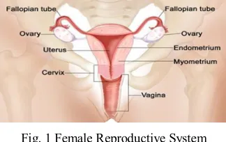
[image:2.612.241.403.576.678.2]The cervix is the lower part of the uterus (womb). It is sometimes called the uterine cervix. The fetus grows in the body of the uterus (the upper part). The cervix connects the body of the uterus to the vagina (birth canal). The part of the cervix closest to the body of the uterus is called the endocervix. The part next to the vagina is the exocervix (or ectocervix). The two main types of cells covering the cervix are squamous cells (on the exocervix) and glandular cells (on the endocervix). These 2 cell types meet at a place called the transformation zone. Most cervical cancers start in the transformation zone.
Fig. 1 Female Reproductive System
Fig. 2 Pap Test
B. Pap Test
[image:3.612.206.408.365.466.2]The Pap test (or Pap smear) is a procedure used to collect cells from the cervix for cervical cytology testing. The health care professional first places a speculum inside the vagina. A speculum is a metal or plastic instrument that keeps the vagina open so that the cervix can be seen clearly. Next, using a small spatula, a sample of cells and mucus is lightly scraped from the exocervix (the surface of the cervix that is closest to the vagina). A small brush or a cotton-tipped swab is then inserted into the cervical opening to take a sample from the endocervix (the inside part of the cervix that is closest to the body of the uterus). The cell samples are then prepared so that they can be examined under a microscope in the laboratory.
Fig. 3 Cervical cells on Pap smear
II. SOFTWARE TOOL FOR PROCESSING OF PAP SMEAR IMAGE
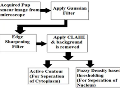
[image:3.612.209.402.558.701.2]The fig.4 shows the block diagram of the proposed system. In this system, the acquired Pap smear image is in .bmp or .jpg file format. Here, MATLAB software is used for processing of the images.
143
A. Pre-Processing of the Image
The Pap Smear image in RGB format is given as input for processing. The input image may have non-uniform distribution of intensity due to staining. Gaussian filter is applied to normally distribute the intensity over the image. Application of Gaussian filter has a smoothening effect on the image. To sharpen the boundaries of cytoplasm and nuclei, an edge sharpening filter is used. This is achieved by subtracting a scaled unsharp version of the image from the original. The resultant image is split up into its RGB components. Contrast Limited Adaptive Histogram Equalization (CLAHE) is applied to each component. CLAHE divides the image into smaller tiles. Histogram of intensity values is found for each tile. The histogram values are then used to redistribute the lightness values of the image. The local contrast of the image is improved. The features of the image are enhanced by CLAHE. The cytoplasm and nuclei can be differentiated from the background and the image has been pre-processed.
B. Extraction of Nucleus
Fuzzy density based thresholding is used to separate the nuclei from the Background removed Pap smear image. Fuzzy density model has a high density in its fuzzy region. The character is described with the help of fuzzy membership function, that is, a closer distance to the centroid would get a higher fuzzy value so its fuzzy average would also be higher. Let U be a data space, and fdm(r,p) be the function to calculate the fuzzy density, where r denotes the region and p is the points within this region of boundary. The higher the fdm(r,p), the more similar these points are. In Pap Smear image, the gray scale is divided into two parts by a selected value X. X is the boundary to both parts and we could get two regions at the same time, i.e. [Min,X], [X,Max], where Min, Max denotes the minimum and maximum gray scale respectively. Pixels under this partition are these points in p. We could use gray level to express the distance of pixels. As a whole, we calculate fdm(r,p) at the selected value of X to choose a proper threshold.
Here, three different types of formula are used to calculate the membership function according to the distance in its fuzzy density region.
1) Zadeh’s S-membership function 2) Gamma membership function 3) Gaussian membership function
By combining fuzzy set theory, fuzzy density model is used to calculate the character of image.
Fig.5 Basic Idea of Fuzzy Density Model Thresholding Method
Xmin, Xmax denotes the minimum and maximum gray level that has pixels in the histogram of the image respectively.
The histogram consists by two groups of pixels, dark part and light part. The target is to split the image histogram into two crisp subsets, namely, object subset O and background subset B, O ∪ B=A. Every time we calculate the fuzzy density of
[Xl,Xi],[Xi,Xr] respectively, then a comparison between them is made. IF (fdm(Xmin,Xi) > fdm(Xi,Xmax)), THEN Xi C L
IF (fdm(Xmin,Xi) < fdm(Xi,Xmax)), THEN Xi C R
density based thresholding, the nuclei are extracted from the image.
C. Extraction of cytoplasm
A snake (active contour) is a flexible 2D line, which is moved around the image to minimize energy functional. The line is parameterized by through a variable p that goes from 0 to 1.
The cytoplasm is extracted from the background removed image using active contours. An Active Contour or Snake is a deformable continuous closed spline curve. The deformation of active contour is determined by internal and external forces of gradient vectors.
Esnake=∫(Eint(V(s))+Eimage(V(s))+Econ(V(s))ds --(1)
The internal force (Ein t(V(s)) acts as constraint on smoothness of snake. Eimage(V(s))and Econ(V(s)) are the external forces.
Econ(V(s)) is the constraint energy that drives the active contour towards the features of the image. Eimage(V(s)) represents the
energy function of image as a combination of energies of lines, edges and terminations in the image. Active contours perform global thresholding and edge detection by assessment of continuity, curvature and strength of local edge. A conventional active contour has important setbacks like initial contour positioning and initial values of parameters. A grid of multiple small circles of radii depending on the size of image is used as initial contour. The initial contour deforms to the cytoplasm boundary in a number of iterations. Thus the cytoplasm is to be extracted from the Pap smear images by this method.
III.CONCLUSION
An effective method to identify and classify cervical cancer is becoming increasingly needed due to the fact that early detection and a decision of correct therapy may save the patient. Medical images have various limitations such as low quality, presence of noise and human error interpretation. Digital image processing can help the pathologists to a great extent.
In this proposed method, the segmentation of nucleus from the Pap smear image is done by fuzzy density based thresholding which is not applicable for low contrast images. So, Gaussian filter and edge sharpening filter is used in the Pap smear image to preserve the edges and to improve the contrast of the image.
Based on the fuzzy set theory, the concept of fuzzy density model is developed to choose the proper threshold for feature extraction. It is fully automatic and no prior knowledge of the image is required. Thus, the nuclei and cytoplasm to be extracted from the Pap smear images can be classified as normal, mild, moderate and severe dysplasia based on nuclei- cytoplasm ratio.
REFERENCES
[1] Cervical cancer – Overview and Incidence website available at:
http://www.medindia.net/patients/patientinfo/cervicalcancer-incidence.htm#ixzz1oDusUgV1
[2] G. Karthigai Lakshmi and K. Krishnaveni (2013), “Automated Extraction of Cytoplasm and Nuclei from Cervical Cytology Images by Fuzzy Thresholding and Active Contours”, International Journal of Computer Applications (0975-8887), Vol. 73, No.15.
[3] Ell Hassanien et al., (2012), “Density Based Fuzzy Thresholding for Image Segmentation”, AMLTA 2012, CCIS 322, pp. 118–127.
[4] M. T. Sreedevi, B. S. Usha, S. Sandya (2012), “Pap smear Image based Detection of Cervical Cancer”, International Journal of Computer Applications (0975 – 8887), Vol. 45, No. 20.
[5] L. B. Mahanta and K. Bora (2012), “Analysis of Malignant Cervical Cells Based on N/C Ratio Using Pap Smear Images”, International Journal of Advanced Research in Computer Science and Software Engineering, Vol. 2, No. 11, ISSN: 2277 128X, pp. 341-346.
[6] L. B. Mahanta, D. C. Nath and C. K. Nath (2012), “Cervix Cancer Diagnosis from Pap Smear Images Using Structure Based Segmentation and Shape Analysis”, Journal Of Emerging Trends in Computing And Information Sciences, Vol. 3, No. 2, pp. 312-325.
[7] C. H. Lin, Y. K. Chan, and C. C. Chen (2009), “Detection and segmentation of cervical cell cytoplast and nucleus”, International Journal of Imaging Systems Technology, Vol. 19, No. 3, pp. 260–270.