Tumor neoantigens: building a framework for personalized
cancer immunotherapy
Matthew M. Gubin, … , Elaine R. Mardis, Robert D. Schreiber
J Clin Invest. 2015;125(9):3413-3421. https://doi.org/10.1172/JCI80008.
It is now well established that the immune system can recognize developing cancers and that therapeutic manipulation of immunity can induce tumor regression. The capacity to manifest remarkably durable responses in some patients has been ascribed in part to T cells that can (a) kill tumor cells directly, (b) orchestrate diverse antitumor immune responses, (c) manifest long-lasting memory, and (d) display remarkable specificity for tumor-derived proteins. This specificity stems from fundamental differences between cancer cells and their normal counterparts in that the former develop protein-altering mutations and undergo epigenetic and genetic alterations, resulting in aberrant protein expression. These events can result in formation of tumor antigens. The identification of mutated and aberrantly expressed self-tumor antigens has historically been time consuming and laborious. While mutant antigens are usually expressed in a tumor-specific manner, aberrantly expressed antigens are often shared between cancers and, therefore, in the past, have been the major focus of therapeutic cancer vaccines. However, advances in next-generation sequencing and epitope prediction now permit the rapid identification of mutant tumor neoantigens. This review focuses on a discussion of mutant tumor neoantigens and their use in personalizing cancer immunotherapies.
Review Series
Find the latest version:
Series Editor: Yiping Yang
Introduction
The idea that tumors of nonviral origin possess unique, tumor-specific antigens (TSAs) arose from work reported in the first half of the twentieth century by several groups, particularly those led by Gross, Foley, Prehn, and Old (1–4). These studies showed that when inbred mice bearing carcinogen-induced tumors were cured of their cancers by surgical resection, they were immune to sub-sequent rechallenge with the same tumor cells, but not with other distinct tumor cells, even those derived in the same manner from different hosts. In the ensuing next quarter century, the roles of the MHC proteins in antigen presentation were discovered (5, 6), methods were developed to propagate antigen-specific cytolytic T lymphocytes (CTL) in culture (7, 8), and it became possible to clone and express gene products using molecular biology tech-niques. Together, these developments provided the elements needed for the first molecular identification of TSAs. Using muta-genized forms of preexisting P815 mastocytoma tumor cells, Boon and colleagues isolated a highly immunogenic, nontumorigenic tumor cell variant (tum–) and used CTL-based expression
clon-ing approaches to show that a point mutation in a ubiquitously expressed protein (P91A) was responsible for the immune rejec-tion of these tumor cells in naive, syngeneic WT mice (9). Shortly thereafter, Hans Schreiber and colleagues showed that tumor-spe-cific mutant proteins could also function as tumor neoantigens in a set of naturally arising, highly immunogenic, primary UV–induced mouse tumors (10, 11).
During this time, independent efforts by Knuth and Old and the Rosenberg group identified T cells in the peripheral blood and tumors of melanoma patients that predominately reacted with melanoma cells but not normal cells, suggesting that human can-cers also possessed tumor antigens whose expression was either tumor specific or showed limited expression in normal cells (12, 13). In 1991, Boon and colleagues used their T cell–based approach to clone the first human tumor antigen (MAGEA1) (14), then sub-sequently cloned a range of different human tumor antigens that included, among others, those derived from tumor-specific mutant genes, alternatively initiated proteins, normal proteins that displayed aberrant quantitative or qualitative expression in tumor cells, and proteins expressed only in germ cells and tumor cells (15). At about the same time, Sahin et al. used an autologous antibody-based cloning approach (SEREX) and also identified dif-ferent types of human tumor antigens (16). Together, these pio-neering efforts led to the acknowledgement that the tumor anti-genome comprised both tumor-specific and tumor-associated antigens (TAAs).
Tumor antigen classification
Today we recognize three broad classifications of tumor antigens: TSAs, TAAs, and cancer-germline/cancer testis antigens (CTAs) (15, 17–19). TSAs are antigens that are not encoded in the normal host genome and may represent either oncogenic viral proteins or abnormal proteins that arise as a consequence of somatic muta-tions (i.e., neoantigens). During cancer initiation and progres-sion, tumor cells acquire protein-altering mutations that are either responsible for transformation (driver mutations) or are a byproduct of the genomic instability that accompanies cellular transformation (passenger mutations) (20–23). Some of these
It is now well established that the immune system can recognize developing cancers and that therapeutic manipulation of immunity can induce tumor regression. The capacity to manifest remarkably durable responses in some patients has been ascribed in part to T cells that can (a) kill tumor cells directly, (b) orchestrate diverse antitumor immune responses, (c) manifest long-lasting memory, and (d) display remarkable specificity for tumor-derived proteins. This specificity stems from fundamental differences between cancer cells and their normal counterparts in that the former develop protein-altering mutations and undergo epigenetic and genetic alterations, resulting in aberrant protein expression. These events can result in formation of tumor antigens. The identification of mutated and aberrantly expressed self-tumor antigens has historically been time consuming and laborious. While mutant antigens are usually expressed in a tumor-specific manner, aberrantly expressed antigens are often shared between cancers and, therefore, in the past, have been the major focus of therapeutic cancer vaccines. However, advances in next-generation sequencing and epitope prediction now permit the rapid identification of mutant tumor neoantigens. This review focuses on a discussion of mutant tumor neoantigens and their use in personalizing cancer immunotherapies.
Tumor neoantigens: building a framework for
personalized cancer immunotherapy
Matthew M. Gubin,1,2 Maxim N. Artyomov,1,2 Elaine R. Mardis,3,4 and Robert D. Schreiber1,2
1Department of Pathology and Immunology, 2Center for Human Immunology and Immunotherapy Programs, 3Department of Genetics, and 4The Genome Institute, Washington University School of
Medicine, St. Louis, Missouri, USA.
Conflict of interest: Robert D. Schreiber is a co-founder of Igenica Biotherapeutics and
Jounce Therapeutics. He has a paid advisory relationship with Third Rock Ventures and received research support from Bristol-Myers Squibb.
TSAs. Using a combination of next generation sequencing, in sili-co epitope prediction, and immunological approaches, Sahin and colleagues (38) and our group (39) independently identified and validated distinct TSAs in murine B16-F10 melanoma tumor cells and in highly immunogenic methylcholanthrene-induced (MCA-induced) sarcoma cells, respectively. Importantly, these studies showed that the time frame needed to identify TSAs could be shortened to only a few weeks as opposed to the months required using conventional antigen-cloning approaches. Moreover, our study (39) and an independent study by Dupage and Jacks (40) showed that TSAs were also key targets of cancer immunoediting — the immunological process that not only protects against cancer development, but also sculpts the immunogenicity of cancers that form in an immunocompetent individual (41–43).
The following year, this work was extended to human cancers. Robbins and Rosenberg showed that exome sequencing could be used to identify mutated human TSAs recognized by adoptively transferred tumor-reactive T cells (44). Concomitantly, Schumacher used a combination of exome sequencing and high-throughput MHC tetramer screening to show that checkpoint blockade immu-notherapy facilitated expansion of preexisting T cells specific for tumor neoantigens in a human melanoma patient (45). Checkpoint blockade therapy, originally developed by Allison and colleagues, is based on the capacity of antibodies to block inhibitory receptors (such as cytoxic T lymphocyte–associated protein 4 [CTLA-4]) on T cells or deplete inhibitory receptor–bearing T cells, thereby unleashing suppressed T cell–dependent antitumor effector func-tions in tumor-bearing hosts (46–48). The capacity of epitope pre-diction algorithms to identify human TSAs was further demon-strated in both prospective and retrospective analyses performed by Fritsch, Hacohen, and Wu (49, 50), Schumacher and colleagues (51), and Nelson et al. (52). Together, the mouse and human studies suggested that it was indeed possible to use genomics and bioin-formatics approaches to rapidly identify mutant proteins expressed exclusively in cancer cells that function as tumor neoantigens.
Identifying tumor-specific mutations and
predicting their capacity to function as TSAs
Whereas these initial studies revealed the power of combining genomics, bioinformatics, and immunological approaches to iden-tify mutant TSAs, additional refinements have been made over the last few years that further improved the accuracy of the process.Next generation sequencing. Advances in sequencing technol-ogy have transformed our ability to decode cancer-specific muta-tions by coupling the sequencing reaction with detection of nucle-otide incorporation events for hundreds of millions of genomic fragments in the same instrument run (53). In particular, tumor-specific or “somatic” mutations can be identified using massively parallel sequencing (MPS) (17) approaches to compare DNA iso-lated from tumor versus normal sources. Similar to DNA-based assays using MPS, RNA from tumors can be analyzed by conver-sion to cDNA and construction of a library suitable for sequenc-ing. Since the genome is large (3 billion base pairs) and its analysis complex, the advent of hybrid capture technology has permitted investigators to focus only on the 1% of the genome that compris-es the coding exons of known gencompris-es, (i.e., the “exome”) (54–56). Here, probes designed to bind the exon sequences of annotated alterations may result in expression of mutant proteins that are
perceived as foreign proteins by the immune system. This class of antigens is likely to be less susceptible to mechanisms of immu-nological tolerance and therefore may represent more visible tar-gets for immune-mediated tumor control (19, 24). TAAs include proteins encoded in the normal genome and may be either normal differentiation antigens or aberrantly expressed normal proteins. Overexpressed normal proteins that possess growth/survival-promoting functions, such as Wilms tumor 1 (WT1) (25) or Her2/ neu (26), represent TAAs that directly participate in the oncogenic process. Posttranslational modifications of proteins such as phos-phorylation may also lead to formation of TAAs (27, 28). Because TAAs are normal proteins, their antigenicity depends on abnor-mal expression levels or context to circumvent naturally occurring mechanisms of immunological tolerance (29, 30). Along these lines, TAAs usually have lower T cell receptor (TCR) affinity pared with TSAs or foreign antigens (31). The third category com-prises CTAs, which are normally expressed in testis, fetal ovaries, and trophoblasts, but can also be expressed in cancer cells (17). Because they are encoded in the normal genome but display high-ly restricted tissue expression, CTAs have received considerable attention as attractive targets for immunotherapy (32).
Paving the way for TSA-based cancer
immunotherapy
In 2005, two important human studies stimulated increased interest in tumor neoantigens as therapeutic targets for cancer immunotherapy. First, using expression-cloning approaches, T. Wölfel et al. showed that the naturally occurring antitumor T cell response in a melanoma patient was directed toward neoantigens formed by somatic point mutations in five distinct genes and that T cell responses against these TSAs prevailed over responses to TAAs (33). Second, Rosenberg and Robbins showed that ex vivo– expanded tumor-infiltrating lymphocytes (TILs), when adoptively transferred into a melanoma patient who subsequently underwent a complete tumor regression, contained T cells that were specif-ic for two mutant tumor antigens (34). T cells specifspecif-ic for these neoantigens persisted in the blood and tumor of the patient after adoptive transfer. Together, these data provided support for the concept that T cells recognizing tumor neoantigens could indeed provide substantial therapeutic benefit to human cancer patients.
These results came at a time when the majority of efforts in the field were focused on identifying TAAs and CTAs for use in cancer immunotherapy. During this period, limited effort was expended on TSAs because their identification was so difficult and represented a case of too much effort for too little gain for too few individuals. However, by 2008, cancer genome sequenc-ing had clearly established that all cancers express somatic muta-tions (35, 36). These observamuta-tions led Allison and Vogelstein to conduct in silico analyses of exome-sequencing data from breast and colorectal cancers, where they found several mutations that were predicted to form tumor-specific mutant antigens for CD8+ T
cells (37), but it would take four years before experimental support could be obtained to corroborate their computational predictions.
some (59) and the resulting 8–11 amino acid peptides transported into the ER by the transporter associated with antigen processing (TAP) (60), where they are loaded onto newly synthesized class I molecules and the stabilized peptide–MHCI (p–MHCI) complexes are transported to the cell surface. MHCI alleles are remarkably polymorphic, and the number of potential self and foreign pep-tides processed by normal, infected, or transformed cells is very large (61). There are nearly 2,500 human MHCI allelic sequences and, because human cells can express as many as six distinct MHCI alleles, the capacity to accurately predict which tumor-derived mutant peptide will bind a particular MHCI is challenging.
Multiple tools exist to predict peptide binding to MHCI. A comprehensive list of prediction tools is available (http://can-cerimmunity.org/resources/webtools/), and the bioinformatics and biochemical aspects of these programs have been extensively reviewed elsewhere (62–64). Whereas SYFPEITHI (65), Rankpep (66), and BIMAS (67) were the first such tools to be developed, more accurate prediction algorithms have now come on line, and some have been incorporated into the Immune Epitope Database and Analysis Resource (IEDB) (68). A subset of these algorithms predicts peptide binding to different MHCI variants based on artificial neural networks (ANN), providing predicted IC50 as an output (69). In this category, NetMHC (70) is one of the most commonly used and best validated prediction programs (71, 72). Neural network–based approaches depend on the quality and size of the training set and therefore are likely to be more accurate for the more common alleles. A modified form of NetMHC, NetM-HCpan (73, 74), expands the training set by including data from other species, leading to improved accuracy for rare MHC alleles. SMM (75) and SMMPMBEC (76) are examples of a second sub-set of prediction algorithms that use position-weight matrices to describe statistical preferences from p–MHCI binding data. This approach suppresses noise caused by both experimental error and a limited number of data points present in the training set.
Neoepitope prioritization. For identifying tumor-derived mutant epitopes, most studies use predicted p–MHCI binding affinity as the primary criterion for generating an initial prioritized list of candidate epitopes. Most of the reported studies indicate that natural immune responses to tumor neoantigens are selective-ly directed to epitopes within a group predicted to have the stron-gest MHCI binding affinities (44, 77, 78). Peptide/MHCI binding is influenced by two additional parameters — epitope abundance and antigen processing (i.e., protein degradation and peptide transport). Although mass spectrometry could potentially provide information on epitope abundance and is rapidly becoming more sensitive, the current sensitivity and requirement for large num-bers of tumor cells remain as important impediments in employ-ing this approach to identifyemploy-ing the entire peptidome expressed on MHCI (79). Therefore, epitope abundance is currently estimated indirectly by quantitating RNA expression levels. In one approach, mutations defined by tumor-to-normal DNA comparisons are subjected to bioinformatic analysis to predict their immunogenic-ity and the levels of candidate immune stimulatory peptides are estimated by RNA-Seq. RNA evaluation provides information regarding (a) whether the variant is expressed in the RNA and (b) the mutant allele’s expression level relative to other genes. In a second approach, cDNA capture is performed from tumor RNA genes are synthesized, biotinylated, and hybridized in
solu-tion with a fragmented whole genome library. The probe-bound library fragments are subsequently captured and isolated using streptavidin-coated magnetic beads. After release from the beads by denaturation, the library fragments are amplified, quantitated, and sequenced.
Exome-capture can be used in a clinical setting, but challeng-es include (a) obtaining information in a clinically relevant time frame, (b) the small amounts of DNA/RNA available from a core biopsy procedure, (c) tissue preservation in formalin and paraffin (formalin-fixed paraffin embedded [FFPE]), which promotes the degradation of nucleic acids via backbone crosslinking, and (d) data interpretation. Recent technical innovations have reduced the time for this approach from approximately one week to around two hours for hybrid capture. It is now feasible to generate exome-capture data and produce a list of somatic mutations in about three days. Hybrid capture also enhances the sequencing data quality obtained from tumor RNA (cDNA) sequencing, especially for low yield and/or FFPE-derived samples (57).
Detecting somatic mutations. Mutation calling from exome-capture sequencing data is achieved by aligning sequence reads to reference genomes, which serve as the keystone for analyzing the short read lengths (~100 bp) produced by MPS platforms. Once reads are aligned to the genome, variants are identified using several algorithms to interpret different types of mutations, including point mutations (or single nucleotide variants [SNVs]) and focused insertion or deletion variants (indels). Tumor variant calls are then compared with data from a matched normal tissue DNA obtained using a similar capture reagent in order to iden-tify tumor-unique (“somatic”) mutations. Subsequent annotation steps convert variations in nucleic acid sequence to changes in amino acid sequence, thereby providing the initial data required to identify and rank order tumor neoantigens.
One aspect of this process that needs further work is the abili-ty to predict neoantigens arising from more “extreme” mutations. In principle, variants that add or delete an amino acid or truncate or extend open reading frames or fusion genes arising from trans-locations or inversions could be a source of highly antigenic novel epitopes; however, indel variants have, in the past, been difficult to detect with high certainty, even when algorithms are employed that are specifically tuned to detect this type of mutation. Howev-er, recent advances now permit the identification of some indels with a fairly high degree of certainty, although indels contain-ing highly repetitive regions remain a difficulty. Structural vari-ants also are difficult to identify, especially from exome-capture data, and hence are not likely to be easily detected unless RNA sequencing (RNA-Seq) data can be evaluated for fusion tran-scripts (which has a correlative high false-positive rate). In all cas-es, the use of RNA data from cDNA capture sequencing (cDNA Cap-Seq) or RNA-Seq to identify and/or confirm such variants is critically important.
protea-and compared with normal DNA to provide a list of mutated pep-tides. Due to the error rate of reverse transcriptase and sources of false positivity in variant calling in RNA versus DNA, the pre-dicted mutations are refined further by applying a series of filters to remove known sources of false-positive variants (e.g., low cov-erage in tumor or normal, or low numbers of variant-containing reads) and to eliminate nonexpressed genes and/or alleles. A final filter eliminates genes with low expression that have a frag-ment per kilobase of million mapped reads (FPKM) of less than one. Downstream steps of immunogenicity prediction can then be made for this filtered set of mutant peptides. Additional algo-rithms exist to refine epitope predictions including those focused on defining proteasomal cleavage (i.e., NetChop) (80) and/or TAP transport (i.e., NetCTL and NetCTLpan) (81, 82). However, because the latter are currently not as reliable as the MHC-affinity prediction algorithms, they should be used with caution.
In contrast to predicting MHCI epitopes, the accuracy of pre-dicting MHCII epitopes has been problematic. Whereas the MHCI binding groove is closed at both ends, MHCII has a peptide-bind-ing groove that is open, leadpeptide-bind-ing to considerable variation in both the length of peptides that can bind to MHCII and the location of the binding core. Multiple MHCII binding prediction algorithms are available (such as TEPITOPE, ref. 83; netMHCII, ref. 84; and SMM-align, ref. 85); however, they have not been extensively used in the past to identify tumor-specific mutant MHCII epitopes.
Recent preclinical studies illustrating the use of
personalized TSAs in immunotherapy
MHCI neoepitopes. Two recent preclinical studies demonstrated the feasibility of developing personalized neoantigen-based can-cer immunotherapies. Using a combination of mass spectrom-etry, next generation sequencing, and bioinformatics approaches, Yadav, Lill, Delamarre, and colleagues identified seven mutant neoepitopes bound to MHCI expressed on MC38 mouse colon adenocarcinoma cells (78). Two of the stronger H-2Db–binding
epi-topes, derived from point mutations of Reps1 and Adpgk, induced antigen-specific CD8+ T cell responses in naive WT syngeneic
mice when administered together with polyinosinic:polycytidylic acid (poly I:C) and agonistic anti-CD40. One H-2Kb epitope,
derived from a point mutation of Dpagt1, also induced a weak T cell response. Therapeutic vaccines comprising synthetic long peptides (SLP) containing the three positive epitopes provided mice bearing established MC38 tumors with a significant degree of therapeutic protection.
[image:5.585.101.214.44.698.2]neoepitope binding to MHCI alleles while discounting mutations that affect TCR binding, which identified unique tumor neoepitopes that provided protection in a prophylactic vaccine setting (86). Intrigu-ingly, many of these displayed MHCI binding affinities that were orders of mag-nitude weaker than other validated MHCI– restricted epitopes. It remains unclear how such weak epitopes could function in a therapeutic setting, and additional work is needed to explore this issue.
MHCII neoepitopes. In many mouse tumor models, spontaneous or therapeuti-cally induced tumor rejection is inhibited following depletion of either CD4+ or CD8+
T cells (39, 40, 77). Moreover, the presence of tumor-specific IgG antibodies in cancer patients and tumor-bearing mice implies that CD4+ T cells play important roles in
the antitumor response (87). Using humanized MHC transgenic mice, Platten and colleagues demonstrated that SLP vaccines con-taining a mutant MHCII epitope from isocitrate dehydrogenase 1 (IDH1; a previously identified mutation in many human glio-blastoma patients) controlled outgrowth of mouse tumors engi-neered to express mutant human IDH1 (88). Furthermore, using exome sequencing along with a high-throughput MHCII epit-ope screening method that did not require prediction of MHCII epitopes, Schumacher and colleagues identified and validated mutant MHCII–restricted neoepitopes in melanomas from several patients (89). They further showed that, whereas MHCII epitopes were identified in 4 of 5 different human melanomas, they repre-sented only 0.5% of the melanoma mutanome, thus translating into only one to two MHCII epitopes per tumor.
In a more recent study, the Sahin group vaccinated naive WT mice with synthetic 27 mer peptides containing putative MHCI epitopes they had identified in a previous study from B16-F10 melanoma cells and found that nearly 30% of these peptides induced both strong T cell responses and strong antitumor activi-ty when injected into tumor-bearing mice (90). Surprisingly, 95% of the epitope-specific lymphocytes induced by vaccination were CD4+ rather than CD8+ T cells. A predominance of MHCII
neo-epitopes was also observed for CT26 (a colorectal adenocarcino-ma) and 4T1 (a breast adenocarcinoadenocarcino-ma) (89). In the case of CT26, the neoepitopes were selected using bioinformatics approaches based exclusively on the use of expression level predictions and MHCII–binding capacity. RNA-based vaccines encoding mutant MHCII epitopes induced complete rejection of established, aggressively growing tumors. By applying the same predictive algorithm to human cancers, the investigators observed a high abundance of MHCII neoepitopes. The number of MHCII epi-topes per tumor cell appears to be vastly different between the Schumacher and Sahin studies (89, 90). At least in part, this dif-ference could reflect the methods used to reveal the epitopes (checkpoint blockade versus active vaccination, respectively), but could also reflect the use of paired tumor and normal samples in the former study versus use of a half-century old tumor where no checkpoint blockade immunotherapy (77). In this study, we used
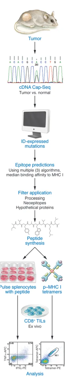
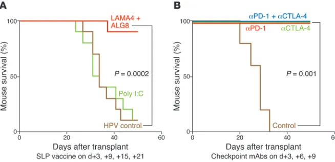
an edited mouse MCA sarcoma line (T3) that formed progressively growing tumors in naive syngeneic WT mice, which were rejected upon treatment with mAbs specific for two different immune check-points — CTLA-4 and programmed death-1 (PD-1) (Figure 1). As assessed by cDNA–Cap-Seq and a recent refinement to calling muta-tions, T3 expressed approximately 2,200 nonsynonymous point mutations. Putative epitopes were predicted using three MHCI bind-ing algorithms (SMM, NetMHC, and NetMHCpan) and calculatbind-ing the median binding affinities for each epitope. Additionally, we applied filters for proteasomal processing (NetChop), neoepitopes (i.e., where the mutant epitope bound to MHCI in a manner equal or greater to WT sequence), and deprioritization of hypothetical pro-teins. Using this approach, two MHCI H-2Kb–restricted tumor
neoan-tigens were identified: point mutations in α-1,3-glucosyltransferase (Alg8) and laminin α subunit 4 (Lama4). These predictions were validated by the following observations. First, primary TILs from T3 tumors were specifically stained by H-2Kb tetramers carrying
either ALG8 or LAMA4 epitopes (but not other predicted epitopes), accumulated in tumors in a time-dependent manner, and produced IFN-γ and/or TNF-α when stimulated with irradiated splenocytes pulsed with either ALG8 or LAMA4 (but not other predicted epi-topes). Second, the same two mutant epitopes were recognized by cloned T cells from mice that had rejected T3 sarcomas following anti–PD-1 treatment. Third, ALG8 and LAMA4 were detected by mass spectrometry bound to H-2Kb on T3 sarcoma cells. Fourth,
ALG8 or LAMA4 peptide vaccines induced T cell responses in naive syngeneic WT mice. Fifth, vaccines comprising ALG8/LAMA4 SLPs plus poly I:C prophylactically protected mice against subsequent challenge with T3 tumor cells and therapeutically induced rejection in mice bearing established T3 tumors. Importantly, the degree of therapeutic protection by the combined neoantigen SLP vaccine was comparable to that afforded by checkpoint blockade therapy (Figure 2). Together, these studies revealed the therapeutic potential of TSA-based personalized cancer immunotherapy.
[image:6.585.44.371.56.214.2]Srivastava and colleagues concomitantly proposed an alterna-tive prediction method based exclusively on the enhancement of
corresponding normal tissue of similar age was available. Never-theless, the two studies support the conclusion that T cell–depen-dent immune control of cancer is depencell–depen-dent on the presence of both mutant MHCII and mutant MHCI tumor neoepitopes.
Therapeutic use of tumor-specific mutant
antigens in human cancer
In the past year, the first examples of TSA-based personalized cancer immunotherapies have begun to emerge. In 2014, the Rosenberg group used a novel TSA-based personalized adop-tive cell therapy (ACT) method to treat a patient with metastatic cholangiocarcinoma (91). Specifically, 26 nonsynonymous
muta-tions were identified in a lung metastasis from the patient by whole-exome sequencing, and candidate minigene constructs for each mutation were transfected into patient-derived APCs. TILs from lung metastases were screened for reactivity with trans-fected APCs, leading to identification of a point mutant ERBB2-interacting protein as a neoantigen recognized by CD4+ T cells
within the TIL population. TILs enriched to 25% for reactivity to the neoantigen were then infused back into the patient, who showed a partial response to the first ACT treatment and showed improved responses to subsequent treatments with TIL prepara-tions enriched to 95% of neoantigen-specific CD4+ T cells. These
results not only demonstrated a therapeutic advantage to person-alizing ACT therapy, but also specifically documented the thera-peutic efficacy of neoantigen-specific CD4+ T cells.
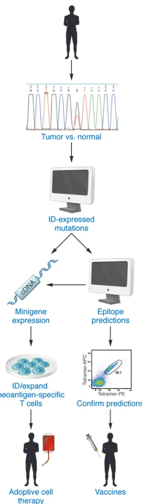
Using more classical vaccine approaches, Carreno et al. recently showed that tumor neoantigens, when administered as a vaccine to three melanoma patients, can enhance both preexisting anti-tumor T cell responses and induce responses to neoepitopes that were undetectable prior to vaccination (92). Thus, these studies support the preclinical work that preceded them and show that vaccines comprising IFN-γ/CD40 ligand–activated dendritic cells pulsed with tumor neoepitopes represent yet another method to deliver per-sonalized cancer vaccines to tumor-bearing individuals (Figure 3).
[image:7.585.87.233.53.570.2]form a hallmark of immunogenic epitopes (95). Thus it is clear that more work is needed to clarify whether there are amino acid motifs that play important roles in determining neoantigenicity.
Concluding remarks
The last decade has seen an explosive growth in our appreciation of the role of immunity in cancer. We now know that the immune system recognizes, shapes, and/or promotes cancer develop-ment or outgrowth (22, 41–43, 96, 97). Based on our enhanced insight into immune system/cancer interactions, new immu-nologic approaches to treating cancer have been developed, often leading to durable clinical responses in a subset of cancer patients (47, 98–100).
Two particular characteristics have brought cancer immuno-therapy to the forefront of modern molecular medicine (101). First, this therapy can be staged such that it induces complete responses or stable disease with less collateral damage than conventional chemotherapy or radiation therapy. Second, because the immune response can continuously evolve new specificities, cancer immu-notherapy has the capacity to respond reactively to genetically unstable, continuously changing cancer cell target populations. However, current cancer immunotherapies still display inherent toxicities because they function within the narrow therapeutic space between tumor immunity and autoimmunity and induce responses only in a subset of patients. The use of TSAs as the basis for personalized cancer immunotherapy offers the potential to make the therapy more specific, more effective, and safer com-pared with the cancer immunotherapies that we have available today. However, key questions remain to be answered. Although we have achieved a level of competency in identifying point muta-tions and indels that can form MHCI epitopes, there remain sev-eral other potential tumor neoantigens (such as products of gene fusions, proteins arising from mistakes in translation, or mutant proteins that form MHCII antigens) that are currently not easy to predict with bioinformatics tools. We need to resolve the important questions of whether tumor-specific mutant antigens (or the close-ly related group of shared driver neoantigens that could reduce the chances of escape variant formation) really offer unique oppor-tunities to target tumor cells compared with shared, nonmutated TAAs. Furthermore, we need to determine what minimum
num-ber of neoepitopes must be employed to reduce the likelihood of formation of antigen-loss variants. Finally, we need to experimen-tally establish that therapeutically targeting tumor neoantigens does not lead to unanticipated autoimmune consequences. Even after we resolve these issues, we still need to determine how best to use this information therapeutically. Is it through defining the best types of personalized cancer vaccines, individualized adoptive cell therapies, some combination of these together with checkpoint blockade immunotherapy, or even combinations of immunologic and standard cancer immunotherapies? Strategies targeting dif-ferent aspects of the “cancer-immunity cycle” (102) (i.e., priming and/or effector immunity) with checkpoint blockade may be help-ful in contextualizing the role of targeting TSAs within the multi-step process of effective antitumor immunity. Will the methods that are ultimately used need to be employed in a manner that is based on the type of cancer being treated? We envisage that these types of questions will be answered in the next decade of cancer immunology, genomics, and immunotherapy research. It is clear that we now stand at an important crossroads of opportunities to enhance our ability to use the immune system to make cancer a controllable and, in some cases, curable disease. We predict that the capacity to personalize cancer immunotherapy will contribute significantly to this important endeavor.
Acknowledgments
We are grateful to K. Sheehan, G. Dunn, A. Miceli, T. Noguchi, J. Ward, and E. Alspach for constructive criticism and comments. R.D. Schreiber receives research support from the National Cancer Institute (RO1 CA043059, RO1 CA190700, U01 CA141541), the Cancer Research Institute, the WWWW Foundation, the Siteman Cancer Center/Barnes-Jewish Hospital (Cancer Frontier Fund), Bristol-Myers Squibb Inc., and Stand Up to Cancer. E.R. Mardis is supported by an NIH large-scale centers grant (U54 HG003079). M.M. Gubin is supported by a postdoctoral training grant (Irving-ton Postdoctoral Fellowship) from the Cancer Research Institute.
Address correspondence to: Robert D. Schreiber, Department of Pathology and Immunology, 660 South Euclid Ave., St Louis, Missouri 63110, USA. Phone: 314.362.8787; E-mail: Schreiber@ immunology.wustl.edu.
1. Gross L. Intradermal immunization of C3H mice against a sarcoma that originated in an animal of the same line. Cancer Res. 1943;3(5):326–333. 2. Foley EJ. Antigenic properties of
methylcholan-threne-induced tumors in mice of the strain of origin. Cancer Res. 1953;13(12):835–837. 3. Prehn RT, Main JM. Immunity to
methylcholan-threne-induced sarcomas. J Natl Cancer Inst. 1957;18(6):769–778.
4. Old LJ. Cancer immunology: the search for speci-ficity. Natl Cancer Inst Monogr. 1982;60:193–209. 5. Babbitt BP, Allen PM, Matsueda G, Haber E,
Unanue ER. Binding of immunogenic peptides to Ia histocompatibility molecules. Nature. 1985;317(6035):359–361.
6. Bjorkman PJ, Saper MA, Samraoui B, Bennett WS, Strominger JL, Wiley DC. Structure of the human class I histocompatibility antigen, HLA-A2.
Nature. 1987;329(6139):506–512.
7. Cerottini JC, Engers HD, Macdonald HR, Brunner T. Generation of cytotoxic T lymphocytes in vitro. I. Response of normal and immune mouse spleen cells in mixed leukocyte cultures. J Exp Med. 1974;140(3):703–717.
8. Gillis S, Smith KA. Long term culture of tumour-specific cytotoxic T cells. Nature. 1977;268(5616):154–156.
9. De Plaen E, et al. Immunogenic (tum-) vari-ants of mouse tumor P815: cloning of the gene of tum- antigen P91A and identification of the tum- mutation. Proc Natl Acad Sci U S A. 1988;85(7):2274–2278.
10. Monach PA, Meredith SC, Siegel CT, Sch-reiber H. A unique tumor antigen produced by a single amino acid substitution. Immunity. 1995;2(1):45–59.
11. Dubey P, et al. The immunodominant antigen of an ultraviolet-induced regressor tumor is generat-ed by a somatic point mutation in the DEAD box helicase p68. J Exp Med. 1997;185(4):695–705. 12. Knuth A, Danowski B, Oettgen HF, Old LJ.
T-cell-mediated cytotoxicity against autologous malignant melanoma: analysis with interleukin 2-dependent T-cell cultures. Proc Natl Acad Sci
U S A. 1984;81(11):3511–3515.
13. Robbins PF, et al. A mutated β-catenin gene encodes a melanoma-specific antigen recognized by tumor infiltrating lymphocytes. J Exp Med. 1996;183(3):1185–1192.
14. van der Bruggen P, et al. A gene encoding an antigen recognized by cytolytic T lym-phocytes on a human melanoma. Science. 1991;254(5038):1643–1647.
Boon T. Tumour antigens recognized by T lym-phocytes: at the core of cancer immunotherapy.
Nat Rev Cancer. 2014;14(2):135–146.
16. Sahin U, et al. Human neoplasms elicit multiple specific immune responses in the autologous host. Proc Natl Acad Sci U S A. 1995;92(25):11810–11813.
17. Simpson AJ, Caballero OL, Jungbluth A, Chen YT, Old LJ. Cancer/testis antigens, gametogenesis and cancer. Nat Rev Cancer. 2005;5(8):615–625. 18. Vigneron N, Stroobant V, Van den Eynde BJ, van der Bruggen P. Database of T cell-defined human tumor antigens: the 2013 update. Cancer Immun. 2013;13:15.
19. Heemskerk B, Kvistborg P, Schumacher TN. The cancer antigenome. EMBO J. 2013;32(2):194–203. 20. Hanahan D, Weinberg RA. The hallmarks of
cancer. Cell. 2000;100(1):57–70.
21. Greenman C, et al. Patterns of somatic muta-tion in human cancer genomes. Nature. 2007;446(7132):153–158.
22. Hanahan D, Weinberg RA. Hallmarks of cancer: the next generation. Cell. 2011;144(5):646–674. 23. Vogelstein B, Papadopoulos N, Velculescu VE,
Zhou S, Diaz LA Jr, Kinzler KW. Cancer genome landscapes. Science. 2013;339(6127):1546–1558. 24. Schumacher TN, Schreiber RD.
Neoanti-gens in cancer immunotherapy. Science. 2015;348(6230):69–74.
25. Ohminami H, Yasukawa M, Fujita S. HLA class I-restricted lysis of leukemia cells by a CD8(+) cytotoxic T-lymphocyte clone specific for WT1 peptide. Blood. 2000;95(1):286–293. 26. Kawashima I, Tsai V, Southwood S, Takesako K,
Sette A, Celis E. Identification of HLA-A3- restricted cytotoxic T lymphocyte epitopes from carcinoembryonic antigen and HER-2/neu by pri-mary in vitro immunization with peptide-pulsed dendritic cells. Cancer Res. 1999;59(2):431–435. 27. Doyle HA, et al. Isoaspartyl
post-transla-tional modification triggers anti-tumor T and B lymphocyte immunity. J Biol Chem. 2006;281(43):32676–32683.
28. Cobbold M, et al. MHC class I-associated phosphopeptides are the targets of memory-like immunity in leukemia. Sci Transl Med. 2013;5(203):203ra125.
29. Pardoll D. Does the immune system see tumors as foreign or self? Annu Rev Immunol. 2003;21:807–839.
30. Hogquist KA, Baldwin TA, Jameson SC. Central tolerance: learning self-control in the thymus.
Nat Rev Immunol. 2005;5(10):772–782.
31. Stone JD, Harris DT, Kranz DM. TCR affinity for p/MHC formed by tumor antigens that are self-proteins: impact on efficacy and toxicity. Curr
Opin Immunol. 2015;33:16–22.
32. Scanlan MJ, Gure AO, Jungbluth AA, Old LJ, Chen YT. Cancer/testis antigens: an expanding family of targets for cancer immunotherapy. Immunol
Rev. 2002;188:22–32.
33. Lennerz V, et al. The response of autologous T cells to a human melanoma is dominated by mutated neoantigens. Proc Natl Acad Sci U S A. 2005;102(44):16013–16018.
34. Zhou J, Dudley ME, Rosenberg SA, Robbins PF. Persistence of multiple tumor-specific T-cell clones is associated with complete tumor
regression in a melanoma patient receiving adoptive cell transfer therapy. J Immunother. 2005;28(1):53–62.
35. Wood LD, et al. The genomic landscapes of human breast and colorectal cancers. Science. 2007;318(5853):1108–1113.
36. Ley TJ, et al. DNA sequencing of a cytogeneti-cally normal acute myeloid leukaemia genome.
Nature. 2008;456(7218):66–72.
37. Segal NH, et al. Epitope landscape in breast and colorectal cancer. Cancer Res. 2008;68(3):889–892. 38. Castle JC, et al. Exploiting the mutanome for tumor
vaccination. Cancer Res. 2012;72(5):1081–1091. 39. Matsushita H, et al. Cancer exome analysis reveals
a T-cell-dependent mechanism of cancer immu-noediting. Nature. 2012;482(7385):400–404. 40. DuPage M, Mazumdar C, Schmidt LM, Cheung
AF, Jacks T. Expression of tumour-specific anti-gens underlies cancer immunoediting. Nature. 2012;482(7385):405–409.
41. Shankaran V, et al. IFNγ and lymphocytes prevent primary tumour development and shape tumour immunogenicity. Nature. 2001;410(6832):1107–1111.
42. Dunn GP, Bruce AT, Ikeda H, Old LJ, Schreiber RD. Cancer immunoediting: from immuno-surveillance to tumor escape. Nat Immunol. 2002;3(11):991–998.
43. Schreiber RD, Old LJ, Smyth MJ. Cancer immu-noediting: integrating immunity’s roles in cancer suppression and promotion. Science. 2011;331(6024):1565–1570.
44. Robbins PF, et al. Mining exomic sequencing data to identify mutated antigens recognized by adoptively transferred tumor-reactive T cells.
Nat Med. 2013;19(6):747–752.
45. van Rooij N, et al. Tumor exome analysis reveals neoantigen-specific T-cell reactivity in an ipilimumab-responsive melanoma. J Clin Oncol. 2013;31(32):e439–e442.
46. Leach DR, Krummel MF, Allison JP. Enhancement of antitumor immunity by CTLA-4 blockade.
Science. 1996;271(5256):1734–1736.
47. Pardoll DM. The blockade of immune check-points in cancer immunotherapy. Nat Rev Cancer. 2012;12(4):252–264.
48. Sharma P, Allison JP. Immune checkpoint targeting in cancer therapy: toward combina-tion strategies with curative potential. Cell. 2015;161(2):205–214.
49. Fritsch EF, Rajasagi M, Ott PA, Brusic V, Haco-hen N, Wu CJ. HLA-binding properties of tumor neoepitopes in humans. Cancer Immunol Res. 2014;2(6):522–529.
50. Rajasagi M, et al. Systematic identification of per-sonal tumor-specific neoantigens in chronic lym-phocytic leukemia. Blood. 2014;124(3):453–462. 51. van Buuren MM, Calis JJ, Schumacher TN. High sensitivity of cancer exome-based CD8 T cell neo-antigen identification. Oncoimmunology. 2014;3:e28836.
52. Wick DA, et al. Surveillance of the tumor muta-nome by T cells during progression from primary to recurrent ovarian cancer. Clin Cancer Res. 2014;20(5):1125–1134.
53. Mardis ER. Next-generation sequencing plat-forms. Annu Rev Anal Chem (Palo Alto Calif). 2013;6:287–303.
54. Albert TJ, et al. Direct selection of human genom-ic loci by mgenom-icroarray hybridization. Nat Methods. 2007;4(11):903–905.
55. Hodges E, et al. Hybrid selection of discrete genomic intervals on custom-designed microar-rays for massively parallel sequencing. Nat Protoc. 2009;4(6):960–974.
56. Blumenstiel B, et al. Targeted exon sequencing by in-solution hybrid selection. Curr Protoc Hum
Genet. 2010;Chapter 18:Unit 18.4.
57. Cabanski CR, et al. cDNA hybrid capture improves transcriptome analysis on low-input and archived samples. J Mol Diagn. 2014;16(4):440–451.
58. Blum JS, Wearsch PA, Cresswell P. Pathways of antigen processing. Annu Rev Immunol. 2013;31:443–473.
59. Rock KL, et al. Inhibitors of the proteasome block the degradation of most cell proteins and the generation of peptides presented on MHC class I molecules. Cell. 1994;78(5):761–771.
60. Van Kaer L, Ashton-Rickardt PG, Ploegh HL, Tonegawa S. TAP1 mutant mice are deficient in antigen presentation, surface class I molecules, and CD4-8+ T cells. Cell. 1992;71(7):1205–1214.
61. Robinson J, Halliwell JA, Hayhurst JD, Flicek P, Parham P, Marsh SG. The IPD and IMGT/HLA database: allele variant databases. Nucleic Acids
Res. 2015;43(Database issue):D423–D431.
62. Lundegaard C, Lund O, Kesmir C, Brunak S, Nielsen M. Modeling the adaptive immune sys-tem: predictions and simulations. Bioinformatics. 2007;23(24):3265–3275.
63. Lin HH, Ray S, Tongchusak S, Reinherz EL, Brusic V. Evaluation of MHC class I peptide bind-ing prediction servers: applications for vaccine research. BMC Immunol. 2008;9:8.
64. Zhang L, Udaka K, Mamitsuka H, Zhu S. Toward more accurate pan-specific MHC-peptide bind-ing prediction: a review of current methods and tools. Brief Bioinform. 2012;13(3):350–364. 65. Rammensee H, Bachmann J, Emmerich NP,
Bachor OA, Stevanovic S. SYFPEITHI: database for MHC ligands and peptide motifs. Immunogenetics. 1999;50(3–4):213–219.
66. Reche PA, Glutting JP, Reinherz EL. Prediction of MHC class I binding peptides using profile motifs. Hum Immunol. 2002;63(9):701–709. 67. Parker KC, Bednarek MA, Coligan JE. Scheme
for ranking potential HLA-A2 binding peptides based on independent binding of individual pep-tide side-chains. J Immunol. 1994;152(1):163–175. 68. Vita R, et al. The immune epitope database
(IEDB) 3.0. Nucleic Acids Res. 2015;43(Database issue):D405–D412.
69. Lundegaard C, Lund O, Nielsen M. Prediction of epitopes using neural network based methods.
J Immunol Methods. 2011;374(1–2):26–34.
70. Lundegaard C, Lamberth K, Harndahl M, Buus S, Lund O, Nielsen M. NetMHC-3.0: accurate web accessible predictions of human, mouse and monkey MHC class I affinities for peptides of length 8-11. Nucleic Acids Res. 2008;36(Web Server issue):W509–W512.
72. Lundegaard C, Nielsen M, Lund O. The validity of predicted T-cell epitopes. Trends Biotechnol. 2006;24(12):537–538.
73. Nielsen M, et al. NetMHCpan, a method for quantitative predictions of peptide binding to any HLA-A and -B locus protein of known sequence.
PLoS One. 2007;2(8):e796.
74. Hoof I, et al. NetMHCpan, a method for MHC class I binding prediction beyond humans.
Immu-nogenetics. 2009;61(1):1–13.
75. Peters B, Sette A. Generating quantitative models describing the sequence specificity of biological processes with the stabilized matrix method.
BMC Bioinformatics. 2005;6:132.
76. Kim Y, Sidney J, Pinilla C, Sette A, Peters B. Deri-vation of an amino acid similarity matrix for pep-tide: MHC binding and its application as a Bayes-ian prior. BMC Bioinformatics. 2009;10:394. 77. Gubin MM, et al. Checkpoint blockade cancer
immunotherapy targets tumour-specific mutant antigens. Nature. 2014;515(7528):577–581. 78. Yadav M, et al. Predicting immunogenic
tumour mutations by combining mass spec-trometry and exome sequencing. Nature. 2014;515(7528):572–576.
79. Rammensee HG, Singh-Jasuja H. HLA ligan-dome tumor antigen discovery for personal-ized vaccine approach. Expert Rev Vaccines. 2013;12(10):1211–1217.
80. Nielsen M, Lundegaard C, Lund O, Kesmir C. The role of the proteasome in generating cyto-toxic T-cell epitopes: insights obtained from improved predictions of proteasomal cleavage.
Immunogenetics. 2005;57(1–2):33–41.
81. Peters B, Bulik S, Tampe R, Van Endert PM, Hol-zhutter HG. Identifying MHC class I epitopes by predicting the TAP transport efficiency of epitope
precursors. J Immunol. 2003;171(4):1741–1749. 82. Larsen MV, Lundegaard C, Lamberth K, Buus S, Lund O, Nielsen M. Large-scale validation of methods for cytotoxic T-lymphocyte epitope pre-diction. BMC Bioinformatics. 2007;8:424. 83. Hammer J, Bono E, Gallazzi F, Belunis C, Nagy
Z, Sinigaglia F. Precise prediction of major histo-compatibility complex class II-peptide interac-tion based on peptide side chain scanning. J Exp
Med. 1994;180(6):2353–2358.
84. Nielsen M, Lund O. NN-align. An artificial neural network-based alignment algorithm for MHC class II peptide binding prediction. BMC
Bioinfor-matics. 2009;10:296.
85. Nielsen M, Lundegaard C, Lund O. Prediction of MHC class II binding affinity using SMM-align, a novel stabilization matrix alignment method.
BMC Bioinformatics. 2007;8:238.
86. Duan F, et al. Genomic and bioinformatic profil-ing of mutational neoepitopes reveals new rules to predict anticancer immunogenicity. J Exp Med. 2014;211(11):2231–2248.
87. Gnjatic S, et al. Survey of naturally occurring CD4+
T cell responses against NY-ESO-1 in cancer patients: correlation with antibody responses. Proc
Natl Acad Sci U S A. 2003;100(15):8862–8867.
88. Schumacher T, et al. A vaccine targeting mutant IDH1 induces antitumour immunity. Nature. 2014;512(7514):324–327.
89. Linnemann C, et al. High-throughput epitope discovery reveals frequent recognition of neo-antigens by CD4+ T cells in human melanoma.
Nat Med. 2015;21(1):81–85.
90. Kreiter S, et al. Mutant MHC class II epitopes drive therapeutic immune responses to cancer.
Nature. 2015;520(7549):692–696.
91. Tran E, et al. Cancer immunotherapy based on
mutation-specific CD4+ T cells in a patient with
epithelial cancer. Science. 2014;344(6184):641–645. 92. Carreno BM, et al. A dendritic cell vaccine
increases the breadth diversity of mela-noma neoantigen-specific T cells. Science. 2015;348(6236):803–808.
93. Snyder A, et al. Genetic basis for clinical response to CTLA-4 blockade in melanoma. N Engl J Med. 2014;371(23):2189–2199.
94. Rizvi NA, et al. Cancer immunology. Muta-tional landscape determines sensitivity to PD-1 blockade in non-small cell lung cancer. Science. 2015;348(6230):124–128.
95. Chowell D, et al. TCR contact residue hydro-phobicity is a hallmark of immunogenic CD8+ T cell epitopes. Proc Natl Acad Sci U S A.
2015;112(14):E1754–E1762. 96. Grivennikov SI, Greten FR, Karin M.
Immunity, inflammation, and cancer. Cell. 2010;140(6):883–899.
97. Trinchieri G. Cancer and inflammation: an old intuition with rapidly evolving new concepts.
Annu Rev Immunol. 2012;30:677–706.
98. Hodi FS, et al. Improved survival with ipilimumab in patients with metastatic melanoma. N Engl J
Med. 2010;363(8):711–723.
99. Topalian SL, et al. Safety, activity, and immune correlates of anti-PD-1 antibody in cancer. N Engl
J Med. 2012;366(26):2443–2454.
100. Sharma P, Allison JP. The future of immune check-point therapy. Science. 2015;348(6230):56–61. 101. Mellman I, Coukos G, Dranoff G.
Can-cer immunotherapy comes of age. Nature. 2011;480(7378):480–489.