oncogenesis, and therapy
Dirk P. Dittmer, Blossom Damania
J Clin Invest. 2016;
126(9)
:3165-3175.
https://doi.org/10.1172/JCI84418
.
Kaposi sarcoma–associated herpesvirus (KSHV), also known as human herpesvirus 8, is the etiologic agent underlying
Kaposi sarcoma, primary effusion lymphoma, and multicentric Castleman’s disease. This human gammaherpesvirus was
discovered in 1994 by Drs. Yuan Chang and Patrick Moore. Today, there are over five thousand publications on KSHV
and its associated malignancies. In this article, we review recent and ongoing developments in the KSHV field, including
molecular mechanisms of KSHV pathogenesis, clinical aspects of KSHV-associated diseases, and current treatments for
cancers associated with this virus.
Review
Find the latest version:
Introduction
Kaposi sarcoma (KS) is the most common cancer in individuals
living with HIV/AIDS today (1, 2). While the introduction of
effec-tive HIV therapy was concurrent with a decline in the incidence of
KS in the United States, KS incidence has stabilized and remained
essentially level since 2000. In Africa, where KSHV and HIV
infec-tions are highly prevalent, KS is among the most common cancer
type in men overall. In some sub-Saharan countries, KS is more
prevalent than prostate cancer is in the US (3). As the expected
lifespan of individuals living with HIV/AIDS increases, we foresee
an increase in all cancers in this population, including KS.
Kaposi sarcoma–associated herpesvirus (KSHV) is necessary
for KS development. KSHV DNA is found in all KS lesions (4, 5). KS
prevalence follows KSHV seroprevalence, and in most cases
fulmi-nant KS is accompanied and preceded by a rise in KSHV viral load
in blood. In addition to KS, KSHV is also the etiologic agent of the
plasmablastic variant of multicentric Castleman’s disease (MCD)
(6) and primary effusion lymphoma (PEL) (7, 8). Moreover, KSHV
is the causative agent of KS-immune reconstitution syndrome
(KS-IRIS) (9, 10) and KSHV-inflammatory cytokine syndrome
(KICS) (11). However, not all KSHV infections lead to
KSHV-asso-ciated conditions. The majority of primary KSHV infections have
no clinical symptoms and, as with other human oncogenic viruses,
cancer emerges only after decades of dormancy. KSHV can be
transmitted via asymptomatic oral shedding as well as through
bodily fluids (12–14). KSHV can infect many different types of cells
including endothelial cells, B lymphocytes, monocytes, dendritic
cells (DCs), and epithelial cells. KSHV provides a growth
advan-tage to infected endothelial cells. The virus consistently
immor-talizes, but rarely transforms, primary cells in culture (15–19). It is
only under special circumstances and perhaps upon infection of
rare progenitor cells with stem cell properties that the interplay
between virus and host leads to a fully transformed state.
Why is the human immune system so powerful in
suppress-ing disease, yet can never eliminate this pathogen? Like all
her-pesviruses, KSHV establishes lifelong infection in the host and
molecular latency in cells in culture. KS is primarily the
conse-quence of systemic viral reactivation from a latent reservoir, most
likely a lymph node–resident B cell (20–23). Prior to the
emer-gence of HIV, endemic KS in sub-Saharan Africa was a disease of
both children and adults, and classic KS was a disease of elderly
men in the Mediterranean region. Today, KS also develops with
higher frequency in HIV-infected individuals (HIV-associated
KS) compared with HIV-negative individuals, as well as in solid
organ transplant recipients (transplant KS). Thus, it appears that
KS develops in response to severe T cell depletion or inactivation.
Infant, aging-, chemical-, or HIV-induced immune deficiency is
an essential cofactor for the development of KS.
Primary infection and the innate immune
response to KSHV
KSHV is thought to enter cells predominantly through the
endo-cytic pathway. Viral attachment involves several different
recep-tor binding proteins on the virion. KSHV can infect multiple cell
types, including B cells, endothelial cells, monocytes, and DCs,
and hence uses multiple viral receptors to enter the host cell.
One such receptor is the gB glycoprotein, which contains an
inte-grin-binding RGD (Arg-Gly-Asp) motif that plays a role in virion
binding and entry of endothelial cells (24–26). Activated B cells,
macrophages, and DCs express a DC-specific ICAM-3-grabbing
non-integrin (DC-SIGN; CD209) that facilitates KSHV infection
in these cell types (27, 28). The cysteine transporter (xCT) can also
serve as a receptor for the virus (29).
KSHV is thought to enter cells predominantly through the
endocytic pathway (30–32). During its entry into the host cell, the
virus encounters multiple innate immune sensors that activate
an antiviral response. It is likely that the activation of such innate
immune responses during primary infection induces the virus to
enter molecular latency, which is a more quiescent and less
immu-nogenic phase of the lifecycle.
Kaposi sarcoma–associated herpesvirus (KSHV), also known as human herpesvirus 8, is the etiologic agent underlying
Kaposi sarcoma, primary effusion lymphoma, and multicentric Castleman’s disease. This human gammaherpesvirus was
discovered in 1994 by Drs. Yuan Chang and Patrick Moore. Today, there are over five thousand publications on KSHV and its
associated malignancies. In this article, we review recent and ongoing developments in the KSHV field, including molecular
mechanisms of KSHV pathogenesis, clinical aspects of KSHV-associated diseases, and current treatments for cancers
associated with this virus.
Kaposi sarcoma–associated herpesvirus:
immunobiology, oncogenesis, and therapy
Dirk P. Dittmer and Blossom Damania
Lineberger Comprehensive Cancer Center Program in Global Oncology, Department of Microbiology and Immunology, Center for AIDS Research, School of Medicine, University of North Carolina at Chapel Hill,
Chapel Hill, North Carolina, USA.
Conflict of interest: The authors have declared that no conflict of interest exists.
production and suppresses viral gene
expres-sion following de novo infection with KSHV
as well as during viral reactivation (39, 40).
NLRs. NLR family members can form
inflammasomes, a complex comprised of an
NLR protein, ASC, and pro-caspase-1. NLRs
sense PAMPs, and activation of the NLR
inflammasomes results in cleavage and
production of active IL-1
β
and IL-18, which
are proinflammatory cytokines. Primary
infection with KSHV activates NLRP1 and
NLRP3 (41, 42) and potentially other NLRs.
ALRs. Like NLRs, ALR family
mem-bers can also form inflammasomes to
acti-vate proinflammatory cytokine signaling.
Primary infection with KSHV has been
shown to activate the ALR family member
interferon gamma–inducible protein 16
(IFI16) (43, 44). It was additionally
report-ed that IFI16 can detect KSHV in latently
infected cells (43).
cGAS-STING. Cyclic GMP-AMP
(cGAMP) synthase (cGAS) and STING
are members of the cytosolic
DNA-sens-ing pathway. This cGAS-STING pathway
appears to sense KSHV during both primary
infection and reactivation from latency in
multiple cell types (45–47).
There seems to exist a delicate
equilib-rium between the virus and host response
to infection. Although innate immune
activation might help KSHV enter a latent,
quiescent phase inside the infected cell and
induce expansion of latently infected cells,
a high degree of innate immune response
facilitates killing of the infected cell and ultimately prevents the
establishment of latency. To counter the host response to viral
infection and reactivation, KSHV encodes many viral genes that
blunt innate immune signaling pathways. Some of these viral
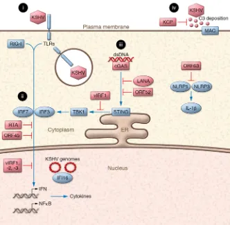
products are summarized in Figure 1 and are described below.
Viral interferon regulatory factors. KSHV encodes four viral
interferon regulatory factors (vIRFs), three of which ablate
cellu-lar IRF signaling and inhibit the production of type I IFNs,
includ-ing IFN-
α
and IFN-
β
(reviewed in ref. 48). KSHV vIRFs have been
shown to inhibit IFN production that lies downstream of TLR3
activation (49), MAVS activation (50), and cGAS-STING
activa-tion (45). KSHV vIRF1 also inhibits the funcactiva-tion of IFN-induced
genes such as ISG15 (51) and the transcription of TLR4 (37).
Complement regulatory proteins. KSHV encodes the
comple-ment regulatory protein KCP, which is encoded by ORF4. KCP is
part of the virion and functions as a cofactor for factor I–mediated
cleavage of C3b and C4b, the complement system’s opsonizing
factors (52, 53). KSHV has also been reported to exploit the host
complement system to promote viral persistent infection (54).
Tegument proteins. Tegument proteins are a characteristic
fea-ture of all herpesviruses, and a large number of them are
depos-ited into the cytoplasm following virion fusion and capsid release.
Cells infected with viruses such as KSHV trigger an innate
immune response through pattern recognition receptors (PRRs)
that recognize pathogen-associated molecular patterns (PAMPs)
and lead to the production of interferon and proinflammatory
cyto-kines. It is important to note that each cell type expresses its own
unique set of PRRs. There are many different PRRs including TLRs,
retinoic acid–like receptors (RLRs), NLRs, absent in melanoma 2
(AIM2)-like receptors (ALRs), and cytosolic DNA sensors (reviewed
in ref. 33). Members of the NLR, ALR, and RLR families can form
inflammasomes that, upon activation, lead to the production of
IL-1
β
and IL-18 (34). KSHV infection and/or reactivation activates a
multitude of PRRs in different cell types; these are described below.
TLRs. Following primary infection, KSHV has been shown to
activate the RNA sensor TLR3 in monocytes, (35) and the DNA
sensor TLR9 in plasmacytoid DCs (pDCs) (36). Activation of
either TLR results in interferon production and upregulation of
cytokines and chemokines. KSHV also activates TLR4 signaling
that likely occurs through recognition of the viral glycoproteins gB
and K8.1 (37). Stimulation of TLR7/8 in PEL cells has been shown
to lead to reactivation from latency (38).
RLRs. The cytosolic RNA sensor RIG-I and its adaptor protein,
[image:3.585.37.369.53.378.2]mitochondrial antiviral signaling protein (MAVS), induces IFN-
β
vMIP-II is an agonist for CCR8, a chemokine receptor that is
preferentially expressed on polarized Th2 T cells (83). Similarly,
KSHV vMIP-III serves as an agonist for the cellular chemokine
receptor CCR4, which is also expressed by Th2 T cells (84).
Hence, the KSHV vMIPs skew T cell responses towards a
Th2-type lymphocytic response, and this may play a role in subverting
the host immune response.
Latent KSHV infection and reactivation
KSHV successfully subverts the cellular innate immune response
to establish a lifelong latent reservoir in the infected host,
primar-ily in B cells. The virus has evolved a number of mechanisms to
ensure that virally infected B cells outcompete their uninfected
counterparts, which in the extreme leads to B cell hyperplasia,
such as MCD (6, 21), or B cell neoplasia, such as PEL (7). These
include inhibiting apoptosis, overcoming G1 phase arrest,
lower-ing the threshold for B cell receptor (BCR) activation, and
provid-ing ligand-independent progrowth signals. In addition to B cells,
this virus can also enter CD34 cells, T cells, monocytes, and pDCs
(36, 85–88), though it is unclear if these cell types contribute to
sys-temic persistence or serve as sentinels to detect infection.
Epstein-Barr virus (EBV) also uses B cells as the predominant latent
reser-voir, as does murine herpesvirus 68 (reviewed in ref. 89); however,
there are important biological differences between latent
infec-tion in B cells in KSHV and other herpesviruses. EBV is easily
detected in blood in circulating CD38
+memory B cells, which
typi-cally emerge from the germinal center. In contrast, KSHV is not
readily detectable in circulating B cells (23), and KSHV viral loads
in blood are 10- to 100-fold lower than those of EBV or human
CMV. These observations suggest that tissue-resident B cells are
the predominant latent reservoir for KSHV.
The deepest insights about the biology of KSHV prior to
dis-ease come from studying the related murine gammaherpesvirus
68 (MHV-68) and from genetically engineered mouse models.
In mouse models, it was possible to define B cell tropism through
functional phenotypes (21). Studies in MHV-68 defined the
dis-tinction between establishment of latency, which drives the size
of the latent reservoir, and persistence of the latent reservoir, i.e.,
long-term survival of infected cells that are still capable of
reacti-vation (for recent examples see refs. 90–92). We do not know the
dynamics of latently infected cells in humans and have only just
begun to decipher the physiological signals that modulate
reacti-vation events and thereby transmission and disease in patients.
Histone deacetylation reactivates KSHV. Vorinostat (also
known as SAHA) and valproic acid induce reactivation in culture
and in patients (93–95). Sodium butyrate and phorbol esters
reac-tivate KSHV from PEL, though only a fraction of episomes is
com-petent for reactivation at any given time (96, 97). The majority of
the KSHV episome is methylated, transcriptionally silent, and
dec-orated with histone markers, indicative of inactive chromatin (96,
98–101). These markers of methylation are established early in
infection and maintained by cellular chromatin remodelers, and
organized by CCCTC-binding factor (a zinc finger protein, also
known as CTCF) recognition elements (100). During latency, the
virus actively engages host chromatin modulators (Figure 2). For
instance, LANA binds to histones H2A and H2B as well as MECP2,
and to the BET family proteins BRD2 and BRD3 (102–104).
KSHV ORF45 is a tegument protein that blocks IRF7
phosphory-lation and activation of type I IFN responses (55, 56). ORF64 is
another conserved herpesviral tegument protein that encodes
potent deubiquitinating activity (57). ORF64 can reduce
TRIM25-dependent ubiquitination and activation of RIG-I, thereby
inhibit-ing this sensinhibit-ing pathway in KSHV-infected cells (40).
DNA-binding proteins. Although it is primarily a DNA-binding
protein and transcription factor, KSHV Rta/ORF50 can also induce
the degradation of innate immune sensors such as IRF7, TLR3, and
myeloid differentiation factor 88 (MyD88) (58–61). The
latency-associated nuclear antigen (LANA) is another DNA-binding protein
that inhibits IFN-
β
induction (62) and the transcription of IFN-
γ
–
inducible genes (63). Recently, it was shown that cytoplasmic
vari-ants of LANA can inhibit the cGAS-STING DNA-sensing pathway
by directly binding to cGAS (47). Interestingly, another KSHV open
reading frame, ORF52, was similarly shown to bind and inhibit
cGAS enzymatic activity. Infection with an ORF52-deficient virus
in endothelial cells resulted in increased cGAS signaling (46).
Furthermore, NLRP1 and NLRP3 inflammasome activation is
inhibited by the tegument protein KSHV ORF63 during de novo
infection, resulting in reduced IL-1
β
and IL-18 production. ORF63
binds to NLRP1 and interferes with the interaction between
NLRP1 and pro-caspase-1 (42).
In addition to the innate immune responses described above,
adaptive immune responses also play an important role in KSHV
pathogenesis. KSHV expresses proteins that affect antigen
presen-tation, B cell targeting, MHC class I display, and neutrophil and
basophil activation. The KSHV-infected cell presents antigenic
peptides from the virus in complex with MHC class I to
cytotox-ic T lymphocytes (CTLs) (64–66). Additionally, KSHV-infected
B cells stimulate activation-induced cytidine deaminase (AID)
expression and are targeted for elimination by NK cells through
upregulation of NKG2D ligands (67). KSHV also encodes genes
that inhibit these immune responses. KSHV K3/MIR1 and K5/
MIR2 are ubiquitin ligases that inhibit MHC class I display (68,
69). K3/MIR 1 downregulates four HLA allotypes (HLA-A, B, C,
and E), while K5/MIR2 downregulates HLA-A and HLA-B (70, 71).
K3 and K5 can also downregulate CD1d (72) and IFN-
γ
receptor 1
(IFNGR1) (73). K5 hinders expression of ICAM-1 and the
costimu-latory molecule B7-2 (CD86) (74, 75). It also downregulates the
NKG2D ligands, MHC class I–related chain A (MICA), MICB, and
the NKp80 ligand, activation-induced C-type lectin (AICL) (76).
KSHV vCD200, also known as viral OX2, is a homolog of cellular
CD200 that is broadly expressed and suppresses neutrophil and
basophil activation (77) as well as activation of macrophages (78).
vCD200 can also function as a negative regulator of
antigen-spe-cific T cell responses, including inhibition of IFN-
γ
production and
CD107a mobilization (79).
length is variable and nonessential for LANA’s nuclear functions,
as a direct N-to-C terminal domain fusion retains the
latency-supporting functions. Whereas the C-terminal end of LANA binds
KSHV DNA directly, the N-terminus (and perhaps regions in the
C-terminus as well) contact cellular chromosome-associated
pro-teins, such as histones H2A and H2B and others (104). The crystal
structures of the KSHV LANA and MHV-68 LANA DNA-binding
domains were solved (102, 103, 116, 117). This work identifies the
DNA contact residues and reveals a folding pattern analogous to
EBV EBNA1 and HPV E2.
LANA-episome complexes adopt higher-order structures in
the nucleus of infected cells and appear as a characteristic punctate
pattern by immunofluorescence (114, 118, 119). Initially considered
a somewhat underwhelming feature, these “LANA dots” have
emerged as the diagnostic gold standard to identify KSHV-infected
cells and to make the diagnosis of KS and PEL (114, 118, 119). The
number of LANA dots correlates with the number of KSHV
plas-mids in an infected cell. During mitosis, LANA, and by inference
KSHV plasmids, decorates condensed chromosomes, thereby
facil-itating proper and equal partitioning of the latent viral genomes
into daughter cells. Loss-of-function LANA mutants in the context
of the viral genome remain competent for lytic replication, but fail
to establish and maintain latency in KSHV and the related
MHV-68. Ablation of LANA in PEL is incompatible with growth. Thus,
LANA can be considered essential for KSHV-associated
lympho-magenesis. However, interpreting genetic experiments for LANA
is rather complex, since tethering the KSHV plasmids to the host
genome is not the only function of LANA. LANA also binds a large
number of cellular proteins to modulate their functions, including
p53 and many other proteins with specialized functions (120, 121).
Most recently, cytoplasmic variants of LANA have been described
(122), and whole-genome screens have highlighted the importance
of LANA during KSHV primary infection (45, 47).
The KSHV Rta protein (also known as ORF50) is necessary
and sufficient to initiate KSHV reactivation (105, 106). Rta is a
potent transcriptional activator that can bind DNA directly or
through RBP-J
κ
(107). Rta reverses and overrides
chromatin-silencing modifications, and deletion of Rta renders MHV-68
unable to reactivate from latency. In subsequent steps, other viral
proteins such as K-bZIP augment the action of Rta to ensure robust
and complete viral replication and virion formation. If Rta is the
master regulator of reactivation from latency, what regulates Rta
expression and Rta function? Here the experimental evidence is
murkier. KSHV LANA and viral miRNAs counteract Rta and
rap-idly drive the virus into latency upon infection of primary
endothe-lial cells, whereas in other environments Rta prevails (108–110).
It is also worth mentioning that viral reactivation can occur in an
Rta-independent fashion (111).
More research is needed to identify physiological triggers
of KSHV reactivation as potential targets of disease prevention.
These are likely to depend on conserved as well as cell- and
micro-environment-specific signaling pathways (112). KSHV
reactiva-tion can be induced by IFN-
γ
, but not IFN-
α
. KSHV reactivation
is induced by TLR7/8 signaling, and reactivation is enhanced by
deletion of RIG-I and MAVS (38, 39). In artificially infected Burkitt
lymphoma B cells (BJAB cells), B cell receptor crosslinking can
reactivate KSHV (113), though PELs are BCR negative. Different
sets of events may trigger KSHV reactivation in the oral cavity
ver-sus endothelial cells.
Update on LANA function and structure
[image:5.585.42.538.55.253.2]LANA binds the viral terminal repeats, specifically two
sequence-conserved, high-affinity binding sites (LBS1 and LBS2) and a
more divergent third, low-affinity site (114–116). LANA can be
thought of as a dumbbell-like structure in which a stalk of
inter-nal repeats separates the two globular termiinter-nal regions. The stalk
is associated with hyperproliferation and lymphoma. EBV relies on
endogenous miRNA-155 to drive lymphoblastoid cell line
immor-talization (129, 135). Similarly, KSHV encodes an ortholog of
miRNA-155 named miRNA-K12-11 that contains 100% seed
sequence identity (136, 137). This KSHV ortholog complements the
proliferative deficits observed in miR-155-deficient mice and drives
lymphoma in a CD34 reconstitution model (138, 139). While
miR-155 is the first and best-studied viral ortholog of a cellular miRNA,
it is not the only one. As our appreciation for the complexities of
host cell miRNA function and regulation grows, we can expect to
gain new insights into the biology of KSHV miRNAs as well.
Genomic explorations of KS and PEL
Only a small fraction of KSHV-infected children develop KS, just
as only a small fraction of EBV-infected children develop Burkitt
lymphoma. In the context of solid organ transplantation, only a
fraction of KSHV-seropositive transplant recipients develop KS,
similar to the small fraction of EBV-seropositive transplant
recipi-ents that develop posttransplant lymphoproliferative disease,
a condition associated with EBV infection of B cells after
thera-peutic immunosuppression. Currently, there is no screening of
organ donors for KSHV positivity, although screening for KSHV in
donors is warranted. KS that develops in transplant patients is
usu-ally a late complication, developing several months after the onset
of immune suppression therapy. By contrast, the onset of herpes
simplex and CMV reactivation disease is more immediate, often
necessitating acyclovir prophylaxis for the first 6 months after
transplantation. The delayed emergence of KS vis-à-vis clinical
diseases associated with these other herpesviruses suggests that in
addition to KSHV and in addition to immune deficiency, genomic
alterations may contribute to KSHV-associated neoplasia.
Family linkage studies in classic KS support the notion of
susceptibility loci for KS (140–142). Whereas t(8;14) and related
translocations targeting MYC are the defining genomic event in
EBV-related Burkitt’s lymphoma, MYC translocations are not
present in PEL. Rather, the KSHV viral protein LANA drives
MYC overexpression (143, 144). Comparative genome
hybridiza-tion uncovered fragile histidine triad (FHIT) delehybridiza-tion as
over-represented in PELs, and targeted sequencing studies identified
a polymorphism in IL-1 receptor–associated kinase 1 (IRAK1)
as significantly overrepresented in PELs (145, 146). Moreover,
IRAK1 signaling is required for PEL growth. This observation
parallels Waldenstrom macroglobulinemia and a fraction of
dif-fuse large B cell lymphomas, where gain-of-function mutations
in MyD88, the upstream partner of IRAK1, are present (147, 148).
It is important, however, to recognize that the rarity of PEL and
classic KS incidence hinders genomic explorations, which limits
the statistical significance of any association.
PTEN, p53, and Rb are not deleted in PEL or KS; rather,
they are inactivated posttranslationally, e.g., by direct binding to
LANA, or via expression of the CDK1-resistant viral cyclin
homo-log vCYC (149). This may explain why KS is initially responsive to
DNA-damaging chemotherapy. Susceptibility to etoposide
cor-relates with p53 mutation status in PEL, and p53 activation by
nutlin-3 leads to apoptosis (120, 121). In KS and PEL, the human
genome is dynamic and the host mutational landscape is shaped
by selection during clonal evolution of the tumor just as it is for
Viral miRNAs support viral infection and latent
persistence
A recently emerged common feature among all herpesviruses is
the utilization of virally encoded miRNAs as a means to
modu-late the host cell during modu-latency and primary infection. In Marek’s
disease virus, a B cell–tropic alpha herpesvirus of chickens, viral
miRNAs are the primary driver of oncogenesis. Recently, KSHV
mir-K12-10a was identified as the molecular driver behind the
in vitro transforming phenotype of KSHV Kaposin, since it is
embedded within the open reading frame of this protein (123).
The role of the KSHV miRNAs is often more subtle, but it is
important to bear in mind that virally encoded miRNAs account
for 50% or more of all miRNAs in a KSHV-infected B cell. KSHV
encodes 12 pre-miRNA loci, which can give rise to 24 mature
miRNAs and many more if alternative processing is considered
(124–126). Many of the viral miRNAs are also secreted into
pleu-ral fluid and circulate in the blood of KS patients (127). Thus, they
serve as biomarkers of latent infection.
In general, viral miRNAs target specific cellular mRNAs,
lead-ing to their degradation (via an siRNA-like mechanism) and
inhi-bition of mRNA-directed translation. miRNAs are
developmen-tally regulated and fine-tune lineage differentiation and cellular
signaling. The targets of the KSHV miRNAs have been established
through a series of comprehensive biochemical studies (128–131).
Thus far, miRNA studies have been constrained by sensitivity
lim-its for detection of individual miRNAs and for the discovery of
miRNA-target interactions. Targets with functions that seem to
befit the biology of B cell development, endothelial cell
differen-tiation, and KSHV (such as BACH1, xCT, MAF, and others) have
been individually validated (132–134). These are by no means the
only targets, and it is anticipated that additional targets will be
identified in the future.
[image:6.585.44.284.56.236.2]Cellular miRNA-155 is central to B cell lineage development
in the germinal center. Downregulation of miRNA-155 is
associ-ated with terminal differentiation of plasma cells and loss of
pro-liferative potential. Conversely, ectopic expression of miRNA-155
non–infection-associated cancers. The presence of KSHV
modi-fies a particular pattern of mutations, but these mutations affect
the same progrowth and antiapoptosis pathways as in other
can-cers. However, the interpretation of signature mutations becomes
complicated in light of their role in infection-associated cancers,
such as PEL or KS. Whether a particular event has been selected
for as a driver of tumorigenesis after viral infection or if it
repre-sents a susceptibility allele for the primary infection event (or
asymptomatic, systemic persistence) is not always apparent.
Only recently have whole KSHV genome sequences become
available from patients and primary biopsies (150, 151). These
sequences augment extensive studies that trace the origin and
evolution of KSHV based on single-gene analyses (152). KSHV
sequences show overall structural concordance and limited
varia-tion, as would be expected since viral replication is the result of
error-correcting, DNA-dependent DNA polymerases (the cellular
DNA polymerase during latency and a viral KSHV-encoded DNA
polymerase during lytic replication). During B cell latency,
multi-ple copies of the KSHV plasmid are maintained, replicated by the
host DNA polymerase, and propagated to daughter cells during
host cell division events. As yet, there is no evidence for
integra-tion of the KSHV genome. Nevertheless, defective variants have
been described and are expected to arise in the context of clonal
expansion of PEL or advanced KS. KSHV noncoding regions such
as the miRNA locus show more variation, and differences in
miRNA sequences correlate with processing and function (153–
155). The number of terminal repeats in the KSHV genome is
highly variable and can be used for strain typing (156, 157).
Like-wise, membrane proteins that are subject to immune recognition,
such as K1 and K15, contain hypervariable regions in the
extracel-lular domains (158–160).
Targeted treatment approaches to
KSHV-associated cancers
KS is a disease of endothelial cells, and details of its pathobiology
have been extensively reviewed. KS is among the most angiogenic
cancers known to arise in humans. If we can decipher which factors
drive KS and which treatments interrupt KS angiogenesis, then we
will have potent leads for other cancers that depend on
angiogen-esis. VEGF, stem cell factor (SCF, also known as KIT ligand), and
platelet-derived growth factor (PDGF) are the best-characterized
paracrine drivers of KS angiogenesis (161, 162), and these are
the target of a number of therapeutic approaches for KS.
VEGF-neutralizing antibodies (bevacizumab) and receptor tyrosine
kinase (RTK) inhibitors, such as imatinib, have efficacy in KS (163,
164), although their therapeutic impact as single agents is limited
because of redundancy in the paracrine network. Clinical studies
have also started investigating the role of thalidomide,
lenalido-mide (NCT01057121), and pomalidolenalido-mide (NCT02659930) in KS.
These structurally related compounds are approved for the
treat-ment of multiple myeloma and have antiinflammatory and
anti-angiogenic activities, though the exact molecular mechanisms
underlying their effects have not necessarily been established.
KSHV activates the PI3K/Akt/mTOR signaling pathway at
differ-ent nodes via the viral proteins vGPCR, K1, ORF36/vPK, vIL-6,
and K15, as well as through virus-mediated upregulation of
cel-lular growth factors, e.g., VEGF and PDGF (165–172) (Figure 3).
PI3K activates the cell survival kinase AKT, which subsequently
activates mTOR. Rapamycin (also known as sirolimus) has
clini-cal activity against KS (173, 174). Switching from cyclosporine A
to rapamycin as the primary immunosuppressant has become the
first line of therapy for transplant KS. The clinical phenotype can
be recapitulated in preclinical models of KS and PEL (175–179). In
KS, targeting mTOR was associated with a decrease in VEGF
pro-duction. In PEL, rapamycin reduced IL-6 and IL-10 secretion, and
inactivating PI3K and mTOR together had more potent antitumor
activity than inhibiting mTOR alone (180). The latter findings
pro-vide a guide path for the development of next-generation PI3K/
AKT/mTOR targeting strategies against KS.
Therapies that target immunomodulatory mechanisms also
hold promise for KS and KSHV-associated diseases. Siltuximab, a
humanized anti–IL-6 antibody, has been FDA approved for classic
Castleman’s disease (181), and is likely to also show efficacy against
MCD. A pilot clinical trial of tocilizumab, a humanized antibody
against the IL-6 receptor, is open for MCD (NCT01441063).
Block-ing IL-6 stymies PEL growth in preclinical models (182). Hsp90
inhibitors exhibited nanomolar EC50 against PEL and KS in three
independent studies (183–185). PELs are also extremely sensitive
to NF-
κ
B pathway inhibitors such as bortezomib (186, 187), and a
clinical trial with adjuvant bortezomib is ongoing. Other targets
with encouraging preclinical results are NOTCH (188–192), and the
KSHV receptor, ephrin receptor A2 (EphA2) (193–195).
While some individual AIDS-KS lesions respond to
combi-nation antiretroviral therapy (cART) and the ensuing immune
reconstitution, others do not. In the US, one third of KS cases now
develop in HIV patients with no detectable HIV viral load and
near-normal CD4 counts (196, 197). This type of KS no longer
signifies terminal AIDS. In sub-Saharan Africa, where KS remains
the most common disease among HIV patients and the most
com-mon AIDS-presenting symptom, initiating cART can lead to KS
exacerbation in KS-IRIS (9, 10). At least two large clinical trials are
currently underway to determine if it is better to give cART and
chemotherapy sequentially or together, and which chemotherapy
is best suited for which stage of KS (198, 199). Liposomal
doxoru-bicin, daunorudoxoru-bicin, other anthracycline formulations, and taxol
constitute the mainstay of KS treatment.
The need for a better understanding of KSHV
remains
Is it time for a KSHV vaccine?
We would argue that KSHV vaccine development is needed, and
that both preventative and therapeutic KSHV vaccines would be of
benefit. KSHV transmission among infants is similar to that of all
other herpesviruses; by puberty, greater than 80% of children
sero-convert in KSHV endemic regions. By contrast, transmission among
adults in many parts of the world (excluding Africa and the
Mediter-ranean) is so poor that repeated contact or immunodeficiency, as in
high-risk populations, is needed to sustain the virus at a greater than
5% population-wide prevalence. This suggests that only a fraction
of exposures leads to establishment of latency and eventual disease.
Systemically circulating and salivary levels of KSHV in
asymptom-atic persons are orders of magnitude lower than those of EBV,
her-pes simplex virus, or human CMV (13, 14, 200). Evidence of KSHV
superinfection in immune-competent persons is limited. A little
priming of the immune system by a vaccine prior to establishment
of latency may be all that is needed to eradicate KSHV and
KS-asso-ciated diseases from the human population.
Acknowledgments
We apologize for not citing many publications due to limits on
reference numbers per journal policy. We thank members of
the Dittmer and Damania labs and all our colleagues for
stim-ulating discussions. Our work is supported by Public Health
Service grants CA019014, CA163217, DE018304, DE023946,
DE018281, and CA096500 from the National Cancer Institute
(NCI) and the Institute of Dental and Craniofacial Research
(NIDCR). DPD is an investigator of the AIDS malignancies
clini-cal trial consortium (AMC). BD is a Leukemia and Lymphoma
Society Scholar, and a Burroughs Wellcome Fund Investigator in
Infectious Disease.
Address correspondence to: Blossom Damania, 450 West
Drive, CB# 7295, Rm 32-026, Lineberger Cancer Center,
Uni-versity of North Carolina at Chapel Hill, Chapel Hill, North
Carolina 27599, USA. Phone: 919.843.6011; E-mail: damania@
med.unc.edu.
1. Silverberg MJ, et al. Cumulative incidence of can-cer among persons with HIV in North America: a cohort study. Ann Intern Med. 2015;163(7):507–518. 2. Robbins HA, Pfeiffer RM, Shiels MS, Li J, Hall HI, Engels EA. Excess cancers among HIV-infected people in the United States. J Natl Cancer Inst. 2015;107(4): dju503.
3. Jemal A, et al. Cancer burden in Africa and opportunities for prevention. Cancer. 2012;118(18):4372–4384.
4. Moore PS, Chang Y. Detection of herpesvirus-like DNA sequences in Kaposi’s sarcoma in patients with and without HIV infection. N Engl J Med. 1995;332(18):1181–1185.
5. Chang Y, et al. Identification of herpesvirus-like DNA sequences in AIDS-associated Kaposi’s sar-coma. Science. 1994;266(5192):1865–1869. 6. Soulier J, et al. Kaposi’s sarcoma-associated
her-pesvirus-like DNA sequences in multicentric Cas-tleman’s disease. Blood. 1995;86(4):1276–1280. 7. Nador RG, et al. Primary effusion lymphoma: a distinct clinicopathologic entity associated with the Kaposi’s sarcoma-associated herpes virus.
Blood. 1996;88(2):645–656.
8. Cesarman E, Chang Y, Moore PS, Said JW, Knowles DM. Kaposi’s sarcoma-associated herpesvirus-like DNA sequences in AIDS-related body-cavity-based lymphomas. N Engl J Med. 1995;332(18):1186–1191.
9. Bower M, et al. Immune reconstitution inflam-matory syndrome associated with Kaposi’s sar-coma. J Clin Oncol. 2005;23(22):5224–5228. 10. Connick E, Kane MA, White IE, Ryder J,
Camp-bell TB. Immune reconstitution inflammatory syndrome associated with Kaposi sarcoma dur-ing potent antiretroviral therapy. Clin Infect Dis. 2004;39(12):1852–1855.
11. Polizzotto MN, et al. Clinical features and out-comes of patients with symptomatic Kaposi sarcoma herpesvirus (KSHV)-associated inflam-mation: prospective characterization of KSHV inflammatory cytokine syndrome (KICS). Clin
Infect Dis. 2016;62(6):730–738.
12. Mayama S, et al. Prevalence and transmission
of Kaposi’s sarcoma-associated herpesvirus (human herpesvirus 8) in Ugandan children and adolescents. Int J Cancer. 1998;77(6):817–820. 13. Koelle DM, Huang ML, Chandran B, Vieira
J, Piepkorn M, Corey L. Frequent detection of Kaposi’s sarcoma-associated herpesvi-rus (human herpesviherpesvi-rus 8) DNA in saliva of human immunodeficiency virus-infected men: clinical and immunologic correlates. J Infect Dis. 1997;176(1):94–102.
14. Gantt S, et al. Reduced human herpesvirus-8 oropharyngeal shedding associated with prote-ase inhibitor-bprote-ased antiretroviral therapy. J Clin
Virol. 2014;60(2):127–132.
15. Wang L, Damania B. Kaposi’s sarcoma-associated herpesvirus confers a survival advantage to endo-thelial cells. Cancer Res. 2008;68(12):4640–4648. 16. Mutlu AD, et al. In vivo-restricted and reversible
malignancy induced by human herpesvirus-8 KSHV: a cell and animal model of virally induced Kaposi’s sarcoma. Cancer Cell. 2007;11(3):245–258. 17. Jones T, et al. Direct and efficient cellular
transfor-mation of primary rat mesenchymal precursor cells by KSHV. J Clin Invest. 2012;122(3):1076–1081. 18. An FQ, et al. Long-term-infected
telomerase-immortalized endothelial cells: a model for Kapo-si’s sarcoma-associated herpesvirus latency in vitro and in vivo. J Virol. 2006;80(10):4833–4846. 19. Lee MS, et al. Human mesenchymal stem cells of
diverse origins support persistent infection with Kaposi’s sarcoma-associated herpesvirus and man-ifest distinct angiogenic, invasive, and transforming phenotypes. MBio. 2016;7(1):e02109–e02115. 20. Dittmer D, et al. Experimental transmission of
Kaposi’s sarcoma-associated herpesvirus (KSHV/ HHV-8) to SCID-hu Thy/Liv mice. J Exp Med. 1999;190(12):1857–1868.
21. Sin SH, Dittmer DP. Viral latency locus augments B-cell response in vivo to induce chronic mar-ginal zone enlargement, plasma cell hyperplasia, and lymphoma. Blood. 2013;121(15):2952–2963. 22. Wang LX, et al. Humanized-BLT mouse model of
Kaposi’s sarcoma-associated herpesvirus infection.
Proc Natl Acad Sci U S A. 2014;111(8):3146–3151.
23. Decker LL, et al. The Kaposi sarcoma-associated herpesvirus (KSHV) is present as an intact latent genome in KS tissue but replicates in the periph-eral blood mononuclear cells of KS patients. J Exp
Med. 1996;184(1):283–288.
24. Akula SM, Wang FZ, Vieira J, Chandran B. Human herpesvirus 8 interaction with tar-get cells involves heparan sulfate. Virology. 2001;282(2):245–255.
25. Akula SM, Pramod NP, Wang FZ, Chandran B. Integrin alpha3beta1 (CD 49c/29) is a cellular receptor for Kaposi’s sarcoma-associated herpes-virus (KSHV/HHV-8) entry into the target cells.
Cell. 2002;108(3):407–419.
26. Wang FZ, Akula SM, Sharma-Walia N, Zeng L, Chandran B. Human herpesvirus 8 envelope gly-coprotein B mediates cell adhesion via its RGD sequence. J Virol. 2003;77(5):3131–3147. 27. Rappocciolo G, et al. Human herpesvirus 8
infects and replicates in primary cultures of acti-vated B lymphocytes through DC-SIGN. J Virol. 2008;82(10):4793–4806.
28. Rappocciolo G, et al. DC-SIGN is a receptor for human herpesvirus 8 on dendritic cells and mac-rophages. J Immunol. 2006;176(3):1741–1749. 29. Kaleeba JA, Berger EA. Kaposi’s
sarcoma-associat-ed herpesvirus fusion-entry receptor: cystine trans-porter xCT. Science. 2006;311(5769):1921–1924. 30. Akula SM, Naranatt PP, Walia NS, Wang FZ,
Feg-ley B, Chandran B. Kaposi’s sarcoma-associated herpesvirus (human herpesvirus 8) infection of human fibroblast cells occurs through endocyto-sis. J Virol. 2003;77(14):7978–7990.
31. Chandran B. Early events in Kaposi’s sarcoma-associated herpesvirus infection of target cells.
J Virol. 2010;84(5):2188–2199.
32. Valiya Veettil M, Sadagopan S, Kerur N, Chakraborty S, Chandran B. Interaction of c-Cbl with myosin IIA regulates Bleb associated mac-ropinocytosis of Kaposi’s sarcoma-associated herpesvirus. PLoS Pathog. 2010;6(12):e1001238. 33. Ma Z, Damania B. The cGAS-STING defense
pathway and its counteraction by viruses. Cell
34. Chen IY, Ichinohe T. Response of host inflam-masomes to viral infection. Trends Microbiol. 2015;23(1):55–63.
35. West J, Damania B. Upregulation of the TLR3 pathway by Kaposi’s sarcoma-associated herpesvirus during primary infection. J Virol. 2008;82(11):5440–5449.
36. West JA, Gregory SM, Sivaraman V, Su L, Dama-nia B. Activation of plasmacytoid dendritic cells by Kaposi’s sarcoma-associated herpesvirus.
J Virol. 2011;85(2):895–904.
37. Lagos D, et al. Toll-like receptor 4 mediates innate immunity to Kaposi sarcoma herpesvirus.
Cell Host Microbe. 2008;4(5):470–483.
38. Gregory SM, West JA, Dillon PJ, Hilscher C, Dit-tmer DP, Damania B. Toll-like receptor signaling controls reactivation of KSHV from latency. Proc
Natl Acad Sci U S A. 2009;106(28):11725–11730.
39. West JA, et al. An important role for mitochon-drial antiviral signaling protein in the Kaposi’s sarcoma-associated herpesvirus life cycle. J Virol. 2014;88(10):5778–5787.
40. Inn KS, et al. Inhibition of RIG-I-mediated sig-naling by Kaposi’s sarcoma-associated herpes-virus-encoded deubiquitinase ORF64. J Virol. 2011;85(20):10899–10904.
41. Jacobs SR, Damania B. NLRs, inflammasomes, and viral infection. J Leukoc Biol. 2012;92(3):469–477. 42. Gregory SM, et al. Discovery of a viral NLR
homolog that inhibits the inflammasome.
Science. 2011;331(6015):330–334.
43. Kerur N, et al. IFI16 acts as a nuclear pathogen sensor to induce the inflammasome in response to Kaposi Sarcoma-associated herpesvirus infec-tion. Cell Host Microbe. 2011;9(5):363–375. 44. Singh VV, et al. Kaposi’s sarcoma-associated
herpesvirus latency in endothelial and B cells activates gamma interferon-inducible protein 16-mediated inflammasomes. J Virol. 2013;87(8):4417–4431.
45. Ma Z, et al. Modulation of the cGAS-STING DNA sensing pathway by gammaherpesviruses. Proc
Natl Acad Sci U S A. 2015;112(31):E4306–E4315.
46. Wu JJ, et al. Inhibition of cGAS DNA sensing by a herpesvirus virion protein. Cell Host Microbe. 2015;18(3):333–344.
47. Zhang G, et al. Cytoplasmic isoforms of Kaposi sarcoma herpesvirus LANA recruit and antago-nize the innate immune DNA sensor cGAS. Proc
Natl Acad Sci U S A. 2016;113(8):E1034–E1043.
48. Jacobs SR, Damania B. The viral interferon regu-latory factors of KSHV: immunosuppressors or oncogenes? Front Immunol. 2011;2:19. 49. Jacobs SR, et al. The viral interferon regulatory
factors of kaposi’s sarcoma-associated her-pesvirus differ in their inhibition of interferon activation mediated by toll-like receptor 3. J Virol. 2013;87(2):798–806.
50. Hwang KY, Choi YB. Modulation of mitochon-drial antiviral signaling by human herpesvi-rus 8 interferon regulatory factor 1. J Virol. 2016;90(1):506–520.
51. Jacobs SR, Stopford CM, West JA, Bennett CL, Giffin L, Damania B. Kaposi’s sarcoma-associated herpesvirus viral interferon regula-tory factor 1 interacts with a member of the interferon-stimulated gene 15 pathway. J Virol. 2015;89(22):11572–11583.
52. Spiller OB, Blackbourn DJ, Mark L, Proctor DG, Blom AM. Functional activity of the complement regulator encoded by Kaposi’s sarcoma-associated herpesvirus. J Biol Chem. 2003;278(11):9283–9289. 53. Spiller OB, et al. Complement regulation by
Kaposi’s sarcoma-associated herpesvirus ORF4 protein. J Virol. 2003;77(1):592–599.
54. Lee MS, Jones T, Song DY, Jang JH, Jung JU, Gao SJ. Exploitation of the complement system by oncogenic Kaposi’s sarcoma-associated herpes-virus for cell survival and persistent infection.
PLoS Pathog. 2014;10(9):e1004412.
55. Liang Q, Fu B, Wu F, Li X, Yuan Y, Zhu F. ORF45 of Kaposi’s sarcoma-associated herpesvirus inhibits phosphorylation of interferon regulatory factor 7 by IKKε and TBK1 as an alternative sub-strate. J Virol. 2012;86(18):10162–10172. 56. Zhu FX, Sathish N, Yuan Y. Antagonism of host
antiviral responses by Kaposi’s sarcoma-associ-ated herpesvirus tegument protein ORF45. PLoS
One. 2010;5(5):e10573.
57. González CM, Wang L, Damania B. Kaposi’s sarcoma-associated herpesvirus encodes a viral deubiquitinase. J Virol. 2009;83(19):10224–10233. 58. Zhao Q, et al. Kaposi’s sarcoma-associated
her-pesvirus-encoded replication and transcription activator impairs innate immunity via ubiquitin-mediated degradation of myeloid differentiation factor 88. J Virol. 2015;89(1):415–427.
59. Ahmad H, et al. Kaposi sarcoma-associated herpesvirus degrades cellular Toll-interleu-kin-1 receptor domain-containing adaptor-inducing beta-interferon (TRIF). J Biol Chem. 2011;286(10):7865–7872.
60. Yu Y, Hayward GS. The ubiquitin E3 ligase RAUL negatively regulates type i interferon through ubiquitination of the transcription factors IRF7 and IRF3. Immunity. 2010;33(6):863–877. 61. Lingel A, et al. Kaposi’s sarcoma-associated
herpesvirus reduces cellular myeloid differ-entiation primary-response gene 88 (MyD88) expression via modulation of its RNA. J Virol. 2016;90(1):180–188.
62. Cloutier N, Flamand L. Kaposi sarcoma-asso-ciated herpesvirus latency-assosarcoma-asso-ciated nuclear antigen inhibits interferon (IFN) beta expres-sion by competing with IFN regulatory factor-3 for binding to IFNB promoter. J Biol Chem. 2010;285(10):7208–7221.
63. Lu F, et al. Identification of host-chromosome binding sites and candidate gene targets for Kaposi’s sarcoma-associated herpesvirus LANA.
J Virol. 2012;86(10):5752–5762.
64. Ribechini E, et al. Identification of CD8+ T cell epitopes within lytic antigens of human herpes virus 8. J Immunol. 2006;176(2):923–930. 65. Stebbing J, et al. Kaposi’s sarcoma-associated
herpesvirus cytotoxic T lymphocytes recognize and target Darwinian positively selected autolo-gous K1 epitopes. J Virol. 2003;77(7):4306–4314. 66. Wang QJ, et al. Identification of an HLA A*0201-restricted CD8(+) T-cell epitope for the glycopro-tein B homolog of human herpesvirus 8. Blood. 2002;99(9):3360–3366.
67. Bekerman E, Jeon D, Ardolino M, Coscoy L. A role for host activation-induced cytidine deami-nase in innate immune defense against KSHV.
PLoS Pathog. 2013;9(11):e1003748.
68. Coscoy L, Ganem D. Kaposi’s sarcoma-associat-ed herpesvirus encodes two proteins that block cell surface display of MHC class I chains by enhancing their endocytosis. Proc Natl Acad Sci
U S A. 2000;97(14):8051–8056.
69. Brulois K, et al. Kaposi’s sarcoma-associated herpesvirus K3 and K5 ubiquitin E3 ligases have stage-specific immune evasion roles during lytic replication. J Virol. 2014;88(16):9335–9349. 70. Stevenson PG, Efstathiou S, Doherty PC, Lehner
PJ. Inhibition of MHC class I-restricted antigen presentation by gamma 2-herpesviruses. Proc
Natl Acad Sci U S A. 2000;97(15):8455–8460.
71. Ishido S, Wang C, Lee BS, Cohen GB, Jung JU. Downregulation of major histocompatibility complex class I molecules by Kaposi’s sarcoma-associated herpesvirus K3 and K5 proteins.
J Virol. 2000;74(11):5300–5309.
72. Lindow M, et al. The virus-encoded chemokine vMIP-II inhibits virus-induced Tc1-driven inflammation. J Virol. 2003;77(13):7393–7400. 73. Li Q, Means R, Lang S, Jung JU. Downregulation
of gamma interferon receptor 1 by Kaposi’s sar-coma-associated herpesvirus K3 and K5. J Virol. 2007;81(5):2117–2127.
74. Coscoy L, Ganem D. A viral protein that selec-tively downregulates ICAM-1 and B7-2 and modulates T cell costimulation. J Clin Invest. 2001;107(12):1599–1606.
75. Ishido S, et al. Inhibition of natural killer cell-mediated cytotoxicity by Kaposi’s sarcoma-associated herpesvirus K5 protein. Immunity. 2000;13(3):365–374.
76. Thomas M, et al. Down-regulation of NKG2D and NKp80 ligands by Kaposi’s sarcoma-associated herpesvirus K5 protects against NK cell cytotoxicity. Proc Natl Acad Sci U S A. 2008;105(5):1656–1661.
77. Rezaee SA, Gracie JA, McInnes IB, Blackbourn DJ. Inhibition of neutrophil function by the Kaposi’s sarcoma-associated herpesvirus vOX2 protein. AIDS. 2005;19(16):1907–1910. 78. Foster-Cuevas M, Wright GJ, Puklavec MJ,
Brown MH, Barclay AN. Human herpesvirus 8 K14 protein mimics CD200 in down-regulating macrophage activation through CD200 receptor.
J Virol. 2004;78(14):7667–7676.
79. Misstear K, et al. Suppression of antigen-spe-cific T cell responses by the Kaposi’s sarcoma-associated herpesvirus viral OX2 protein and its cellular orthologue, CD200. J Virol. 2012;86(11):6246–6257.
80. Chatterjee M, Osborne J, Bestetti G, Chang Y, Moore PS. Viral IL-6-induced cell proliferation and immune evasion of interferon activity.
Science. 2002;298(5597):1432–1435.
81. Kledal TN, et al. A broad-spectrum chemokine antagonist encoded by Kaposi’s sarcoma-associat-ed herpesvirus. Science. 1997;277(5332):1656–1659. 82. Yamin R, et al. The viral KSHV chemokine
vMIP-II inhibits the migration of Naive and activated human NK cells by antagonizing two distinct chemokine receptors. PLoS Pathog. 2013;9(8):e1003568.
vMIP-III is a CCR4 agonist, stimulates angio-genesis, and selectively chemoattracts TH2 cells.
Blood. 2000;95(4):1151–1157.
85. Myoung J, Ganem D. Infection of primary human tonsillar lymphoid cells by KSHV reveals fre-quent but abortive infection of T cells. Virology. 2011;413(1):1–11.
86. Gregory SM, Wang L, West JA, Dittmer DP, Damania B. Latent Kaposi’s sarcoma-associated herpesvirus infection of monocytes downregu-lates expression of adaptive immune response costimulatory receptors and proinflammatory cytokines. J Virol. 2012;86(7):3916–3923. 87. Wu W, et al. KSHV/HHV-8 infection of human
hematopoietic progenitor (CD34+) cells: persis-tence of infection during hematopoiesis in vitro and in vivo. Blood. 2006;108(1):141–51. 88. Parsons CH, et al. KSHV targets multiple
leu-kocyte lineages during long-term productive infection in NOD/SCID mice. J Clin Invest. 2006;116(7):1963–1973.
89. Speck SH, Ganem D. Viral latency and its regula-tion: lessons from the gamma-herpesviruses. Cell
Host Microbe. 2010;8(1):100–115.
90. Feldman ER, et al. Virus-encoded microRNAs facilitate gammaherpesvirus latency and patho-genesis in vivo. MBio. 2014;5(3):e00981–e00914. 91. Mboko WP, et al. Tumor suppressor
interferon-regulatory factor 1 counteracts the germinal cen-ter reaction driven by a cancer-associated gam-maherpesvirus. J Virol. 2016;90(6):2818–2829. 92. Diebel KW, et al. Gammaherpesvirus small
noncoding RNAs are bifunctional elements that regulate infection and contribute to virulence in vivo. MBio. 2015;6(1):e01670–e01614. 93. Bhatt S, et al. Efficacious proteasome/HDAC
inhibitor combination therapy for primary effusion lymphoma. J Clin Invest. 2013;123(6):2616–2628. 94. Lechowicz M, et al. Molecular and clinical
assessment in the treatment of AIDS Kaposi sarcoma with valproic acid. Clin Infect Dis. 2009;49(12):1946–1949.
95. Shin HJ, DeCotiis J, Giron M, Palmeri D, Lukac DM. Histone deacetylase classes I and II regulate Kaposi’s sarcoma-associated herpesvirus reacti-vation. J Virol. 2014;88(2):1281–1292.
96. Darst RP, Haecker I, Pardo CE, Renne R, Kladde MP. Epigenetic diversity of Kaposi’s sarcoma-associated herpesvirus. Nucleic Acids Res. 2013;41(5):2993–3009.
97. Renne R, et al. Lytic growth of Kaposi’s sarcoma-associated herpesvirus (human herpesvirus 8) in culture. Nat Med. 1996;2(3):342–346.
98. Günther T, Grundhoff A. The epigen-etic landscape of latent Kaposi sarcoma-associated herpesvirus genomes. PLoS Pathog. 2010;6(6):e1000935.
99. Toth Z, et al. Biphasic euchromatin-to-hetero-chromatin transition on the KSHV genome following de novo infection. PLoS Pathog. 2013;9(12):e1003813.
100. Kang H, Wiedmer A, Yuan Y, Robertson E, Lieberman PM. Coordination of KSHV latent and lytic gene control by CTCF-cohesin medi-ated chromosome conformation. PLoS Pathog. 2011;7(8):e1002140.
101. Hilton IB, Simon JM, Lieb JD, Davis IJ, Damania B, Dittmer DP. The open chromatin landscape of
Kaposi’s sarcoma-associated herpesvirus. J Virol. 2013;87(21):11831–11842.
102. Domsic JF, Chen HS, Lu F, Marmorstein R, Lieberman PM. Molecular basis for oligomeric-DNA binding and episome maintenance by KSHV LANA. PLoS Pathog. 2013;9(10):e1003672.
103. Hellert J, et al. A structural basis for BRD2/4-mediated host chromatin interaction and oligo-mer assembly of Kaposi sarcoma-associated her-pesvirus and murine gammaherher-pesvirus LANA proteins. PLoS Pathog. 2013;9(10):e1003640. 104. Barbera AJ, et al. The nucleosomal surface as a
docking station for Kaposi’s sarcoma herpesvirus LANA. Science. 2006;311(5762):856–861. 105. Lukac DM, Renne R, Kirshner JR, Ganem D.
Reactivation of Kaposi’s sarcoma-associated her-pesvirus infection from latency by expression of the ORF 50 transactivator, a homolog of the EBV R protein. Virology. 1998;252(2):304–312. 106. Sun R, Lin SF, Gradoville L, Yuan Y, Zhu F, Miller
G. A viral gene that activates lytic cycle expression of Kaposi’s sarcoma-associated herpesvirus. Proc
Natl Acad Sci U S A. 1998;95(18):10866–10871.
107. Liang Y, Chang J, Lynch SJ, Lukac DM, Ganem D. The lytic switch protein of KSHV activates gene expression via functional interaction with RBP-Jkappa (CSL), the target of the Notch signaling pathway. Genes Dev. 2002;16(15):1977–1989. 108. Bellare P, Ganem D. Regulation of KSHV lytic
switch protein expression by a virus-encoded microRNA: an evolutionary adaptation that fine-tunes lytic reactivation. Cell Host Microbe. 2009;6(6):570–575.
109. Lan K, Kuppers DA, Verma SC, Robertson ES. Kaposi’s sarcoma-associated herpesvirus-encod-ed latency-associatherpesvirus-encod-ed nuclear antigen inhibits lytic replication by targeting Rta: a potential mechanism for virus-mediated control of latency.
J Virol. 2004;78(12):6585–6594.
110. Jha HC, Lu J, Verma SC, Banerjee S, Mehta D, Robertson ES. Kaposi’s sarcoma-associated her-pesvirus genome programming during the early stages of primary infection of peripheral blood mononuclear cells. MBio. 2014;5(6):e02261-14. 111. Prasad A, Lu M, Lukac DM, Zeichner SL. An
alternative Kaposi’s sarcoma-associated herpes-virus replication program triggered by host cell apoptosis. J Virol. 2012;86(8):4404–4419. 112. Yu F, et al. Systematic identification of cellular
signals reactivating Kaposi sarcoma-associated herpesvirus. PLoS Pathog. 2007;3(3):e44. 113. Kati S, et al. Activation of the B cell antigen
receptor triggers reactivation of latent Kaposi’s sarcoma-associated herpesvirus in B cells. J Virol. 2013;87(14):8004–8016.
114. Ballestas ME, Chatis PA, Kaye KM. Efficient persistence of extrachromosomal KSHV DNA mediated by latency-associated nuclear antigen.
Science. 1999;284(5414):641–644.
115. Hu J, Renne R. Characterization of the minimal replicator of Kaposi’s sarcoma-associated herpes-virus latent origin. J Virol. 2005;79(4):2637–2642. 116. Hellert J, et al. The 3D structure of Kaposi
sar-coma herpesvirus LANA C-terminal domain bound to DNA. Proc Natl Acad Sci U S A. 2015;112(21):6694–6699.
117. Correia B, et al. Crystal structure of the gamma-2
herpesvirus LANA DNA binding domain identi-fies charged surface residues which impact viral latency. PLoS Pathog. 2013;9(10):e1003673. 118. Kedes DH, Lagunoff M, Renne R, Ganem D.
Identification of the gene encoding the major latency-associated nuclear antigen of the Kapo-si’s sarcoma-associated herpesvirus. J Clin Invest. 1997;100(10):2606–2610.
119. Kellam P, Boshoff C, Whitby D, Matthews S, Weiss RA, Talbot SJ. Identification of a major latent nuclear antigen, LNA-1, in the human her-pesvirus 8 genome. J Hum Virol. 1997;1(1):19–29. 120. Sarek G, et al. Reactivation of the p53 pathway as
a treatment modality for KSHV-induced lympho-mas. J Clin Invest. 2007;117(4):1019–1028. 121. Petre CE, Sin SH, Dittmer DP. Functional p53
sig-naling in Kaposi’s sarcoma-associated herpesvi-rus lymphomas: implications for therapy. J Virol. 2007;81(4):1912–1922.
122. Kwun HJ, Toptan T, Ramos da Silva S, Atkins JF, Moore PS, Chang Y. Human DNA tumor viruses generate alternative reading frame proteins through repeat sequence recoding. Proc Natl
Acad Sci U S A. 2014;111(41):E4342–E4349.
123. Forte E, et al. MicroRNA-mediated transforma-tion by the Kaposi’s sarcoma-associated herpesvi-rus Kaposin locus. J Virol. 2015;89(4):2333–2341. 124. Samols MA, Hu J, Skalsky RL, Renne R.
Clon-ing and identification of a microRNA cluster within the latency-associated region of Kaposi’s sarcoma-associated herpesvirus. J Virol. 2005;79(14):9301–9305.
125. Cai X, Lu S, Zhang Z, Gonzalez CM, Damania B, Cullen BR. Kaposi’s sarcoma-associated herpes-virus expresses an array of viral microRNAs in latently infected cells. Proc Natl Acad Sci U S A. 2005;102(15):5570–5575.
126. Pfeffer S, et al. Identification of microRNAs of the herpesvirus family. Nat Methods. 2005;2(4):269–276.
127. Chugh PE, et al. Systemically circulating viral and tumor-derived microRNAs in KSHV-associated malignancies. PLoS Pathog. 2013;9(7):e1003484. 128. Haecker I, et al. Ago HITS-CLIP expands
understanding of Kaposi’s sarcoma-associated herpesvirus miRNA function in primary effusion lymphomas. PLoS Pathog. 2012;8(8):e1002884. 129. Gottwein E, et al. Viral microRNA targetome of KSHV-infected primary effusion lymphoma cell lines. Cell Host Microbe. 2011;10(5):515–526. 130. Ziegelbauer JM, Sullivan CS, Ganem D. Tandem
array-based expression screens identify host mRNA targets of virus-encoded microRNAs. Nat
Genet. 2009;41(1):130–134.
131. Ziegelbauer JM. Viral microRNA genomics and target validation. Curr Opin Virol. 2014;7:33–39. 132. Hansen A, et al. KSHV-encoded miRNAs target MAF to induce endothelial cell reprogramming.
Genes Dev. 2010;24(2):195–205.
133. Qin Z, et al. Upregulation of xCT by KSHV-encoded microRNAs facilitates KSHV dissemina-tion and persistence in an environment of oxida-tive stress. PLoS Pathog. 2010;6(1):e1000742. 134. Hu M, et al. A KSHV microRNA directly targets
G protein-coupled receptor kinase 2 to promote the migration and invasion of endothelial cells by inducing CXCR2 and activating AKT signaling.
135. Linnstaedt SD, Gottwein E, Skalsky RL, Luftig MA, Cullen BR. Virally induced cellular microRNA miR-155 plays a key role in B-cell immortalization by Epstein-Barr virus. J Virol. 2010;84(22):11670–11678.
136. Gottwein E, et al. A viral microRNA functions as an orthologue of cellular miR-155. Nature. 2007;450(7172):1096–1099.
137. Skalsky RL, et al. Kaposi’s sarcoma-associated herpesvirus encodes an ortholog of miR-155.
J Virol. 2007;81(23):12836–12845.
138. Boss IW, Nadeau PE, Abbott JR, Yang Y, Mergia A, Renne R. A Kaposi’s sarcoma-associated herpesvirus-encoded ortholog of microRNA miR-155 induces human splenic B-cell expan-sion in NOD/LtSz-scid IL2Rγnull mice. J Virol. 2011;85(19):9877–9886.
139. Sin SH, Kim YB, Dittmer DP. Latency locus complements MicroRNA 155 deficiency in vivo.
J Virol. 2013;87(21):11908–11911.
140. Guttman-Yassky E, et al. Familial cluster-ing of classic Kaposi sarcoma. J Infect Dis. 2004;189(11):2023–2026.
141. Byun M, et al. Inherited human OX40 deficiency underlying classic Kaposi sarcoma of childhood.
J Exp Med. 2013;210(9):1743–1759.
142. Byun M, et al. Whole-exome sequencing-based discovery of STIM1 deficiency in a child with fatal classic Kaposi sarcoma. J Exp Med. 2010;207(11):2307–2312.
143. Bubman D, Guasparri I, Cesarman E. Deregulation of c-Myc in primary effusion lymphoma by Kapo-si’s sarcoma herpesvirus latency-associated nucle-ar antigen. Oncogene. 2007;26(34):4979–4986. 144. Liu J, Martin HJ, Liao G, Hayward SD. The
Kaposi’s sarcoma-associated herpesvirus LANA protein stabilizes and activates c-Myc. J Virol. 2007;81(19):10451–10459.
145. Yang D, Chen W, Xiong J, Sherrod CJ, Henry DH, Dittmer DP. Interleukin 1 receptor-associated kinase 1 (IRAK1) mutation is a common, essential driver for Kaposi sarcoma herpesvirus lymphoma. Proc Natl Acad Sci U S A. 2014;111(44):E4762–E4768.
146. Roy D, Sin SH, Damania B, Dittmer DP. Tumor suppressor genes FHIT and WWOX are deleted in primary effusion lymphoma (PEL) cell lines.
Blood. 2011;118(7):e32–e39.
147. Treon SP, et al. MYD88 L265P somatic mutation in Waldenström’s macroglobulinemia. N Engl J
Med. 2012;367(9):826–833.
148. Ngo VN, et al. Oncogenically active MYD88 mutations in human lymphoma. Nature. 2011;470(7332):115–119.
149. Swanton C, Mann DJ, Fleckenstein B, Neipel F, Peters G, Jones N. Herpes viral cyclin/Cdk6 complexes evade inhibition by CDK inhibitor proteins. Nature. 1997;390(6656):184–187. 150. Tamburro KM, et al. Vironome of Kaposi
sar-coma associated herpesvirus-inflammatory cytokine syndrome in an AIDS patient reveals co-infection of human herpesvirus 8 and human herpesvirus 6A. Virology. 2012;433(1):220–225. 151. Olp LN, Jeanniard A, Marimo C, West JT, Wood C. Whole-genome sequencing of Kaposi’s sarco-ma-associated herpesvirus from Zambian Kapo-si’s sarcoma biopsy specimens reveals unique viral diversity. J Virol. 2015;89(24):12299–12308.
152. Hayward GS, Zong JC. Modern evolutionary history of the human KSHV genome. Curr Top
Microbiol Immunol. 2007;312:1–42.
153. Han SJ, et al. Kaposi’s sarcoma-associated herpes-virus microRNA single-nucleotide polymorphisms identified in clinical samples can affect microRNA processing, level of expression, and silencing activity. J Virol. 2013;87(22):12237–12248. 154. Gottwein E, Cai X, Cullen BR. A novel assay
for viral microRNA function identifies a single nucleotide polymorphism that affects Drosha processing. J Virol. 2006;80(11):5321–5326. 155. Ray A, et al. Sequence analysis of Kaposi
sarco-ma-associated herpesvirus (KSHV) microRNAs in patients with multicentric Castleman disease and KSHV-associated inflammatory cytokine syndrome. J Infect Dis. 2012;205(11):1665–1676. 156. Lagunoff M, Ganem D. The structure and coding
organization of the genomic termini of Kaposi’s sarcoma-associated herpesvirus. Virology. 1997;236(1):147–154.
157. Boulanger E, Duprez R, Delabesse E, Gabarre J, Macintyre E, Gessain A. Mono/oligoclonal pat-tern of Kaposi Sarcoma-associated herpesvirus (KSHV/HHV-8) episomes in primary effusion lymphoma cells. Int J Cancer. 2005;115(4):511–518. 158. Lacoste V, et al. Molecular epidemiology of
human herpesvirus 8 in Africa: both B and A5 K1 genotypes, as well as the M and P genotypes of K14.1/K15 loci, are frequent and widespread.
Virology. 2000;278(1):60–74.
159. Mbulaiteye S, et al. Molecular evidence for mother-to-child transmission of Kaposi sarcoma-associated herpesvirus in Uganda and K1 gene evolution within the host. J Infect Dis. 2006;193(9):1250–1257.
160. White T, et al. Genetic diversity of the Kaposi’s sarcoma herpesvirus K1 protein in AIDS-KS in Zimbabwe. J Clin Virol. 2008;42(2):165–171. 161. Moses AV, et al. Kaposi’s sarcoma-associated
herpesvirus-induced upregulation of the c-kit proto-oncogene, as identified by gene expression profiling, is essential for the transformation of endothelial cells. J Virol. 2002;76(16):8383–8399. 162. Cavallin LE, Goldschmidt-Clermont P, Mesri EA.
Molecular and cellular mechanisms of KSHV oncogenesis of Kaposi’s sarcoma associated with HIV/AIDS. PLoS Pathog. 2014;10(7):e1004154. 163. Koon HB, et al. Phase II trial of imatinib in
AIDS-associated Kaposi’s sarcoma: AIDS Malig-nancy Consortium Protocol 042. J Clin Oncol. 2014;32(5):402–408.
164. Uldrick TS, et al. Phase II study of bevacizumab in patients with HIV-associated Kaposi’s sarcoma receiving antiretroviral therapy. J Clin Oncol. 2012;30(13):1476–1483.
165. Bais C, et al. G-protein-coupled receptor of Kapo-si’s sarcoma-associated herpesvirus is a viral oncogene and angiogenesis activator. Nature. 1998;391(6662):86–89.
166. Wang L, et al. The Kaposi’s sarcoma-associated herpesvirus (KSHV/HHV-8) K1 protein induces expression of angiogenic and invasion factors.
Cancer Res. 2004;64(8):2774–2781.
167. Wang L, Dittmer DP, Tomlinson CC, Fakhari FD, Damania B. Immortalization of primary endothelial cells by the K1 protein of Kaposi’s sarcoma-associated herpesvirus. Cancer Res.
2006;66(7):3658–3666.
168. Lee H, et al. Deregulation of cell growth by the K1 gene of Kaposi’s sarcoma-associated herpes-virus. Nat Med. 1998;4(4):435–440.
169. Bala K, et al. Kaposi’s sarcoma herpesvirus K15 protein contributes to virus-induced angiogen-esis by recruiting PLCγ1 and activating NFAT1-dependent RCAN1 expression. PLoS Pathog. 2012;8(9):e1002927.
170. Zhang Z, Chen W, Sanders MK, Brulois KF, Dit-tmer DP, Damania B. The K1 protein of Kaposi’s sarcoma-associated herpesvirus (KSHV) aug-ments viral lytic replication [published online ahead of print June 15, 2016]. J Virol. doi:10.1128/ JVI.03102-15.
171. Bhatt AP, et al. A viral kinase mimics S6 kinase to enhance cell proliferation. Proc Natl Acad Sci
U S A. 2016;113(28):7876–7881.
172. Morris VA, Punjabi AS, Wells RC, Wittkopp CJ, Vart R, Lagunoff M. The KSHV viral IL-6 homolog is sufficient to induce blood to lym-phatic endothelial cell differentiation. Virology. 2012;428(2):112–120.
173. Krown SE, et al. Rapamycin with antiretroviral therapy in AIDS-associated Kaposi sarcoma: an AIDS Malignancy Consortium study. J Acquir
Immune Defic Syndr. 2012;59(5):447–454.
174. Stallone G, et al. Sirolimus for Kaposi’s sarcoma in renal-transplant recipients. N Engl J Med. 2005;352(13):1317–1323.
175. Roy D, et al. mTOR inhibitors block Kaposi sar-coma growth by inhibiting essential autocrine growth factors and tumor angiogenesis. Cancer
Res. 2013;73(7):2235–2246.
176. Sodhi A, et al. The TSC2/mTOR pathway drives endothelial cell transformation induced by the Kaposi’s sarcoma-associated herpesvi-rus G protein-coupled receptor. Cancer Cell. 2006;10(2):133–143.
177. Nichols LA, Adang LA, Kedes DH. Rapamycin blocks production of KSHV/HHV8: insights into the anti-tumor activity of an immunosuppressant drug. PLoS One. 2011;6(1):e14535.
178. Chang HH, Ganem D. A unique herpesviral tran-scriptional program in KSHV-infected lymphatic endothelial cells leads to mTORC1 activation and rapamycin sensitivity. Cell Host Microbe. 2013;13(4):429–440.
179. Sin SH, et al. Rapamycin is efficacious against primary effusion lymphoma (PEL) cell lines in vivo by inhibiting autocrine signaling. Blood. 2007;109(5):2165–2173.
180. Bhatt AP, Bhende PM, Sin SH, Roy D, Dittmer DP, Damania B. Dual inhibition of PI3K and mTOR inhibits autocrine and paracrine prolifera-tive loops in PI3K/Akt/mTOR-addicted lympho-mas. Blood. 2010;115(22):4455–4463. 181. van Rhee F, et al. Siltuximab for multicentric
Castleman’s disease: a randomised, double-blind, placebo-controlled trial. Lancet Oncol. 2014;15(9):966–974.
182. Jones KD, Aoki Y, Chang Y, Moore PS, Yarchoan R, Tosato G. Involvement of interleukin-10 (IL-10) and viral IL-6 in the spontaneous growth of Kaposi’s sarcoma herpesvirus-associated infected primary effusion lymphoma cells. Blood. 1999;94(8):2871–2879.