Strategies and Methods for Research on Sex Differences
in Brain and Behavior
Jill B. Becker, Arthur P. Arnold, Karen J. Berkley, Jeffrey D. Blaustein, Lisa A. Eckel,
Elizabeth Hampson, James P. Herman, Sherry Marts, Wolfgang Sadee, Meir Steiner, Jane Taylor, and Elizabeth Young
Department of Psychology (J.B.B.), University of Michigan, and Department of Psychiatry (E.Y.), University of Michigan Medical School, Ann Arbor, Michigan 48109; Department of Physiological Science (A.P.A.), University of California at Los Angeles, Los Angeles, California 90095; Program in Neuroscience (K.J.B.) and Department of Psychology (L.A.E.), Florida State University, Tallahassee, Florida 32306; Center for Neuroendocrine Studies (J.D.B.), University of Massachusetts, Amherst, Massachusetts 01003; Department of Psychology (E.H.), University of Western Ontario, London, Ontario, Canada N6A SC2; Department of Psychiatry (J.P.H.), University of Cincinnati, Cincinnati, Ohio 45237; Society for Women’s Health Research (S.M.), Washington, D.C. 20036; Department of Pharmacology (W.S.), Ohio State University, Columbus, Ohio 43210; Department of Psychiatry and Behavioral Neurosciences (M.S.), McMaster University, Hamilton, Ontario, Canada L8N 4A6 and Department of Psychiatry (J.T.), Yale University School of Medicine, New Haven, Connecticut 06520
Female and male brains differ. Differences begin early during development due to a combination of genetic and hormonal events and continue throughout the lifespan of an individual. Although researchers from a myriad of disciplines are begin-ning to appreciate the importance of considering sex differ-ences in the design and interpretation of their studies, this is an area that is full of potential pitfalls. A female’s reproductive status and ovarian cycle have to be taken into account when studying sex differences in health and disease susceptibility, in the pharmacological effects of drugs, and in the study of brain and behavior. To investigate sex differences in brain and behavior there is a logical series of questions that should be answered in a comprehensive investigation of any trait. First, it is important to determine that there is a sex difference
in the trait in intact males and females, taking into consider-ation the reproductive cycle of the female. Then, one must consider whether the sex difference is attributable to the ac-tions of gonadal steroids at the time of testing and/or is sex-ually differentiated permanently by the action of gonadal steroids during development. To answer these questions re-quires knowledge of how to assess and/or manipulate the hor-monal condition of the subjects in the experiment appropri-ately. This article describes methods and procedures to assist scientists new to the field in designing and conducting exper-iments to investigate sex differences in research involving both laboratory animals and humans. (Endocrinology 146: 1650 –1673, 2005)
W
ITHIN THE BASIC AND CLINICAL scientificcom-munity, there is increasing recognition that differ-ences between males and females, across the lifespan, affect an individual’s health, his/her development of disease, signs and symptoms of pathophysiology, and response to therapy. Scientists new to the field, however, can be daunted by meth-odological concerns, which represent a serious barrier to research. Potential investigators of sex/gender difference want to be know the answers to the following questions. What is a good experimental design for studying sex/gender differences in humans and other animals? What controls do we need? How do we consider hormones? How do we best measure or otherwise assess and manipulate hormonal mi-lieu? How do we incorporate interacting sex/gender factors in our experimental design and in interpreting our data? How will studies that include or add assessments of sex/ gender issues affect costs? Although investigators who are
new to the field can find some answers by scouring the literature or consulting experts who do such research, there are no published sources currently available that provide overall and comprehensive guidelines.
This article was conceived from discussions held among the members of the Isis Fund Network on Sex, Gender, Drugs and the Brain, sponsored by the Society for Women’s Health Research. The Society for Women’s Health Research is ded-icated to building research capacity to study biological dif-ferences between males and females. Our discussions lead us to conclude that the difficulty of working with both male and female animals to scientists not familiar with all of the con-siderations and pitfalls is a major barrier to advances in the field. The purpose of this monograph is to offer guidelines and, in doing so, facilitate the initiation of new research. The intention is to give investigators interested in pursuing re-search on sex differences as many resources as possible to enable development and data interpretation of a well-designed study. It is important to note, however, that this monograph should be viewed simply as a guide, not a pre-scription for the right way to do such work.
The article begins with an overview of mammalian sexual differentiation and ontogeny of sex differences. Then it an-swers questions that new investigators might pose with ad-First Published Online December 23, 2004
Abbreviations: CAH, Congenital adrenal hyperplasia; HPA, hypo-thalamic-pituitary-adrenal; HPG, hypothalamic-pituitary-gonadal; HRT, hormone replacement therapy.
Endocrinologyis published monthly by The Endocrine Society (http:// www.endo-society.org), the foremost professional society serving the endocrine community.
ditional references for more details. Because many of the questions are appropriate for both clinical and basic research, this manuscript provides guidance for both.
Introduction
Sexual dimorphism in mammals begins early during de-velopment as a result of a combination of genetic and hor-monal events and continues throughout the lifespan. Phys-iological sex differences account for marked differences in disease incidence, manifestation, prognosis, and treatment observed between the sexes. Differences in disease suscep-tibility, time of onset of symptoms, and drug response have become evident in diseases such as cancer, obesity, coronary heart disease, and autoimmune and mental health disorders, among others. In 2001, the Institute of Medicine (IOM) Com-mittee on Understanding the Biology of Sex and Gender Differences published its findings in a landmark report, “Ex-ploring the Biological Contributions to Human Health: Does Sex Matter?” (1). This report highlights the need to conduct hypothesis-driven biomedical studies that take into account sex as a basic human variable. The IOM report validates the need for research on sex-based differences at the molecular, cellular, and whole organism levels and at different stages of the life span. The IOM committee further recommends the use of animal models that best mimic conditions being addressed.
Although researchers from a myriad of disciplines are beginning to appreciate the importance of considering sex differences in the design and interpretation of their studies, this is an area that is full of potential pitfalls. A female’s reproductive status and ovarian cycle have to be taken into account when studying sex differences in health and disease susceptibility or in the pharmacological effects of drugs. To facilitate the consideration of these variables in work in dif-ferent fields, it is our goal to discuss many of the issues that are important for proper design of studies on sex differences.
Definition of terms (see also the glossary at the end of this article)
A sex difference in a trait occurs when normal males and females of a species differ in that trait. The trait is said to be sexually dimorphic because it occurs in two forms, one form typical of males and the other typical of females. Traits may differ in their mean, variability, temporal progression, or developmental profile. Sex differences may be transient (re-versible) or permanent.
Masculine and feminine are used to describe traits that are typical of males and females of a species, respectively.
Masculinization and feminization refer to any change that makes an animal more like typical males or females.
Demasculinization and defeminization refer to any change that makes an animal less like typical males or females.
Masculinization and defeminization are not synonymous because the differentiation of tissues subserving feminine functions can be independent of differentiation of tissues subserving masculine functions (2). For example, some treat-ments may masculinize, but not defeminize.
Although these definitions are rather general, it should be noted that in the behavioral neuroendocrine literature,
mas-culinization and feminization typically refer to permanent (organizational) effects of testosterone and its metabolites, usually not to the more transient (activational) effects of hormones in adulthood. The present review emphasizes work on mammals, but many of the conceptual issues and experimental designs can be applied to other species, espe-cially vertebrates.
Conceptual framework: the origins of sex differences
All biological sex differences are initiated by genes en-coded on the sex chromosomes. All other known inherited factors (e.g.autosomal genes, mitochondrial genes, and cy-toplasmic factors derived from the egg) on average are thought to be inherited equally by males and females. Al-though two individuals may differ in any of these inherited factors, the only factors that are consistently inherited in a sex-specific fashion are those on the sex chromosomes. The difference between XY and XX cells is potentially attributed to 1) the presence of Y genes only in male cells, 2) the po-tentially higher dose of X genes in XX cells than in XY cells; and 3) the presence of a paternal genomic imprint on the X chromosome that occurs only in females (see Ref. 3 for fur-ther discussion).
A critical gene on the mammalian Y chromosome isSRY, the testis-determining gene. This gene causes the embryonic undifferentiated gonad to develop into a testis rather than an ovary (4). This developmental decision is the primary event determining whether an individual is phenotypically male or female. The embryonic testes secrete three hormones: tes-tosterone, Mu¨llerian-inhibiting hormone, and IGF-3. Mu¨lle-rian-inhibiting hormone causes regression of the Mu¨llerian ducts, which are otherwise destined to form the oviducts and upper vagina. IGF-3 is required for descent of the testes. These hormones act on specific cells throughout the body and brain and cause them to develop in a masculine (male-typical) fashion (5, 6). For example, testosterone enters the brain during early critical periods of development and causes the formation of neuronal cell groups and synaptic connec-tions that control functional and behavioral traits that are more common in males than in females. In short, testosterone (and depending on the species, its metabolites dihydrotes-tosterone and/or 17-estradiol) masculinizes the brain per-manently by acting during early critical periods of neuronal development (7–10). When testosterone concentrations are low, as occurs typically in genetic females, the development of the brain is feminine. Testosterone also acts elsewhere in the body to cause masculine patterns of development. It acts on the urogenital primordia to cause formation of a penis rather than clitoris, and a scrotum rather than vaginal labia. Thus, the initial commitment of the primitive gonad to a testicular fate results in widespread hormone-driven mas-culinization of numerous tissues.
of her sons more than those of her daughters (11–13). These differences in environment have profound effects on the development of gender-specific behavioral repertoires. The sexual differentiation of the body also means that males and females have different bodily sensations (e.g.those arising from malevs.female reproductive functions) and thus can be conditioned differently by those experiences. Although the effects ofSRYand testosterone on formation of the urogenital system are events that happen prenatally in humans and other mammals, the effects of hormones on the developing central nervous system extend into early postnatal life in many species, including rats and mice.
Long after the fetal and postnatal periods of sexual dif-ferentiation of the brain and body, the gonads continue to be a major contributor to sex differences in brain and behavioral traits. In laboratory rodents, for example, males are exposed to high concentrations of testosterone, which is secreted by the testes more or less continuously. In contrast, the major secretions of the ovary are estrogens (particularly the potent estrogen, 17-estradiol) and progestins (particularly proges-terone), which fluctuate in concentration during the estrous cycle and pregnancy. These circulating gonadal steroid hor-mones act to influence brain activity differently in males and females. (Large fluctuations in hormone concentrations oc-cur in both sexes in seasonally breeding mammals, because of the period of reproductive quiescence tied to specific en-vironmental conditions.) Traditionally these activational ef-fects are transient (they do not last long after steroid con-centrations decline) and are contrasted with the permanent organizing or differentiating effects of early gonadal secre-tions (14, 15).
Virtually all sex differences in brain and behavioral phe-notypes are attributed to the actions of gonadal steroids, in combination with environmental events, on the brain and other target tissues. The genetic sex of brain cells is also theoretically a source of sexually dimorphic information. For example, genes encoded on the Y chromosome are expressed in the brains of males and could have a male-specific effect if other genes in females do not mimic their role. Moreover, genes encoded on the X chromosome may be expressed at a different level in the brains of females and males. In a few cases, these putative sex-specific, cell-autonomous actions of sex chromosome genes have been implicated as the cause of some sex differences in the brain and other tissues in mam-mals and birds (16). Beyond the roles of gonadal steroids and genes, behavioral phenotypes in humans may also be influ-enced by sex differences in experience or cultural factors.
This brief review of the causes of sex differences leads to a logical series of questions that should be answered in a comprehensive investigation of the origins of sex differences in any brain or behavioral trait in mammals.
Designs and Methods for Research on Sex Differences
The same fundamental questions and experimental design issues apply across a broad range of content areas that re-searchers might choose to investigate (see Fig. 1). Theoretical considerations as well as practical methods for the competent study of sex differences are discussed in the following
sec-tions. For each question we give examples of studies that have taken the approach or used the treatments being discussed.
Question 1: is there a sex difference in the trait?
One might think that to study sex differences, a two-group design in which one simply measures the trait in a group of males and a group of females and tests whether the two groups differ is sufficient. To compare males and females, however, one must determine the age at which testing will occur, consider the environments in which they have devel-oped, the time of day of the testing, the appropriate method of measurement of the trait,etc.In short, knowledge of the individual’s biology is important as well as a knowledge of the environments and testing situations that would be op-timal for testing for a sex difference. Some sex differences occur only in certain environments, at specific ages, or at particular stages of the reproductive cycle. If one finds a group difference in a trait in males vs. females, one can conclude that the there is a sex difference in the trait at least when it is measured under those conditions. For practical purposes, it is often of interest to determine whether there is a sex difference in adults of reproductive age. It would be an error, however, to simply assume the observed sex difference will generalize to other ages or conditions.
where the stage of the ovarian cycle is known. A three-group design in rats, for example, could compare males with fe-males on two specific days of the estrous cycle. A five-group design would compare males with females on each of the four days of the rat estrous cycle (e.g.Ref. 17). The results of the study would show whether males and females differ on the trait and, if so, on which estrous cycle days (see Question 5). This experimental design has the potential to indicate whether actions of gonadal steroids at the time of testing could influence the dependent measure. Because other vari-ables co-vary with the ovarian cycle, any estrous or men-strual cycle effects that are found need to be verified in follow-up studies in which hormone concentrations are ex-perimentally manipulated to determine the role that the spe-cific hormones play in affecting the dependent variable (see Questions 2 and 6).
If a trait varies with the ovarian cycle, it is by definition a sexually dimorphic trait because males will not show the same cyclical variation (because they don’t have an ovarian cycle). With a simple two-group design discussed in the last paragraph, if females differ from males on some, but not all, days of the estrous or menstrual cycle when females are selected randomly, they may not be randomly distributed across the cycle, and a sex difference could be missed. This is particularly true if the sample size is small or if all females are measured on the same day, because female rodents, hu-mans, and other mammals that spend a lot of time together may synchronize their cycles (18 –20). Even if females used in an experiment are at different stages of the ovarian cycle, a sex difference could be missed because of the variability among the animals in the group.
Measuring a trait in gonadally intact males and females is important because it gives information on the trait measured under natural conditions, with all endocrine feedback sys-tems operating and all natural gonadal secretions operating. The presence of a sex difference, however, does not tell you which hormones, if any, are important for their effects on the trait being measured.
Question 2: is the sex difference attributable to the actions of gonadal steroids at the time of testing?
The following sections discuss methods used, first in lab-oratory animals (primarily rats and mice) and then in humans.
Laboratory animals.Once a sex difference is detected, the next step is to determine whether gonadal secretions cause or affect the difference by acting just before or during the time of testing (see Fig. 1, Q2). In studies with laboratory animals, the classical method to answer this question has two stages, endocrine ablation and hormone replacement therapy. First, the gonads of males and females are removed (controls are operated on and the gonads are externalized and then re-placed, a procedure called sham gonadectomy), and the trait is measured. Depending on the trait of interest, it may be necessary to wait days or weeks for the effects of gonadal secretions to wear off. Indeed, the time after gonadectomy can be varied to determine how fast the effects subside, if this is relevant. If gonadectomy changes the trait in one sex, then one concludes that the gonads are important for the trait in
that sex (e.g.Ref.21). One cannot conclude strictly at this stage that gonadal secretions influence the trait because other fac-tors (e.g.neural afferents from the gonad) are also manipu-lated by gonadectomy. If gonadectomy abolishes the sex difference, then by Occam’s razor there is no need to invoke any other factors (e.g. permanent masculinizing effects of testosterone during fetal life) as the source of the sex differ-ence. If gonadectomy abolishes the sexual dimorphism, it is still possible that two sex-specific factors, such as hormonal and genetic factors, operate in a sex-specific fashion to coun-teract each other, reducing rather than producing sex dif-ferences in phenotype (22).
If gonadectomy in nonhuman animals changes the trait in one sex, the second step is to administer gonadal steroids to gonadectomized animals (controls receive placebo treat-ment) to determine whether the gonadal steroids cause the trait to return to its natural state (i.e. the state typical of gonadally intact animals). This experiment can be performed in a three-group design: gonadectomized animals given go-nadal steroid, gonadectomized given placebo, and intact con-trols. If testosterone treatment in castrated males returns their phenotype to that of an intact male after a given time period, the result strongly supports the idea that testosterone from the testes influences the trait, directly or indirectly (e.g. Ref. 23). To reach this conclusion it is important that the concentrations of gonadal steroid be within the physiological range; otherwise, pharmacological effects of the hormone can be observed. It should also be noted that the effect of a hormone treatment can vary with the time after treatment, because steroid hormones can have both rapid effects as well as effects that are not seen for days or months. So, choosing appropriate time points is another thing to think about when designing the experimental protocol. Another important de-cision is which gonadal hormones to replace. Depending on the specific parameters of the experiment, one might wish to replace the male’s hormones (e.g. testosterone) or the fe-male’s (e.g.estradiol or progesterone alone or in combina-tion) (see Question 6).
There are numerous variations on the classic two-step endocrine experiment. Various drugs are available to block specific gonadal steroid receptors or the synthesis of steroids to test the influence of steroid hormones on the trait. Exam-ples are flutamide, a well-established and effective androgen receptor blocker (e.g.Ref. 24); tamoxifen, an estrogen receptor antagonist (e.g.Ref. 25), and RU486, an effective anti-pro-gestin (e.g.Ref. 26, 27). Fadrozole is an effective inhibitor of aromatase, the enzyme catalyzing 17-estradiol synthesis from testosterone (e.g.Ref. 28). Discussion of the huge variety of steroid hormone agonists and antagonists is beyond the scope of this paper. It should be noted, however, that use of these drugs requires some research on the specific applica-tion before beginning; we have given examples of studies that have used these agents as a guide. For example, tamox-ifen is an estrogen receptor antagonist in some systems (breast and uterus) and an agonist in others (bone), and RU-486 is active at glucocorticoid receptors as well as at progestin receptors.
genetic strain, the influence of a specific steroid receptor can be measured (e.g.the Tfm mouse that is lacking androgen receptors and so appears feminine even if the animal has testes). Knockout mice have allowed the identification, for example, of previously undetected estrogen receptor-depen-dent effects on specific traits (e.g.Ref. 29). The knockout mice, however, do not represent animals in whom all effects of a specific hormone have been eliminated, because other ste-roid receptors are often functional in these animals, and nearly all of the gonadal steroids also act via mechanisms other than those mediated by their classic receptors. More-over, when comparing knockout with wild-type mice, the differences are attributable to the loss of hormone action in adulthood and/or throughout development, because recep-tors are absent throughout life. With the advent of condi-tional knockout mice, in which a receptor can be inactivated at specific life stages and/or in selected tissues, it should become possible to specify more clearly the site of action of the hormone and the life stage at which the trait is influenced by the hormone acting through specific receptors.
Humans.In humans, one can reduce the variability that stems from fluctuating hormone concentrations associated with the menstrual cycle by measuring ovarian steroids at the time of study and classifying women into groups accordingly (e.g. (30). Classifying women by stage of the menstrual cycle defined on the basis of timing or indirect criteria such as changes in basal body temperature is a less desirable alter-native, because there are large interindividual differences in the hormone concentrations attained at each stage. In addi-tion, self-reports of menstrual cycle stage are notoriously unreliable. Because the menstrual cycle is only a rough index of the hormonal profile, it is therefore better methodology to directly measure ovarian hormones, unless for theoretical reasons the menstrual cycle itself is of interest (e.g. when studying the timing of mood changes linked to the cycle) or when there is a need to prospectively estimate the timing of future endocrine events.
Women using hormonal contraceptives must be consid-ered separately, because the contraceptives greatly alter the endocrine environment in ways that depend on the specific contraceptive formulation. Studying females in different en-docrine states or choosing one enen-docrine state and compar-ing it with males matched on specific criteria (e.g. age, education, socioeconomic status, race,etc.) will aid in deter-mining factors that contribute to variability between women and within a woman as well as between women and men (see Questions 7–9).
As in females, hormone concentrations differ systemati-cally in males as a result of puberty, adolescence, adulthood, and aging. In men, total testosterone concentrations in serum drop by approximately 35% and bioavailable testosterone by 50% between ages 30 and 80 (31). There are also circadian and seasonal variations in testosterone concentrations [this is true in many species including humans where testosterone is highest in the early morning and in autumn (32, 33)]. These biological rhythms need to be taken into account in designing studies, because the actions of testosterone at the time of testing might influence the sex differences observed. For example, it may be necessary to control the time of day at
which measurements are taken or to use stratified sampling techniques.
In humans, trying to determine the cause of a sex differ-ence is much more difficult. The gonads cannot be removed for experimental purposes. Therefore, we must rely on other methods. These might involve observing the effects of sur-gical removal of the gonads [e.g. surgical menopause (34), suppression of gonadal secretions as in men being treated with antiandrogens for prostate cancer (35), or the effects of replacement hormones, e.g.hormone replacement therapy (HRT) in postmenopausal women (36, 37)]. One issue is that such clinical manipulations are typically carried out in older persons. Whether aging is accompanied by any changes in receptors that might affect hormone responsivity is not known. Although still a valuable strategy, the applicability of the findings to pubertal girls and young women of re-productive age is unknown. Another issue is that pure hor-monal preparations (i.e.those that contain a single hormone or that are the same as the hormone produced by the body) are not always used. Exposure to a mixture of estrogens, for example, is common in HRT. We will further discuss the methods used in human studies below (see Question 8).
Question 3: is the trait sexually differentiated permanently by the action of gonadal steroids during pre- and/or postnatal periods of development?
Laboratory animals.If gonadectomy around the time of testing (e.g.in adulthood) does not eliminate the sex difference in the trait, one then suspects that testosterone (or its metabolites 17-estradiol or dihydrotestosterone) might have acted dur-ing early stages of development to cause permanent sex differences in development of the tissues mediating the trait (see Fig. 1, Q3). Another outcome suggesting this hypothesis is if gonadectomized males and females treated identically with gonadal steroid(s) show differences in the trait. The latter situation holds, for example, when the trait occurs only in the presence of gonadal steroids in one sex. For example, female sexual behavior in guinea pigs is abolished by ovari-ectomy but is reinstated in females much more than males when both are given 17-estradiol followed by progesterone (38). In sum, permanent sex differences are suspected when-ever males and females show a difference in a trait under conditions of equivalent concentrations of circulating go-nadal steroids. So, if an investigator is following the sequence of experiments depicted in Fig. 1, there are instances when there will be a role for hormones at the time of testing (Q2), and one proceeds to Q3 to investigate whether there are permanent differentiating effects of gonadal steroids, be-cause equivalent hormone treatments produced different ef-fects in males and females.
control group for comparison. If testosterone treatment causes females to become more like males, it is possible to conclude that females can respond to testosterone and that testosterone can cause masculinization of females. To deter-mine whether testosterone normally masculinizes the trait in genetically male animals, the classic endocrine experiment is performed (endocrine gland removal followed by hormone replacement) by manipulating exposure to testosterone be-fore and/or just after birth. A typical approach to reduce the action of testosterone in prenatal males is to administer an androgen receptor blocker to the pregnant mother (24). These drugs pass through the placenta to the fetus. Because many of the masculinizing actions of testosterone in rodents re-quire conversion of the testosterone to 17-estradiol in the brain, estrogen antagonists or aromatase inhibitors are also given to block normal masculinization of traits in males. Controls receive placebo. After birth, the hormones are ma-nipulated by removal of the testes (controls are sham gon-adectomized). In rats, from gestational d 18 through the first 10 d of postnatal life encompass the critical periods where several aspects of brain masculinization and defeminization can occur (9, 10). The trait of interest is often measured some weeks or months later, under conditions appropriate to the trait.
If blocking or removing testicular secretions during fetal or postnatal periods prevents the development of a mascu-line trait in males, and testosterone (or 17-estradiol) treat-ment prevents this demasculinization of males and causes masculinization of females, then the trait is sexually differ-entiated by the actions of testosterone during the early de-velopmental period tested. The effects of testosterone are typically permanent. They can be measured many months after the endocrine manipulation, long after the steroid hor-mone itself is no longer present (9, 10).
Often administering testosterone to females causes incom-plete masculinization, in that the trait in testosterone- or 17-estradiol-treated females is intermediate to that of nor-mal nor-males and fenor-males. The explanation for the incomplete masculinization of females usually is that the testosterone treatment does not fully mimic the normal patterns of con-centrations or timing of hormone exposure found in males. For example, testosterone treatment during the perinatal pe-riod does not produce complete masculinization of the phal-lus in female rodents (7, 10). Similarly, if antisteroids and/or postnatal castration do not completely prevent masculine development in males, the investigator might conclude that the reduction in testicular secretions does not fully mimic the endocrine state of the developing female. In each case, the failure to fully cause or prevent masculine development is not necessarily taken as strong evidence for the importance of other factors (other hormones or nonhormonal factors) to cause the sex difference.
Experiments in which the serum concentrations of gonadal steroids are manipulated allow strong conclusions about the effects of gonadal steroids during specific developmental periods but do not necessarily answer the question of whether the sex difference is entirely explained by gonadal secretions. In cases in which treatment of females with phys-iological concentrations of testosterone cause complete sex
reversal (e.g.Ref. 40), there is no need to invoke other possible causes of the sex difference (41).
In general, experiments such as these show strong effects of testosterone or in some species its metabolite 17 -estra-diol, which act on the brain to cause masculine patterns of development. A role for ovarian secretions in bringing about feminine development of the brain has also been suggested, but the effects of ovarian secretions appear to be more subtle, perhaps more limited, and to act in a different time frame during development (42). The same experimental ap-proaches described for testosterone above are appropriate for manipulating the concentrations of ovarian hormones during various phases of development to determine their importance in causing sex differences.
Humans. The classic experimental approach involving go-nadectomy and manipulation of hormones cannot be applied in human studies. However, sexual differentiation of the human nervous system and other tissues under the influence of testosterone might well occur. One approach used in hu-mans is to study the behavior and physiology of females who were exposed to high concentrations of androgens prenatally as a result of endogenous overproduction of androgens by the adrenal glands. This occurs in genetic females with the classic form of 21-hydroxylase deficiency, or congenital ad-renal hyperplasia (CAH). In this condition, excess androgens are producedin utero, but once cortisone replacement ther-apy is initiated, usually in the first few weeks after birth, androgen concentrations become normalized. Therefore, fe-males with CAH are exposed to high concentrations of an-drogens during prenatal but not postnatal development. Ac-cordingly, this is the best available model in humans for studying the effects of prenatal androgens on sexual differ-entiation. Psychosexual development and gender role be-haviors have been the focus of most studies (43, 44), but the effects of CAH on patterns of gonadotropin secretion, men-strual cyclicity, and other features of female reproductive physiology could also be studied.
Although such individuals are rare, it is also possible to study genetic males who have androgen insensitivity syn-drome for insights into the role of androgens in sexual dif-ferentiation of the nervous system. As in other species, the syndrome is caused by absence or malformation of the an-drogen receptor, resulting in an inability of the tissues to respond to androgens. The insensitivity may be partial or complete. In practice, the study of males with androgen insensitivity syndrome is complicated by the fact that they are raised as females and typically take estrogen supple-mentation (10).
Question 4: do sex chromosome genes act in a cell-autonomous fashion to cause the sex difference?
responsible for the sex difference, but they act at a develop-mental stage that has not yet been tested. In some cases, for example, steroids acting during puberty may have persistent effects on traits measured later in life (45, 46). So far, little is known about the potential for pubertal effects in humans. Of course, the lack of effect of a hormonal manipulation in animal studies does not prove that hormones are not in-volved, because the experiments may have failed to manip-ulate the hormones effectively (e.g.dose too high or too low, wrong metabolite used, insensitive measurement of the de-pendent variable,etc.).
Laboratory animals. Another potential explanation is that genes encoded on the sex chromosomes (X-linked or Y-linked genes) act within cells to sexually differentiate XX and XY cells (Fig. 1, Q4). In this article, this kind of effect is defined as a sex chromosome effect. This mode of sexual differentiation is well established in many tissues in inver-tebrates such as Drosophila melanogaster and Caenorhabditis elegans(47) and in the mammalian gonad (48). Specific X and Y genes are implicated in each of these cases. In a few cases, this mechanism also operates in nongonadal tissues in mam-mals (49 –53). In all of these mammalian cases in which a sex chromosome effect is inferred, the sex difference can be de-tected before differentiation of the gonads. Hence gonadal secretions (e.g.testosterone, Mu¨llerian-inhibiting factor, IGF-3), which are first secreted after the gonads differentiate, cannot be responsible for the initial stages of sexual differ-entiation of the tissue, and a cell-autonomous sex chromo-some effect is implicated. In each mammalian case, however, the specific X- or Y-linked genes responsible have yet to be identified.
To test for a sex chromosome effect to explain sex differ-ences in a trait that develops after gonadal differentiation, when hormones cannot easily be dismissed as a contributing factor, the basic approach would be to manipulate the dose of X and/or Y genes (or allelic differences in those genes) while keeping the concentrations of sex hormones as con-stant as possible. For example, one could detect a sex chro-mosome effect by finding a sex difference in a trait in XXvs. XY animals if the gonadal secretions were either entirely absent throughout life or were held scrupulously constant. In practice, this has been achieved rarely if at all. Manipulation of gene expression in an intact mammal is currently eco-nomical and routinely feasible only in mice and is much more difficult than manipulation of hormone concentrations. Here we describe one experimental approach pioneered by Bur-goyne, Lovell-Badge, and colleagues (49, 54, 55).
A strain of mouse exists in whichSry, the testis-determin-ing gene, is deleted on the Y chromosome. This Y⫺ chromo-some therefore does not induce testis development. Instead, XY⫺animals have ovaries and are called females (male and female here are defined by gonadal phenotype). Some mice carry anSrytransgene inserted into an autosome making the mouse XY⫺Sry. Phenotypically, the animal is male (with testes) and breeds normally. When XY⫺Srymales are mated to XY⫺females, four types of progeny result: XY⫺Srymales, XXSrymales, XY⫺females, and XX females. These are called the four core genotypes. If one measures traits in these four genotypes, the comparison is 2⫻2 (the factors are gonadal
sex and sex chromosome complement). One can discover phenotypes that are influenced by hormonal secretions of the gonads by comparing females and males (either XX females vs. XXSrymales, or XY⫺females vs.XY⫺Srymales). These two comparisons keep the genetic sex of cells constant but vary the sexual phenotype of the gonads (ovariesvs.testes). The other comparison keeps the sexual phenotype of the gonads constant (almost constant, see caveat below) while varying the genetic sex of cells throughout the body: XY⫺ femalesvs.XX females; XY⫺Srymalesvs.XXSrymales (56). For example, if one measures the size of brain nucleus A in the four core genotypes and finds that A is larger in male groups than in female groups (XY⫺Sry⬎XY⫺and/or XX-Sry⬎XX), the difference is attributable to the action ofSry (41). The most likely sites of action ofSryto cause a difference in region A are the gonads because of the well-known role of gonadal steroids in causing sex differences in the brain and other somatic tissues. As a shortcut, these comparisons are said to test for a role of gonadal hormones. However, this experiment does not rule out an effect ofSryelsewhere in the body, for example in the brain itself. (Logically, such an effect is possible, although presently we know of no such actions ofSryoutside the gonad.) On the other hand, if one finds that region A is (for example) larger in XY⫺mice than XX mice within sex (XY⫺Sry⬎XXSryand/or XY⫺⬎XX), then the conclusion is that the complement of sex chromosomes of cells causes the group differences. For a more specific ex-ample of this type of cell autonomous sexual differentiation of the brain, see Ref. 41. This result does not indicate which genes are responsible or where they act. They might act, for example, inside or outside of the brain. The result does not prove that the sex chromosome effect is nonhormonal be-cause some hormonal mechanisms could conceivably be in-volved. Hypothetically, if the adrenals of XY⫺females se-creted more androgen than those of XX females, the sex chromosome effect could be mediated by androgens. In the long run, the mechanisms of action caused by sex chromo-somes can be elucidated by identifying the genes responsible and studying their cellular site and mechanisms of action. Finally, although the two male groups will differ strikingly from the two female groups in their gonadal secretions, the two male groups (or two female groups) may differ more subtly from each other in the concentrations of hormones secreted by the gonads. Despite this possibility, the two male groups have been found not to be different from each other, and the two female groups are not different from each other, as assessed by measuring several brain regions and behav-iors that are sensitive to threshold concentrations of gonadal hormones (41, 57). Therefore, the within-sex differences in gonadal secretions are likely to be minor.
allow measurement of responses in mice that lack gonads entirely.
Research Issues in the Study of Hormone-Behavior Relations in Adults
This section includes more specific questions and issues that investigators face when studying hormone-behavior re-lations within the context of sex differences research.
Question 5: how is the stage of the estrous cycle determined for the female rat?
Ovarian cyclicity has profound effects on most, if not all, of the nervous system. In rats, as well as other species, these effects produce transient changes in behavior. For example, reproduc-tive behavior, food intake, fluid intake, and locomotor activity vary markedly with stage of the estrous cycle in female rats (59–61). The study of estrous cycle effects represents an op-portunity to study neuronal plasticity and its physiological and behavioral consequences, yet many investigators remain wary because published protocols for assessing estrous stage seem inconsistent and unclear. This issue is addressed by describing two of the main strategies used for assigning the rat’s estrous stage in relation to a light-dark cycle. Behavioral endocrinolo-gists frequently use a 14-h light, 10-h dark cycle or a 12-h light, 12-h dark cycle (see below).
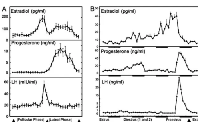
The female rat is a spontaneously ovulating polyestrous mammal. Ovulation occurs at 4- or 5-d intervals throughout the reproductive life of the rat, except during pregnancy, pseudopregnancy, and lactation (60 – 62). 17-Estradiol and progesterone are the hormones produced in the largest quan-tity by the ovaries. During the estrous cycle, 17-estradiol and progesterone act on the brain to stimulate the hormonal events that result in ovulation and sexual behaviors (63). These hormones also act on peripheral receptors and glands to induce the production of pheromones by the female rat. Therefore, these hormones will produce qualitative changes in the value of a female as a stimulus in addition to changing her behavior. For example, 17-estradiol enhances whereas progesterone decreases attractiveness of the female rat to a male rat because of changes in the female’s pheromone
pro-duction (64). For sexual behavior, 17-estradiol primes the brain for progesterone by inducing the production of pro-gesterone receptors, and then propro-gesterone activates sexual receptivity (63).
As mentioned above, female rats have only a 4- or 5-d estrous cycle (Fig. 2B). This is one of the most rapid ovarian cycles among mammals, and it is made possible by truncat-ing the cycle after ovulation. In other female mammals, ovu-lation is followed by a luteal phase, which is maintained by hormones produced by the corpus luteum (a transient en-docrine gland comprising the residual components of the follicle that remain after ovulation) (63). In rats, the corpus luteum becomes functional only when the female engages in sexual behaviors that activate a progestational reflex that prolongs the secretion of progesterone from the corpus lu-teum. This reflex induces the release of prolactin from the anterior pituitary. Prolactin in turn stimulates the corpus luteum, which in turn secretes progesterone to prepare the uterus for implantation, and this increases the probability that pregnancy will occur. Stimulation of the vaginocervical area by the male rat or the experimenter can induce the progestational reflex, resulting in a period of corpus luteum activity that lasts 12–14 d. This condition is known as pseu-dopregnancy (60, 65).
The estrous cycle. When the aim of an experiment is to de-termine whether an outcome measure varies with the stage of the estrous cycle it is important to determine whether animals are cycling normally. Thus, it is prudent to establish that at least two complete cycles have occurred before ini-tiating testing.
[image:8.594.231.552.528.725.2]Follicular phase.The ovarian cycle begins with the devel-opment of follicles from oocytes in the ovary. Low concen-trations of FSH from the pituitary stimulate follicular devel-opment. There is also increased steroidogenesis at this phase caused by stimulation by LH. 17-Estradiol secretion in-creases gradually during this phase. In the rat, this stage is 2 d long. The first day is called diestrus 1 or metestrus, and the second day is diestrus 2 or just diestrus (Fig. 2B) (63).
Periovulatory period.The time just before and after ovula-tion is dynamic. 17-Estradiol increases dramatically, acting on the brain to trigger GnRH release, which induces a surge of LH from the pituitary that induces ovulation. Progester-one rises a few hours before ovulation and contributes to this process. In the rat, this phase is called proestrus (63). Max-imum 17-estradiol release from the ovary starts 18 h before ovulation with serum 17-estradiol reaching a peak of 50 – 150 pg/ml around 6 –12 h before ovulation (60, 65). A sig-nificant increase in progesterone occurs 4 – 6 h after the 17 -estradiol surge, during the afternoon of proestrus. The serum concentration of progesterone at the proestrus peak is ap-proximately 25–50 ng/ml (60, 65). Once LH and progester-one are released into the circulation, ovulation occurs 10 –12 h later (Fig. 2B).
Estrous phase.Estrus is the period of sexual receptivity and the actual day of ovulation. Sexual receptivity onset occurs shortly after the onset of the dark phase of the light-dark cycle and precedes ovulation by a few hours in most animals (Fig. 3). Ovulation, induced by the LH surge on proestrus, occurs 10 –13 h after the surge (66), and sexual receptivity persists for 12–20 h (depending on whether the female mates or not). Note that behavioral receptivity occurs 36 – 48 h after the initial increase in 17-estradiol and 4 – 6 h after
proges-terone. Baseline serum concentrations of 17-estradiol at vaginal estrus or behavioral estrus are approximately 3–12 pg/ml (60, 65).
Further details of the stages of the estrous cycle and the endocrine changes that accompany them can be found in Ref. 67 for the rat, hamster, guinea pig, sheep, dog, and rhesus monkey.
Nomenclature.Clearly, the estrous cycle is a dynamic pro-cess with changes happening rapidly, particularly during proestrus and estrus. This can lead to some confusion and misunderstanding about findings if the dynamic nature of the process is not taken into consideration. For example, the hormonal profile during the morning of proestrus is very different from on the afternoon of proestrus (60). Further-more, confusion can occur because of different nomencla-tures that can be used to determine when a day begins and ends. Behavioral neuroendocrinologists, for example, fre-quently define a day of the cycle as beginning with the time that lights go off in the animal colony so that the entire active period of the animal’s subjective day falls on one day of the estrous cycle (see Fig. 3). Other scientists may define a day as beginning at midnight to be consistent with the scientist’s subjective day. Given the dynamic nature of the cycle, it doesn’t really matter which nomenclature is used as long as
it is defined and results are interpreted relative to the en-docrine events happening at the time of the experimental manipulations.
Determining estrous cycle phase by vaginal lavage.For research purposes, it is generally preferable to measure hormone con-centrations in the serum directly if inferences are to be made about hormone concentrations. However, hormonal changes associated with the ovarian cycle result in cellular changes in vaginal cytology. For some research purposes (e.g.verifying whether an experimental treatment has disrupted the estrous cycle) it may be sufficient to monitor changes in hormones indirectly by monitoring changes in vaginal epithelial cells. Most investigators assign estrous stage using the relatively noninvasive technique of examining vaginal cytology at 24-h intervals. Using this technique, it has been determined that the estrous cycle consists of four stages named diestrus 1 (D1, sometimes referred to as metestrus), diestrus 2 (D2), proestrus (P), and estrus (E). Each of these stages has varying
lengths (D1, ⬃6 – 8 h; D2, ⬃55–57 h; P, ⬃12–14 h; and E, ⬃25–27 h), and the specific changes depend on the species and the light-dark schedule. If samples are obtained at 24-h intervals, not all of these stages may be sequentially visible in a set of smears from an individual rat, because some stages are less than 24 h (60). Note, also, that even though the nomenclature is the same, these terms do not refer to the same 24-h periods identified in Fig. 3 and in the paragraphs above. For example, the vaginal cytology characteristic of proestrus lasts 12–14 h, not the 24 h associated with the day of proestrus as identified above. Finally, the vaginal cytology does not immediately reflect changes in hormonal secretion, because there is a delay between hormonal secretion and the morphological changes in the vaginal target cells.
As illustrated in Fig. 4, vaginal lavage is usually done using an eyedropper and 0.9% saline. The tip of the eye-dropper is filled with a small amount (one or two drops) of saline and then inserted into the vagina of the female rat (Fig.
4, A–D). The fluid is expelled into the vagina and then col-lected two to three times, or until the saline becomes opaque, and then the fluid is placed onto a microscope slide. The eyedropper is thoroughly rinsed with distilled water, and the next rat is sampled. The resulting sample is examined under a light microscope (⫻40 –100 magnification), while still wet. Alternatively, a cotton swab, moistened with 0.9% saline, may be inserted into the vaginal canal and then rapidly withdrawn (Fig. 4, E–F). The resulting vaginal smear is trans-ferred to a glass slide and examined under a light microscope at the same magnification (⫻40 –100). Figure 3 describes the changes in cell types characteristic of the four estrous cycle stages in the rat.
For both strategies it is important that care is taken to avoid stimulating the cervix. If the eyedropper or cotton swab stimulates the cervix with enough pressure during proestrus or estrus, it can induce pseudopregnancy and, therefore, disrupt the rat’s estrous cycle for approximately 12 d. Vaginal cytology should be examined daily, at the same time each day. Examination of these cells can be used to determine the cycle length and regularity of multiple cycles for each rat.
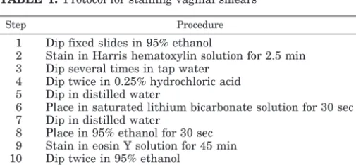
Reading the slides. It is not necessary to stain the cells, because the cells are sufficiently opaque to be visualized under a microscope with a blue or green filter. Alternatively, cells obtained via the vaginal smear technique may be fixed with ethanol (e.g.Surgipath cytology spray; Surgipath Med-ical Industries, Inc., Richmond, IL) to preserve the sample. Table 1 provides a protocol for staining the preserved sam-ples for photographic purposes. The cytology sample can be viewed under a⫻40 –100 objective with a blue or green filter. Interpreting the stage of estrous cycle from the cell mor-phology requires practice and experience with a particular rat. The precise cellular morphology on a particular day of the cycle varies with the time of day (especially on proestrus). Rather than trying to guess what stage of the estrous cycle a female rat is in, describe the cell types that appear in the smear. After 5– 6 d, it is usually possible to determine what that rat’s smear looks like on estrus because of the abundance of cells usually obtained from lavage and their unmistakable appearance. Collect data for at least two complete cycles before determining where a female rat is in the cycle. This same method may be used to monitor the estrous cycle of mice, but the cells have slightly different characteristics (68 –70).
Question 6: which forms of the hormones should be given to rodents and by which route of administration?
Just as there are numerous choices in hormone replace-ment therapy for women lacking ovarian steroid hormone secretions, there is a wide variety of choices and variables (form of hormone, time, route, length of administration,etc.) for hormone replacement in research animals, whose ovaries have been removed experimentally. In this section, we dis-cuss some of the hormone replacement procedures that have been used in rodents, and the rationales for selecting among them are discussed. Hormones administered to induce sex-ual receptivity and other physiological responses in female rats are used as examples. In the many studies on the hor-monal regulation of sexual behaviors, the many facets of hormone administration have been extensively studied.
The choice for hormone replacement after gonadectomy must be guided by consideration of a variety of factors de-pending on the goals of the study. Is the goal of the study to 1) determine the hormones involved in a particular response during the estrous cycle or during pregnancy, 2) determine whether there is a sex difference in a particular response that is already known to be influenced by the estrous cycle, 3) determine whether gonadectomized females respond to a treatment that has a particular effect in gonadectomized males, or 4) assess whether a particular cellular mechanism of action of a hormone shows a sex difference in response?
Nomenclature (see also the glossary at the end of this article).There are two primary classes of ovarian hormones. Estradiol (also known as 17-estradiol) is the principal circulating hormone of the class of hormones known as estrogens, and proges-terone (also known as pregn-4-ene-3,20, dione) is the prin-cipal circulating hormone of the class known as progestins (or gestagens). Although 17-estradiol is often referred to as estrogen, this usage is not technically correct, because 17 -estradiol is a specific hormone, whereas estrogen is a class of hormones. 17-Estradiol is the most abundant circulating estrogen in vertebrates, so this is the particular estrogen that is usually administered in estrogen replacement in research animals. In males, the testicular hormone is primarily tes-tosterone, an androgen.
Different estrogens. Although 17-estradiol is typically the most physiologically active estrogen, this is not the only estrogen that has been used in endocrine or neuroendocrine research. For example, in a variety ofin vitrosystems, estriol has been compared in effectiveness to 17-estradiol (71). In some behavioral experiments, estrone has been used rather than 17-estradiol (72), perhaps in part because it is less effective than 17-estradiol and less prone to inducing progesterone-independent feminine sexual behavior.
[image:11.594.33.286.609.726.2]Routes of administration.17-Estradiol and progesterone can be administered by a wide variety of routes of administra-tion. These include sc, iv, im, ip, and intracranial routes of administration. In some experiments, to alleviate the incon-venience of daily injections for long-term treatments, testos-terone, 17-estradiol, or progesterone, either in crystalline form (73, 74) or dissolved in peanut oil, has been enclosed in a small length of silicone tubing. It can also be delivered by an Alzet mini-pump (75) that delivers a consistent dose of TABLE 1. Protocol for staining vaginal smears
Step Procedure
1 Dip fixed slides in 95% ethanol
2 Stain in Harris hematoxylin solution for 2.5 min 3 Dip several times in tap water
4 Dip twice in 0.25% hydrochloric acid 5 Dip in distilled water
6 Place in saturated lithium bicarbonate solution for 30 sec 7 Dip in distilled water
8 Place in 95% ethanol for 30 sec 9 Stain in eosin Y solution for 45 min 10 Dip twice in 95% ethanol
hormone for days, or by pellets supplied to deliver a par-ticular dose of steroid hormone daily when implanted sc (76). Each mode of hormone replacement has its appropriate place, but different routes and regimens may provide dis-crepant results. In some cases, these differences in response to different treatments have been exploited to understand the hormones-behavior relationships better.
Esterified vs. free hormone.Because the circulating amount of 17-estradiol does not remain elevated for very long after systemic injection of 17-estradiol, slower-release, esterified forms of 17-estradiol are often used in physiological re-search. A variety of esters and other modifications have been used, such as ethynylestradiol (77), estradiol valerate (78), or estradiol dipropionate (79). However, the most commonly used form is estradiol-3-benzoate, which is 17-estradiol with a benzoic acid esterified in the third carbon position. This form is hydrolyzedin vivoto the physiologically active 17-estradiol. Progesterone is injected only in an unmodified form (80). Testosterone is usually administered as testoster-one or testostertestoster-one propionate (81).
In much of the early work in behavioral endocrinology, estradiol benzoate was the most effective form of estrogen found to induce sexual receptivity in females when followed by progesterone (80), so this became standard in the field of hormonal regulation of feminine sexual behaviors. It is likely that this form was chosen over free 17-estradiol or estrone because it is generally effective in lower doses than either of these free, unesterified estrogens. Interestingly, though, many years after the pioneering work, others reported that free 17-estradiol is more effective if given in a pulsatile fashion (82), for example, with two injections separated by at least 3–36 h (83).
The most typical mode of administration when attempting to induce lordosis behavior is one, two, or three daily injec-tions of estradiol benzoate followed by progesterone. These treatments reliably induce the expression of feminine sexual behavior. In some cases, chronic daily injections of estradiol benzoate have been given without progesterone (84), al-though progesterone is essential for the facilitation of sexual receptivity during the estrous cycle (85) and for the full complement of sexual behaviors in rats (86). Although 17 -estradiol alone may be effective in inducing sexual behavior and in causing many of the behavioral changes seen during the estrous stage of the estrous cycle, the injection of daily 17-estradiol or estradiol benzoate does not duplicate the preovulatory pattern of ovarian steroid hormones seen dur-ing the estrous cycle for two reasons. The preovulatory 17 -estradiol rises and falls over the course of a day and a half (See Fig. 2B), and the preovulatory 17-estradiol is also fol-lowed by a surge in progesterone from the follicle and in-terstitium (87) (see Fig. 2B). Recently, Asarian and Geary (88) used a procedure in which a low dose of estradiol benzoate was injected sc every 4 d in ovariectomized rats. This treat-ment, which closely mimics the changes in 17-estradiol concentrations over the estrous cycle, was effective in main-taining a variety of estrogen-dependent responses including responsiveness to progesterone facilitation of sexual behav-ior every 4 d, at rates similar to that seen during the rat’s 4-d
estrous cycle, suggesting that this regimen might be a very useful strategy for hormone replacement.
Silicone tubing capsules.When the treatment regimen requires or can accommodate prolonged exposure to the hormones, 17-estradiol, progesterone, and testosterone have often been administered in the form of crystalline hormone im-plants in silicone tubing implanted sc (74, 89). Lipophilic steroid hormones dissolve through the wall of the silicone tubing and are released at a constant rate that depends on the surface area (length⫻diameter) of the capsule and the thick-ness of the capsule wall. Resulting hormone concentrations can be titrated by dissolving the hormone in oil vehicle (90) by diluting it with crystalline cholesterol (91) or by varying the size of the tubing itself. In addition, concentrations of circulating hormone remain rather consistent for an ex-tended period of time (92) compared with injection of 17 -estradiol benzoate or another estrogen (93). To implant and remove silicone tubing capsules, the animals have to be anes-thetized. However, they do not need to be handled daily as would be the case with injections. In some cases, silicone tubing capsules are inserted and removed at particular in-tervals, either to approximate hormone concentrations seen during the estrous cycle (90) or to provide the hormone in a more-or-less discontinuous pattern (94).
A similar mode of administration that has been used in mice is chronic implantation of silicone tubing capsules con-taining 17-estradiol dissolved in oil (95). Often progester-one is injected weekly before tests for sexual receptivity. On the background of the chronic 17-estradiol exposure, the weekly progesterone is quite effective. It must be noted, however, that although this might be useful in certain types of studies when prolonged elevation of 17-estradiol con-centrations is desired, the treatments bear no similarity to the patterns seen during the estrous cycle, as discussed earlier.
Pharmacokinetics. When replacing hormone by any of the modes of administration discussed here, pharmacokinetics must be taken into account. As would be expected, an iv, unesterified 17-estradiol injection results in an immediate massive increase in blood 17-estradiol concentrations, which decline with a time course dependent upon dose of hormone injected (96). On the other hand, a sc injection of the esterified estradiol benzoate induces a slower rise and fall of blood 17-estradiol concentrations, typically lasting for one (97) or two (88, 98) days. A silicone tubing capsule implanted sc results in more of a square wave in 17-estradiol concen-trations with insertion and removal of the capsule (92), al-though the dynamics can be greatly influenced by whether crystalline 17-estradiol (91) is used or 17-estradiol dis-solved in peanut oil vehicle (99, 100). Likewise, the hormone peak can be influenced by preincubation of the capsules before insertion (74, 101), the percentage of 17-estradiol relative to cholesterol in the implants (91), and other factors.
cell nuclear estrogen receptors (96), functioning as transcrip-tion factors, long after circulating concentratranscrip-tions of 17 -estradiol have declined. Likewise, 17-estradiol remains bound to estrogen receptors for at least 2 or 3 d after injection of a low dose of estradiol benzoate in rats (102) and guinea pigs (103), and the cellular and behavioral consequences of the hormone injection may persist for several days (102, 104), long after circulating concentrations of 17-estradiol have decreased. In the case of another estrogen, 17␣ -ethynylestra-diol, residual effects can be observed more than 2 wk after cessation of treatment (77). Although 17-estradiol concen-trations circulating in the blood may decline quite rapidly after removal of a sc silicone tubing implant containing 17 -estradiol (92), the behavioral response persists for at least a day (94), as does 17-estradiol remaining bound in cells (105).
Intracranial and iv administration. Sex steroid hormones are extensively metabolized by the liver. To bypass this metab-olism or the delay in delivery of the hormone to the neural site of action, hormones can be infused by cannula directly into the cerebral ventricles (106). If the neuroanatomical site of action of the hormones is being investigated, the hormone can be implanted directly into specific neuroanatomical areas (107). Similarly, both 17-estradiol (96, 108) and progester-one (109 –111) have been administered iv either to increase the amount of unmetabolized hormone reaching the brain or to deliver the hormone to the brain as rapidly as possible. In some cases, these modes of administration have been used when the particular compound being administered is costly (e.g. radioactively labeled estrogen or a difficult-to-isolate metabolite) or when a route that optimizes efficient delivery is desirable.
Pulses of hormones and sex differences. The most compelling evidence that different types of hormone replacement can produce different results comes from studies of sex differ-ences in hormone-induced feminine sexual behavior in rats and guinea pigs. It is well known and accepted that there is a dramatic sex difference in response of many species to hormonal induction of feminine sexual behavior; after ovar-ian hormone treatment, the vast majority of gonadectomized female rats (112, 113) or guinea pigs (14, 114) express sexual behaviors, whereas most males do not (for review see Ref. 115). In contrast, if low-dose pulses of free 17-estradiol are injected in an episodic manner separated by about 12–24 h and then are followed by progesterone injection, male rats (116, 117) and guinea pigs (118) express high rates of lordosis, sometimes as much as females. Although the reason that the pulses eliminate a quite robust sex difference seen with the more-typical single injections remains a fascinating mystery, it exemplifies the importance of considering the many op-tions of hormonal replacement regimen for a particular study. The finding that pulse administration eliminates or attenuates the sex difference in sexual behavior should not be interpreted as a nuisance or an artifact; rather it tells us that the male brain interprets episodic 17-estradiol expo-sure as meaning something different from continuous 17 -estradiol exposure. Because the brain of a female does not respond in a qualitatively different manner to episodic vs.
continuous 17-estradiol exposure, this represent an intrigu-ing sex difference in response to different modes of hormone administration.
Experiments with mice.Mice are not just small rats, and their neuroendocrine systems are regulated in a number of ways that are different from rats. In particular, the hormonal reg-ulation of sexual behaviors is different in mice than in rats, and even strains of mice can differ markedly from each other. This is made more difficult for the laboratory scientist by the relative paucity of information about hormones and brain function and behavior in mice. Researchers working in rats or guinea pigs, on the other hand, have a voluminous liter-ature on the effects of different doses, injection regimens, routes of administration, and particular hormones to draw on. A good deal of this work on rats and guinea pigs came originally from the work of William C. Young’s research group in the 1930s (80) (seee.g.Ref.119). This work has been added to and clarified many times over during the past half-century. This is not the case with mouse reproductive neuroendocrinology. Many of the critical parametric exper-iments have yet to be performed in mice. The result is that mice are used as a model species with which to study mo-lecular neuroendocrine relationships involved in sexual be-havior and other aspects of reproductive neuroendocrinol-ogy; however, unlike the situation in guinea pigs (120) and rats (85), less is known about the factors necessary for the induction of feminine sexual behavior during the estrous cycle. For example, unlike rats and guinea pigs, when female mice are given typical injections of 17-estradiol followed by progesterone (106, 121, 122), or when given implants of a silicone tubing capsule of 17-estradiol followed by pretest injections of progesterone (95), they do not show full sexual response until approximately the fourth weekly behavioral test. Furthermore, chronic estradiol treatment is not well tolerated by male mice (123, 124).
disposition affects not only the peak of the hormone deliv-ered but also the time course of hormone present in the blood system. So, a dose that produces equal concentrations of hormones in males and females at 30 min will probably not result in equal concentrations of hormones in blood at 2 h. Of course, all drugs and hormones are different, so each must be evaluated independently.
Summary.In summary, this section discussed the most com-mon modes of administration and regimens used in ovarian hormone replacement treatment of rats. Each has its advan-tages and disadvanadvan-tages, and each can be applied to the study of sex differences. A great deal of thought must go into choosing the particular hormones administered, their form, and the mode and timing of administration, and a good deal of thought must go into providing equitable treatments in males and females. Fortunately, much is already known about the effects of varying particular parameters on phys-iological responses, so well-informed choices are possible. Although testicular hormone replacement was not specifi-cally discussed, all of the same considerations must be made in administering androgens as in administering estrogens and progestins.
Question 7: how do you study the stage of the menstrual cycle and other hormone variation in men and women?
Menstrual cycle. Just as is the case in laboratory animals, hormonal changes associated with a woman’s reproductive cycle may contribute to sex differences observed. For exam-ple, the hormonal changes occurring during the normal men-strual cycle (i.e.from the follicular phase to the luteal phase; Fig. 2A) must be taken into account in evaluating many physiological or brain-based sex differences. The idea that men are hormonally static is not really true. Men, too, ex-perience biological rhythms in the production of sex steroids, including significant diurnal and seasonal changes in tes-tosterone production (30). A researcher studying sex differ-ences should be aware of variations in endogenous hor-mones that may be pertinent to variables under study and be prepared to monitor them when appropriate. Thus, men-strual cycle stage in female subjects may need to be deter-mined. Other major changes in sex steroids that could have significant implications for sex differences occur at puberty in both sexes, during pregnancy and the postpartum in women, and with aging.
During the typical menstrual cycle, serum concentrations of 17-estradiol range from 30 –300 pg/ml (127, 128), and peak concentrations are achieved directly before ovulation. It is important to realize that there can be large inter- and intra-individual variations in the exact timing of menstrual events and the concentrations of hormones attained at the various stages. Researchers should not assume that the length of a woman’s cycle is 28 da In fact, themedianlength of the menstrual cycle is 29.5 d, and it is common for women to have regular cycles as short as 25 or as long as 35 d (127). Because the length of the luteal phase is relatively fixed at approximately 13–15 d, variations in cycle length largely reflect variation in the length of the follicular phase, and the timing of ovulation will vary accordingly. Concentrations of
progesterone vary from less than 1.0 to around 15 ng/ml during the normal menstrual cycle, and peak progesterone concentrations are reached in the mid-luteal phase (127, 128). Follicular-phase progesterone concentrations are extremely low, along with concentrations in anovulatory and meno-pausal women. The late luteal phase is associated with a reduction in serum concentrations of 17-estradiol, proges-terone, or 17-estradiol and progesterone. FSH concentra-tions range from less than 1.0 to 12 mIU/ml, whereas LH concentrations range from less than 2.0 to around 50 mIU/ml and peak at the time of ovulation (see Fig. 2A). Menstruation onset can be determined by simply asking women them-selves or by analyzing hormone concentrations and com-paring them with a chart similar to Fig. 2A.
Analyzing vaginal secretions (for day of maximum cervi-cal mucus), analyzing urine samples (for daily estrone con-jugates revealing ovarian follicular dynamics), taking basal body temperature, and using ovulation kits are all methods that can provide information regarding the approximate tim-ing of ovulation (129, 130). Not all methods may be equally useful in a given study. For example, the rise in body tem-perature is small (between 0.2 and 0.5 C), occurs only after ovulation has transpired, and does not occur reliably in all women, even if cycles are ovulatory. Knowing the time of ovulation, however, can aid in the determination of the men-strual cycle stage of a female subject. To permit the proper adjustments to be made for menstrual cycle length, women should be asked to chart their cycle if there is doubt about its typical length. Monitoring two or more cycles may be needed to derive a reliable estimate (131). Although more elaborate methods are also available, this could be as simple as re-cording the date of menstrual onset over several cycles.
Puberty.The stages of puberty are conventionally defined in both sexes by the degree of development of secondary sex characteristics and rated on the Tanner scale (132). A com-plete description of the hormonal changes and stages of puberty in humans is beyond the scope of this article. Pu-berty is a time when females undergo major hormonal changes associated with the menstrual cycle (133). There is also a dramatic rise in androgen production in males.
Menopause.Menopause results from a sequence of events that leads to a modification in the programming along the hy-pothalamic-pituitary-gonadal (HPG) axis. After the meno-pause, production of 17-estradiol and estrone by the ovaries declines considerably. A relationship between declining ovarian hormone concentrations and the symptoms of meno-pause has been suggested. The reciprocity between gonadal hormones and the serotonergic system presents some prom-ising clues into the mechanism that initiates the onset of menopausally related neuroregulatory changes (134, 135).