Journal compilation#2006 Blackwell Publishing doi: 10.1111/j.1600-0854.2006.00472.x Traffic 2006;7:1378–1387
Blackwell Munksgaard
Palmitoylation Plays a Role in Targeting Vac8p to
Specific Membrane Subdomains
Yutian Peng1,2, Fusheng Tang2,3and Lois S. Weisman1,2,*
1
Department of Cell & Developmental Biology, Life Sciences Institute, 210 Washtenaw Avenue, Room 6437, University of Michigan, Ann Arbor, MI 48109-2216, USA
2
Department of Biochemistry, University of Iowa, Iowa City, IA 52242, USA
3
Current address: Department of Biology, University of Arkansas, Little Rock, AR 72204-1099, USA
*Corresponding author: Lois S. Weisman, [email protected]
Vac8p is a multifunctional yeast protein involved in several distinct vacuolar events including vacuole inheritance, vacuole homotypic fusion, nucleus–vacuole junction for-mation and the cytoplasm to vacuole protein targeting pathway. Vac8p associates with the vacuole membrane via myristoylation and palmitoylation. Vac8p has three puta-tive palmitoylation sites, at Cys 4, 5 and 7. Here, we show that each of these cysteines may serve as a palmitoylation site. Palmitoylation at Cys 7 alone provides partial function of Vac8p, whereas palmitoylation at either Cys 4 or Cys 5 alone is sufficient for Vac8p function. In the former mutant, there is a severe defect in the localization of Vac8p to the vacuole membrane, while in the latter mutants, there is a partial defect in the localization of Vac8p. In addition, our studies provide evidence that palmitoylation targets Vac8p to specific membrane subdomains.
Key words: membrane subdomain, organelle inherit-ance, palmitoylation, Vac8p, yeast
Received 22 June 2006, revised and accepted for publica-tion 6 July 2006
During cell division, most cytoplasmic organelles are actively distributed to the new daughter cells. In budding yeast, the distribution of vacuoles initiates early in the cell cycle (1); a portion of the mother vacuole most proximal to the incipient bud site moves along actin filaments and is drawn into the emerging bud (2). Vacuole movement requires the class V myosin, Myo2p (2–4). The globular tail of Myo2p binds directly Vac17p, which in turn binds directly to Vac8p (5,6). Vac8p is attached to the vacuole membrane. The Vac17p–Vac8p complex forms the vacuole-specific Myo2p receptor and specifically regulates Myo2p attachment to the vacuole (5,6). Myo2p attach-ment to its other cargoes occurs through interaction with other receptors [reviewed by Pruyne et al. and Pashkova et al. (7,8)].
Vac8p is an armadillo-repeat-containing protein (9–11). In addition to its role in vacuole inheritance, Vac8p is required for at least four other vacuole-related processes: homo-typic vacuole fusion (12–14), the cytoplasm to vacuole protein targeting pathway (15), formation of the nucleus– vacuole junction (16) and caffeine resistance (17). Each of these functions involves a unique Vac8p binding partner; the armadillo repeats of Vac8p serve as docking sites for these proteins (17).
Vac8p is both myristoylated and palmitoylated; the former modification occurs at Gly 2, and the latter at one or more of the nearby residues, Cys 4, Cys 5 and/or Cys 7 (11). Exactly which site(s) is palmitoylated has not yet been determined. The covalent attachment of myristate to the amino-terminal Gly occurs cotranslationally and facil-itates the subsequent post-translational addition of palmi-tate to the nearby Cys residues [reviewed by Linder and Deschenes (18)]. Myristoylation and palmitoylation are critical for Vac8p association with the vacuole membrane (11).
Palmitoylation is a common reversible modification of eukaryotic proteins that increases their membrane af-finity and reversibly affects both protein localization and function (19). In addition to playing a general role in membrane association, studies on proteins such as Src kinases and Ga subunits suggest that palmitoylation specifically targets these proteins to lipid rafts, subdo-mains on the plasma membrane that are enriched in sphingolipids and cholesterol (19). Targeting to a membrane subdomain would increase the local concen-tration and likely facilitates protein sorting and signaling (20–22).
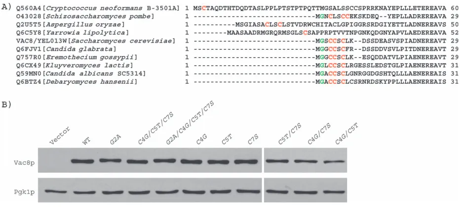
Palmitoylation of Vac8p is important for its localization and its function in vacuole inheritance (11) and homotypic vacuole fusion (12–14). Notably, Vac8p is uncommon among myristoylated and palmitoylated proteins. For example, Vac8p is localized on the yeast vacuole; only one other protein that is myristoylated and palmitoylated, Meh1p, is found on the vacuole. Other known myristoy-lated and palmitoymyristoy-lated proteins are found at the plasma membrane (25). Moreover, Vac8p possesses three cys-teines near its myristoylation site; each cysteine is a poten-tial palmitoylation site. This contrasts with other studied myristoylated and palmitoylated proteins, which have one or at most two palmitoylation sites. Importantly, the three cysteines found at the amino terminus of Vac8p are highly conserved in Vac8p proteins from other yeasts and fungi. So far, Vac8p has been sequenced from ten species. In six of the sequences, the spacing of Gly 2 and the three cysteines is identical (Figure 1A). This degree of conser-vation strongly suggests that the three cysteines are important. These findings raise several questions: (1) Are all three cysteines palmitoylated? (2) How many palmitoy-lation sites are required for full function of Vac8p? (3) Does palmitoylation serve a role in addition to its function to attach Vac8p to the vacuole membrane? (4) Do different sites have different functions?
Here, we show, by analysis of all possible single and double mutants of Cys 4, 5 and 7, that all three of the putative palmitoylation sites in Vac8p play roles in its association with the vacuole membrane and its vacuole-membrane-related functions. In addition, we present evi-dence that palmitoylation may target Vac8p to specialized lipid subdomains.
Results
Palmitoylation of either Cys 4 or Cys 5 is required for Vac8p association with the vacuole membrane Our previous studies demonstrated that Vac8p is myris-toylated and palmimyris-toylated and strongly suggested that Cys 4, 5 and 7 are each sites of palmitoylation (11). In order to determine whether each of these cysteines was a site of palmitoylation and to determine the role of acylation in Vac8p localization, all possible single and double mutants of Cys 4, 5 and 7 were constructed. Analysis of these mutants showed that acylation is not required for Vac8p expression or its stability. As measured by immunoblot analysis, Vac8p mutants that are not myristoylated or not palmitoylated are present in cells at similar steady state levels as wild-type Vac8p (Figure 1B).
[image:2.595.70.536.442.652.2]Acylation of Vac8p is critical to its proper localization. Wild-type Vac8p was readily observed on the vacuole mem-brane by conventional immunofluorescence microscopy (Figure 2). However, using the same technique, neither thevac8-G2Anor thevac8-C4G/C5T/C7Smutants could be detected (data not shown). Therefore, we used a more sen-sitive ‘sandwich’ immunofluorescence microscopy tech-nique (seeMaterials and Methods). Use of this technique revealed that a fraction of the Vac8p mutant that cannot be myristoylated, vac8-G2A, was still associated with the vacuole membrane. This mutant is not palmitoylated to the same extent as wild type (11). Therefore, this finding suggests that myristoylation and/or normal levels of palmi-toylation are required for full association of Vac8p with the vacuole membrane. In contrast, in the absence of any myristoylation and palmitoylation, Vac8p cannot associate
with the vacuole membrane. Thevac8-G2A/C4G/C5T/C7S
mutant, which cannot be acylated, was detected only as small, dispersed spots in the cytoplasm. Loss of only one putative palmitoylation site had little effect on Vac8p localization: all three of the single mutants (vac8-C4G,
C5TandC7S) had levels of vacuole membrane-associated Vac8p that were detectable by conventional immunofluo-rescence microscopy. Loss of two potential palmitoylation sites resulted in decreased localization of Vac8p to the vacuole membrane, such that detection of the protein required the use of a sandwich technique. Two of the double mutants (vac8-C5T/C7Sandvac8-C4G/C7S) were found to be localized to both the vacuole membrane and the cytoplasm. The remaining double mutant (vac8-C4G/ C5T) had a more severe phenotype. Some Vac8p was detected on the vacuole membrane, but most of the protein was present as small spots dispersed in the cytoplasm. This indicates that either Cys 4 or Cys 5 is crucial for the proper localization of Vac8p to the vacuole membrane. These immunofluorescence localization studies parallel those observed using subcellular fractionation (26).
Palmitoylation of either Cys 4 or Cys 5 is required for vacuole inheritance, vacuole fusion and caffeine resistance
Palmitoylation is required for Vac8p function (11). Simulta-neous mutation of both the myristoylation and the palmi-toylation sites abolishes Vac8p function in vacuole inheritance (11), homotypic vacuole fusion (13) (also see
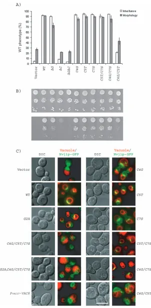
Figure 3A) and caffeine resistance (Figure 3B). Because Vac8p-G2A is not palmitoylated to the same extent as wild-type Vac8p (11), it is difficult to test the role of myristoy-lation in Vac8p function. Consistent with a partial defect in palmitoylation, the vac8-G2A mutant, while capable of supporting vacuole inheritance, had a modest defect in homotypic vacuole fusion as measured by vacuole frag-mentation (11,13) (also see Figure 3A). Moreover, myris-toylation alone is not sufficient for Vac8p function: the palmitoylation-minus mutant vac8-C4G/C5T/C7S was defective in vacuole inheritance, homotypic vacuole fusion and caffeine resistance (11,13) (Figure 3A–B).
Each of the single Cys point mutants functioned similarly to wild-type VAC8 in vacuole inheritance (Figure 3A). These mutants were virtually normal for homotypic vacu-ole fusion (Figure 3A) and caffeine resistance (Figure 3B). Likewise, two of the double mutantsvac8-C5T/C7Sand
[image:3.595.121.465.48.286.2]vac8-C4G/C7Sbehaved the same as the single mutants (Figure 3A–B). Importantly, however, the remaining dou-ble mutantvac8-C4G/C5Twas severely defective in these functions: less than 20% of cells displayed normal vacuole inheritance and less than 40% showed normal vacuole morphology (Figure 3A). This double mutant also was less resistant to caffeine and had a clear growth defect on 0.2% caffeine plates (Figure 3B). These results indicate that palmitoylation at either Cys 4 or Cys 5 is required for vacuole inheritance, homotypic vacuole fusion and caf-feine resistance. Palmitoylation at Cys 7 alone results in a partial defect in these Vac8p functions.
The levels of Vac8p on the vacuole membrane are crucial for formation of the nucleus–vacuole junction Vac8p–green fluorescent protein (GFP) is enriched in at least two regions of the vacuole membrane: the nucleus– vacuole junction (10) and vacuole–vacuole junctions (verti-ces) (10,14). The nucleus–vacuole junction is the site of piecemeal microautophagy of the nucleus, whereby a por-tion of the nuclear membrane is engulfed by the vacuole and degraded by vacuolar hydrolases (27). Nvj1p, an outer nuclear membrane protein, is a component of the nucleus– vacuole junction and interacts with Vac8p in a yeast two-hybrid test (16). The proper localization of Nvj1p at the nucleus–vacuole junction requires enrichment of Vac8p at the same region (16).
Consistent with the above studies, in the presence of wild-type VAC8, Nvj1p–GFP concentrated at the nucleus– vacuole junction (Figure 3C). In thevac8-G2A mutant or in cells containing any of the single Cys mutants, Nvj1p– GFP was predominantly localized to the nucleus–vacuole junctions, with a small amount of Nvj1p–GFP dispersed around the nuclear envelope (Figure 3C). Two of the double mutants (vac8-C5T/C7Sandvac8-C4G/C7S) were also nearly normal with most of the Nvj1p–GFP at the nucleus–vacuole junction. In contrast, in the double mutant
vac8-C4G/C5T and the mutants in which all three Cys residues are replaced, Nvj1–GFP was dispersed through-out the through-outer nuclear membrane (Figure 3C). These find-ings suggest that palmitoylation of Vac8p at either Cys 4 or Cys 5 is required for localization of Vac8p and Nvj1p to the nucleus–vacuole junction.
Defects observed in nucleus–vacuole junction formation in the Vac8p mutant that cannot be palmitoylated at both Cys 4 and Cys 5 could be due directly to a loss of palmitoylation or due to the fact that levels of the mutant on the vacuole membrane are significantly lower than those of wild-type Vac8p. To distinguish between these possibilities, we compared thevac8-C4G/C5Tmutant with a secondvac8
mutantPVAC17-VAC8, which is composed of the wild-type
VAC8open-reading frame driven by theVAC17promoter. The levels ofVAC8expressed from theVAC17promoter are 10-fold lower than those of wild type (17). As measured by immunoblot analysis of isolated vacuoles,
PVAC17-VAC8andvac8-C4G/C5Tmutants have similar levels of vacuole membrane-associated Vac8p (Figure 4A). Notably, this low level of Vac8p is not sufficient for formation of the nucleus–vacuole junction; Nvj1p–GFP was mislocalized in both mutants (Figure 3C). The above results show that the levels of Vac8p on the vacuole membrane are critical for formation of the nucleus–vacuole junction. Thus, we could not test whether palmitoylation plays a direct role.
Palmitoylation plays a role in targeting Vac8p to microdomains on the vacuole membrane
To further test the roles of Vac8p palmitoylation, we compared additional functions of the PVAC17-VAC8 and
vac8-C4G/C5Tmutants. These two mutants demonstrated functional differences in vacuole fusion and vacuole inher-itance.PVAC17-VAC8was significantly better at these two functions thanvac8-C4G/C5T(Figure 4B). To analyze the partial defect ofvac8-C4G/C5Tin vacuole inheritance, we compared the localization of Myo2p relative to the vacuole in wild-type,vac8-C4G/C5T/C7S, vac8-C4G/C5T, PVAC17
-VAC8andvac8Dcells. In wild-type cells, a portion of the vacuole extends to the bud tip, where it colocalizes with Myo2p–GFP. Likewise in thePVAC17-VAC8mutant, which contains all three potential palmitoylation sites, a portion of the vacuole colocalizes with Myo2p–GFP. In contrast, in the absence of Vac8p, the vacuole remains in the mother cell, while Myo2p–GFP is concentrated at the bud tip. The
vac8-C4G/C5T/C7S mutant shows the same phenotype. Notably, the vac8-C4G/C5T mutant has an intermediate phenotype. In approximately 50% of the cells, a small portion of the vacuole moves into the bud and colocalizes with Myo2p–GFP (Figure 4C). These results strongly sup-port the hypothesis that Cys 7 can serve as a palmitoylation site. It is tempting to speculate that a single palmitoyl moiety at Cys 7 is not sufficient to fully anchor Vac8p on the vacuole membrane.
The fact that Vac8p is palmitoylated raises the possibility that vacuole microdomains may exist and, furthermore, may share common properties with the better character-ized plasma membrane microdomains, specifically lipid rafts. Palmitoylated plasma membrane proteins, such as G proteins and Src kinases, reside in lipid rafts that are rich in sphingolipids and cholesterol (18). Sphingolipids and ergosterol (the yeast equivalent of cholesterol) are found in very low levels in the vacuole membrane (28). Intriguingly, a block in the biosynthesis of ergosterol (erg6D) or very long chain fatty acids (sur4D), sphingolipid precursors, results in fragmented vacuoles (29), a phenotype similar to that observed in thevac8Dmutant. Moreover, like Vac8p, ergosterol accumulates at vacuole–vacuole vertices (30). These results suggest that ergosterol, sphingolipids and additional lipids form specific microdomains on the vacuole membrane and that Vac8p is enriched in these micro-domains. Thus, we conducted the following experiments.
Plasma membrane lipid rafts are insoluble in Triton X-100. We tested the behavior of Vac8p in Triton X-100 and in other more mild detergents, Brij 98, Lubrol WX and Tween-20. These detergents have been successfully used to separate distinct types of plasma membrane rafts in mammalian and yeast cells (31–33). Each of these plasma membrane rafts has a unique protein and lipid composition (33). Moreover, ‘Lubrol rafts’ and ‘Brij 98 rafts’ have distinct physiological roles compared to ‘Triton rafts’
in vivo(34–37).
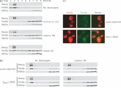
associated with lipid and floated in the low-density (mem-brane-containing) fraction, while Pgk1p appeared with the soluble components (Figure 5A). The behavior of Vac8p in detergent was more complex. In the absence of deter-gent, most of Vac8p floated and cofractionated with Pma1p (Figure 5A). In the presence of Triton X-100 (Fig-ure 5A) or Brij 98 (data not shown), Vac8p was completely soluble. In contrast, in the presence of the milder deter-gents, Lubrol WX or Tween-20, a significant amount of Vac8p cofractionated with Pma1p (Figure 5A), suggesting that a fraction of the Vac8p resides in ‘Lubrol’ rafts or ‘Tween 20 rafts’.
Next, we tested whether the Lubrol WX- or Tween 20-insoluble portion of Vac8p accounted for the functional difference between the vac8-C4G/C5T and the PVAC17
-VAC8mutants. We applied the same gradient centrifuga-tion to the mutants in the presence of Lubrol WX (Figure 5B) or Tween-20 (data not shown). A modest but reproducible difference was observed. We consistently detected more PVAC17-Vac8p in the same fractions as Pma1p.
In order to further test whether fully palmitoylated Vac8p resides in subdomains on the membrane, we compared
the localization of PVAC17-Vac8p with Vac8p-C4G/C5T. By
immunoblot analysis, both proteins were found to be at similar levels on the vacuole membrane (Figure 4A). However, when detected by immunofluorescence micros-copy, PVAC17-Vac8p was observed as many spots on the
vacuole membrane, whereas the Vac8p-C4G/C5T mutant had far fewer spots, presumably because it is more diffusively distributed (Figure 5C). Interestingly, this distri-bution of PVAC17-Vac8p to small spots on the vacuole
membrane is similar to what has been observed for its binding partner Vac17p (6). We postulate that these small spots are Vac8p microdomains that facilitate the interac-tion between Vac8p and Vac17p.
Discussion
[image:6.595.90.517.50.320.2]In this study, a series of Vac8p mutants missing the myristoylation and/or palmitoylation sites were generated and the effects of these mutations on the localization and functions of Vac8p were assessed. Point mutations that abolish myristoylation and/or palmitoylation do not affect the stability of Vac8p but greatly affect its distribution. Importantly, the different behaviors exhibited by point
mutants that affect two of the three palmitoylation sites indicate that these sites are not equivalent. Mutants that have point mutations at two of the three palmitoylation sites, but are palmitoylated either at Cys 4 or Cys 5, localize to the vacuole membrane although partial mislocalization of these mutant proteins to the cytoplasm occurs. In contrast, thevac8mutant that contains Cys 7 as its sole site of palmitoylation localizes to small dots, some on the vacuole, but many are elsewhere in the cell.
The genetic analysis presented here suggests that Cys 4, 5 and 7 are each palmitoylated. Two lines of evidence suggest that Cys 7 is palmitoylated. First, comparison of
vac8-C4G/C5T and vac8-C4G/C5T/C7S shows that the former mutant is more functional than the latter in vacuole inheritance and vacuole fusion (Figure 3A). Second, in the
vac8-C4G/C5T mutant, approximately 50% of the vacuoles colocalize with Myo2p on the bud tip (Figure 4C), suggesting that Vac8p is still associated with vacuole membrane in these cells. In contrast, in the vac8-C4G/ C5T/C7S mutant, vacuoles are not drawn by Myo2p to the bud (Figure 4C). This defect is likely due to the fact that
Vac8p is not retained on the vacuole membrane. Our results contrast with the studies using acyl–biotin exchange, where it had been proposed that Cys 7 is not palmitoylated (26). However, in that analysis, only membrane-bound proteins were included. Because most of the Vac8p-C4G/C5T is soluble, the acyl–biotin exchange method likely is not sensitive enough to detect palmitoylation at Cys 7.
[image:7.595.93.492.51.341.2]Palmitoylation at Cys 7 alone was not sufficient for targeting Vac8p to the nucleus–vacuole junction. It was not possible to test whether palmitoylation of Vac8p is specifically required for Vac8p targeting to the nucleus– vacuole junction because palmitoylation of Vac8p is import-ant for its overall attachment to the vacuole membrane. When compared with wild type, thevac8-C4G/C5Tmutant has 10-fold less Vac8p on the vacuole membrane (Figure 4 A). We found that a 10-fold drop in Vac8p levels, either through lower expression of Vac8p using thePVAC17-VAC8 mutant or through loss of palmitoylation due to the vac8-C4G/C5Tmutant, causes a mislocalization of Nvj1p from the nucleus–vacuole junction (Figure 3C), a sign that Vac8p is also not targeted there.
Figure 5: Palmitoylation plays a role in targeting Vac8p to specific microdomains.A) Solubility of Vac8p in the absence or presence of Triton X-100, Lubrol WX and Tween 20. Wild-type cells were lysed and fractionated in a 0–40% (w/v) Optiprep gradient in the absence or presence of 1% (w/v) Triton X-100, 0.5% (w/v) Lubrol WX or 1% (w/v) Tween 20. Samples were collected from the top (fraction 1) to the bottom (fraction 9) of the gradient, and proteins were subjected to immunoblot analysis for Pma1p, Vac8p and Pgk1p. B) Solubility of Vac8p-C4G/C5T and PVAC17-Vac8p in the absence or presence of 0.5% Lubrol WX. C) The localization of Vac8p-C4G/C5T and PVAC17-Vac8p by immunofluorescence microscopy. Vac8p (green) was detected by a ‘sandwich’ technique. Vac8p spots are much less frequent in the
Our studies show that palmitoylation of Vac8p is directly required for Vac8p function in homotypic vacuole fusion.
PVAC17-VAC8 and vac8-C4G/C5T mutants have similar levels of Vac8p on the vacuole membrane; yet, the former mutant supports vacuole fusion better than the latter (Figure 4B). Vac8p is targeted to vacuole–vacuole vertices (14,27), where it functions in homotypic vacuole fusion (12,13). The localization of Vac8p to vacuole–vacuole vertices is regulated and does not occur at the interface of vacuoles that have been physically forced into contact by sedimentation (14). Along with Vac8p, docked vacuoles are also enriched for ergosterol at the vacuole–vacuole vertices (30). Thus, it is tempting to speculate that reversible palmitoylation of Vac8p regulates its movement into lipid raft-like, ergosterol-containing vacuole–vacuole vertices where fusion occurs. In support of this hypothe-sis, Vac8p is reversibly palmitoylated during vacuole fusion (12,13). Furthermore, ergosterol is required for vacuole– vacuole fusion. Absence ofERG6, one of the ergosterol biosynthetic genes, results in fragmented vacuoles (38), and conversely, overexpression ofERG6increases vacu-ole fusion (39).
Palmitoylation of Vac8p may also localize it to micro-domains related to vacuole inheritance. Similar to vacuole fusion, PVAC17-VAC8 supports vacuole inheritance better thanvac8-C4G/C5T. Myo2p and Vac17p are concentrated at small patches on the vacuole membrane (4,6). Whether ergosterol specifically colocalizes with the Vac8p–Vac17p– Myo2p complexes remains to be tested. Fillipin, which is fluorescent and binds ergosterol-containing membranes
in vivoandin vitro, has been used to observe ergosterol on isolated vacuoles. However, when yeast are treated with fillipinin vivo, the fluorescence at the plasma membrane is too strong to detect the ergosterol that is present on internal membranes (40).
Lipid rafts were identified first on the plasma membrane (41). A number of recent studies have demonstrated that rafts also exist on intracellular membrane organelles along the secretory pathway including the Golgi complex (42), the endoplasmic reticulum (43) and endosomes (34). Likewise, functional lipid microdomains likely exist on the yeast vacuole membrane. It is likely that the lipid compo-nents of these microdomains are different from those of the lipid rafts characterized thus far. Characterization of the putative Vac8p microdomains may require development of new techniques to probe its lipid components.
Materials and Methods
Yeast strains and plasmids
The following strains were used in this study: LWY7235 (MATaleu2, 3-112 ura3-52 his3-D200 trp1-D901 lys2-801 suc2-D9) (44), LWY2887 (MATaleu2, 3-112 ura3-52 his3-D200 trp1-D901 lys2-801 suc2-D9 vac8D::HIS3) (11) and LWY7814 (MATaleu2, 3-112 ura3-52 his3-D200 trp1-D901 lys2-801 suc2-D9 vac8D::HIS3 myo2::MYO2-GFP).
vac8-G2A, vac8-C4G/C5T/C7S, vac8-G2A/C4G/C5T/C7S, NVJ1-GFP, PVAC17-VAC8andVAC17-GFPplasmids were described previously (11,17).
vac8-C4G,vac8-C5Tandvac8-C7Splasmids were constructed using the Transformer Site-Directed Mutagenesis Kit (ClonTech, Mountain View, CA, USA). vac8-C5T/C7S,vac8-C4G/C7S andvac8-C4G/C5T plasmids were constructed using the QuickChange Site-Directed Mutagenesis Kit (Stratagene, La Jolla, CA, USA). The residues substituted at each cysteine were chosen based on the original well-characterized mutants,vac8-C4G/ C5T/C7Sandvac8-G2A/C4G/C5T/C7S(11,17). Comparison of the pheno-types of the above point changes with those reported for mutants where cysteine was substituted with alanine (26) revealed that thevac8-C4G, vac8-C5Tandvac8-C7Sandvac8-C4A,vac8-C5Aandvac8-C7Amutants behave similarly.
Immunoblot analysis
To detect Vac8p in total cell extracts, mid-log phase cells (OD6000.4–0.8) were lysed by vigorous agitation with 0.5-mm glass beads in 100mL of lysis buffer [0.3Msorbitol, 10 mM Tris pH 7.5, 0.1MNaCl, 1 mMMgCl2, 1.0 mM ethylenediaminetetraacetic acid (EDTA) and 1yeast protease inhibitor cocktail (Sigma, St. Louis, MO, USA)]. Cell extracts were mixed with 36mL of 4SDS loading buffer and heated at 708C for 10 min. Five microliters of each sample (equal to 0.037 OD600units of cells) was loaded onto a 10% SDS–PAGE. Proteins were transferred to nitrocellulose membranes and subjected to immunoblotting.
Fluorescence microscopy
Observation of vacuole inheritance and morphology with FM4-64 (N-(3-triethylammoniumpropyl-)-4-(6(4-(diethylamino)phenyl)hexatrienyl) pyridi-nium dibromide) (Molecular Probes, Inc. Eugene, OR, USA) was performed as described (11). Images were obtained using an Axioscope 2 (Carl Zeiss MicroImaging, Jena, Germany) or the DeltaVision RT Restoration Micros-copy System (Applied Precision, Issaquah, WA, USA).
Localization of Vac8p in yeast cells by indirect immunofluorescence microscopy was performed as described (6). Briefly, for cells with Vac8p levels similar to wild type, spheroplasts were labeled with rabbit anti-Vac8p (1:100) and Alexa-488-conjugated goat anti-rabbit immunoglobulin G (IgG) (1:200). For cells with low levels of Vac8p, spheroplasts were labeled with rabbit anti-Vac8p (1:100), goat anti-rabbit IgG (1:240), and Alexa-488-conjugated donkey anti-goat IgG (1:200). Vacuole membranes were visualized with a mouse monoclonal anti-vacuolar adenosine triphospha-tase (60 kDa subunit) antibody (1:100; Molecular Probes) and rhodamine-red-conjugated donkey anti-mouse IgG (1:200; Jackson ImmunoResearch, West Grove, PA, USA). Images were obtained with a laser confocal system (MRC-1024; Bio-Rad, Hercules, CA, USA).
All images were processed by MetaMorph Imaging 4.5 Software (Universal Imaging, Buckinghamshire, UK) or Adobe Photoshop (Adobe, San Jose, CA, USA).
Comparison of vacuole-associated Vac8p in the
vac8-C4G/C5TandPVAC17-VAC8mutants
Vacuoles were isolated (13) fromvac8Dcells containing pRS416 (vector), pVAC8, pvac8-C4G/C5Tor pPVAC17-VAC8. The total protein concentration of the vacuole fraction was measured with a protein assay kit (Bio-Rad) using BSA as a standard. Five micrograms of total proteins from each membrane fraction was analyzed by immunoblotting for Vac8p and Pho8p.
Biochemical isolation of lipid rafts
microliters of the cleared lysate was mixed with 30mL of 10% (w/v) Triton-X-100 (Fisher, Hanover, IL, USA), 5% (w/v) Brij 98 (Sigma), 5% (w/v) Lubrol WX (Serva, Heidelberg, Germany), 10% (w/v) Tween-20 (Fisher) or water. The reactions were incubated at 48C for 30 min. Two hundred and fifty microliters of the treated lysate was adjusted to 40% (w/v) Optiprep by addition of 500mL of 60% Optiprep (Accurate, Westbury, NY, USA). About 1.2 mL of 30% and 200mL of 0% Optiprep were layered on top. The gradients were subjected to centrifugation (2.5 h at 160 000 xgin a TLS-55 rotor) at 48C. Ten fractions (200mL each) were collected from the top. Five microliters of each fraction was subjected to immunoblot analysis for detection of Pma1p, Vac8p and Pgk1p.
Acknowledgments
The authors wish to thank Drs Robert Piper, Marcus Lee and Gopal Thinakaran for sharing reagents. We thank Ting Liu for construction of thevac8-C4G,C5TandC7S.We also thank the members of the Weisman lab for their helpful discussions. This work was supported by a National Institutes of Health grant, R01-GM62261 (to L. S. W), and an American Heart Association Predoctoral Fellowship 0410029Z (to Y. P.).
References
1. Weisman LS. Yeast vacuole inheritance and dynamics. Annu Rev Genet 2003;37:435–460.
2. Hill KL, Catlett NL, Weisman LS. Actin and myosin function in directed vacuole movement during cell division in Saccharomyces cerevisiae. J Cell Biol 1996;135:1535–1549.
3. Catlett NL, Duex JE, Tang F, Weisman LS. Two distinct regions in a yeast myosin-V tail domain are required for the movement of different cargoes. J Cell Biol 2000;150:513–526.
4. Catlett NL, Weisman LS. The terminal tail region of a yeast myosin-V mediates its attachment to vacuole membranes and sites of polarized growth. Proc Natl Acad Sci U S A 1998;95:14799–14804.
5. Ishikawa K, Catlett NL, Novak JL, Tang F, Nau JJ, Weisman LS. Identification of an organelle-specific myosin V receptor. J Cell Biol 2003;160:887–897.
6. Tang F, Kauffman EJ, Novak JL, Nau JJ, Catlett NL, Weisman LS. Regulated degradation of a class V myosin receptor directs movement of the yeast vacuole. Nature 2003;422:87–92.
7. Pruyne D, Legesse-Miller A, Gao L, Dong Y, Bretscher A. Mechanisms of polarized growth and organelle segregation in yeast. Annu Rev Cell Dev Biol 2004;20:559–591.
8. Pashkova NJ, Jin Y, Ramaswamy S, Weisman LS. Structural basis for myosin V discrimination between distinct cargoes. Embo J 2006;25: 693–700.
9. Fleckenstein D, Rohde M, Klionsky DJ, Rudiger M. Yel013p (Vac8p), an armadillo repeat protein related to plakoglobin and importin alpha is associated with the yeast vacuole membrane. J Cell Sci 1998;111: 3109–3118.
10. Pan X, Goldfarb DS. YEB3/VAC8 encodes a myristylated armadillo protein of the Saccharomyces cerevisiae vacuolar membrane that functions in vacuole fusion and inheritance. J Cell Sci 1998;111: 2137–2147.
11. Wang YX, Catlett NL, Weisman LS. Vac8p, a vacuolar protein with armadillo repeats, functions in both vacuole inheritance and protein targeting from the cytoplasm to vacuole. J Cell Biol 1998;140: 1063–1074.
12. Veit M, Laage R, Dietrich L, Wang L, Ungermann C. Vac8p release from the SNARE complex and its palmitoylation are coupled and essential for vacuole fusion. Embo J 2001;20:3145–3155.
13. Wang YX, Kauffman EJ, Duex JE, Weisman LS. Fusion of docked membranes requires the armadillo repeat protein Vac8p. J Biol Chem 2001;276:35133–35140.
14. Wang L, Seeley ES, Wickner W, Merz AJ. Vacuole fusion at a ring of vertex docking sites leaves membrane fragments within the organelle. Cell 2002;108:357–369.
15. Scott SV, Nice DC III, Nau JJ, Weisman LS, Kamada Y, Keizer-Gunnink I, Funakoshi T, Veenhuis M, Ohsumi Y, Klionsky DJ. Apg13p and Vac8p are part of a complex of phosphoproteins that are required for cytoplasm to vacuole targeting. J Biol Chem 2000;275:25840–25849. 16. Pan X, Roberts P, Chen Y, Kvam E, Shulga N, Huang K, Lemmon S,
Goldfarb DS. Nucleus-vacuole junctions in Saccharomyces cerevisiae are formed through the direct interaction of Vac8p with Nvj1p. Mol Biol Cell 2000;11:2445–2457.
17. Tang F, Peng Y, Nau JJ, Kauffman EJ, Weisman LS. Vac8p, an armadillo repeat protein, coordinates vacuole inheritance with multiple vacuolar processes. Traffic 2006; Epub ahead of print. PMID:16824055. 18. Linder ME, Deschenes RJ. New insights into the mechanisms of
protein palmitoylation. Biochemistry 2003;42:4311–4320.
19. Smotrys JE, Linder ME. Palmitoylation of intracellular signaling pro-teins: regulation and function. Annu Rev Biochem 2004;73:559–587. 20. el-Husseini Ael D, Bredt DS. Protein palmitoylation: a regulator of
neuronal development and function. Nat Rev Neurosci 2002;3:791–802. 21. Patterson SI. Posttranslational protein S-palmitoylation and the com-partmentalization of signaling molecules in neurons. Biol Res 2002; 35:139–150.
22. Bijlmakers MJ, Marsh M. The on-off story of protein palmitoylation. Trends Cell Biol 2003;13:32–42.
23. Bijlmakers MJ, Isobe-Nakamura M, Ruddock LJ, Marsh M. Intrinsic signals in the unique domain target p56(lck) to the plasma membrane independently of CD4. J Cell Biol 1997;137:1029–1040.
24. El-Husseini Ael D, Craven SE, Brock SC, Bredt DS. Polarized targeting of peripheral membrane proteins in neurons. J Biol Chem 2001;276: 44984–44992.
25. Roth AF, Wan J, Bailey AO, Sun B, Kuchar JA, Green WN, Phinney BS, Yates JR III, Davis NG. Global analysis of protein palmitoylation in yeast. Cell 2006;125:1003–1013.
26. Subramanian K, Dietrich LE, Hou H, Lagrassa TJ, Meiringer CT, Ungermann C. Palmitoylation determines the function of Vac8 at the yeast vacuole. J Cell Sci 2006;119:2477–2485.
27. Roberts P, Moshitch-Moshkovitz S, Kvam E, O’Toole E, Winey M, Goldfarb DS. Piecemeal microautophagy of nucleus in Saccharomyces cerevisiae. Mol Biol Cell 2003;14:129–141.
28. Bagnat M, Chang A, Simons K. Plasma membrane proton ATPase Pma1p requires raft association for surface delivery in yeast. Mol Biol Cell 2001;12:4129–4138.
29. Seeley ES, Kato M, Margolis N, Wickner W, Eitzen G. Genomic analysis of homotypic vacuole fusion. Mol Biol Cell 2002;13:782–794. 30. Fratti RA, Jun Y, Merz AJ, Margolis N, Wickner W. Interdependent
assembly of specific regulatory lipids and membrane fusion proteins into the vertex ring domain of docked vacuoles. J Cell Biol 2004; 167:1087–1098.
31. Chamberlain LH. Detergents as tools for the purification and classifi-cation of lipid rafts. FEBS Lett 2004;559:1–5.
32. Lucero HA, Robbins PW. Lipid rafts-protein association and the regula-tion of protein activity. Arch Biochem Biophys 2004;426:208–224. 33. Schuck S, Honsho M, Ekroos K, Shevchenko A, Simons K. Resistance
of cell membranes to different detergents. Proc Natl Acad Sci U S A 2003;100:5795–5800.
35. Peng Y, Akmentin W, Connelly MA, Lund-Katz S, Phillips MC, Williams DL. Scavenger receptor BI (SR-BI) clustered on microvillar extensions suggests that this plasma membrane domain is a way station for cholesterol trafficking between cells and high-density lipoprotein. Mol Biol Cell 2004;15:384–396.
36. Drevot P, Langlet C, Guo XJ, Bernard AM, Colard O, Chauvin JP, Lasserre R, He HT. TCR signal initiation machinery is pre-assembled and activated in a subset of membrane rafts. Embo J 2002;21:1899–1908. 37. Roper K, Corbeil D, Huttner WB. Retention of prominin in microvilli
reveals distinct cholesterol-based lipid micro-domains in the apical plasma membrane. Nat Cell Biol 2000;2:582–592.
38. Kato M, Wickner W. Ergosterol is required for the Sec18/ATP-dependent priming step of homotypic vacuole fusion. Embo J 2001;20:4035–4040. 39. Tedrick K, Trischuk T, Lehner R, Eitzen G. Enhanced membrane fusion in sterol-enriched vacuoles bypasses the Vrp1p requirement. Mol Biol Cell 2004;15:4609–4621.
40. Beh CT, Rine J. A role for yeast oxysterol-binding protein homologs in endocytosis and in the maintenance of intracellular sterol-lipid distribu-tion. J Cell Sci 2004;117:2983–2996.
41. Bagnat M, Keranen S, Shevchenko A, Simons K. Lipid rafts function in biosynthetic delivery of proteins to the cell surface in yeast. Proc Natl Acad Sci U S A 2000;97:3254–3259.
42. Gkantiragas I, Brugger B, Stuven E, Kaloyanova D, Li XY, Lohr K, Lottspeich F, Wieland FT, Helms JB. Sphingomyelin-enriched micro-domains at the Golgi complex. Mol Biol Cell 2001;12:1819–1833. 43. Sevlever D, Pickett S, Mann KJ, Sambamurti K, Medof ME, Rosenberry
TL. Glycosylphosphatidylinositol-anchor intermediates associate with triton-insoluble membranes in subcellular compartments that include the endoplasmic reticulum. Biochem J 1999;343:627–635.