0095-1137/95/$04.0010
Copyrightq1995, American Society for Microbiology
Molecular Subtyping of Prevalent M Serotypes of
Streptococcus pyogenes Causing Invasive Disease
JOHN STANLEY,1* DENNIS LINTON,1MEETA DESAI,1ANDROULLA EFSTRATIOU,2
ANDROBERT GEORGE2
Molecular Biology Unit, Virus Reference Division,1and Streptococcus and
Diphtheria Reference Unit, Respiratory and Systemic Infection
Laboratory,2Central Public Health Laboratory,
London NW9 5HT, United Kingdom
Received 14 June 1995/Returned for modification 18 July 1995/Accepted 16 August 1995
Reproducible methodologies and a scheme for high-resolution genotyping of Streptococcus pyogenes were defined with respect to a study of six predominant M serotypes causing invasive group A streptococcal disease in the United Kingdom. Serotype reference strains were compared with nine clinical isolates of each serotype from patients with diseases such as pneumonia, puerperal sepsis, toxic shock-like-syndrome, cellulitis, or necrotizing fasciitis. Four enzymes were evaluated for their discriminatory power in 16S rRNA gene-specific ribotyping. Discriminatory power was greatest withEcoRI, which generated serotype-specific ribotypes, and withSacI, which could subdivide strains of the same M serotype. Twenty-five combined ribotypes were found among the 60 strains, and the indices of discriminatory power (Dvalues) of this method varied from 0.51 within serotype M1 to 0.98 within strains of serotype M5. Macrorestriction with the rarely cutting endonucleaseSmaI and pulsed-field gel electrophoresis gaveDvalues varying from 0.37 within serotype M1 to the maximal 1.0 within serotype M5. Comparison of macrorestriction profiles revealed various degrees of genetic heterogeneity within M serotypes. Strains of M1, M3, M6, and M11 exhibited clonally related macrorestriction profiles, while those of R28 and M5 strains were consistent with polyphyletic origin.
The Lancefield group A streptococci (Streptococcus
pyo-genes) (23) are major causative agents of human disease,
rang-ing from pharyngitis to severe invasive diseases such as toxic shock-like syndrome and necrotizing fasciitis (9, 19, 24). Epi-demiological surveillance has classically been based on pheno-typic (serological) markers. The antiphagocytic M protein, en-coded by the emm gene(s), is the antigenic basis of a serotyping scheme (7), constituting more than 80 M types. However, many (29% of current S. pyogenes isolates in the United Kingdom) isolates of S. pyogenes are nonserotypeable (10). There is con-siderable interest in the development of genotypic methods which can differentiate strains and/or subdivide M serotypes for the purposes of epidemiological typing.
Strains of S. pyogenes have been analyzed by several methods collectively termed genotypic typing (15). They include restric-tion endonuclease analysis of genomic DNA, rRNA gene poly-morphism analysis (ribotyping), pulsed-field gel electrophore-sis (PFGE), and multilocus enzyme electrophoreelectrophore-sis. These studies have had diverse and sometimes conflicting implica-tions. Restriction endonuclease digests of genomic DNA were shown to be M type specific (6) and were found to be more discriminatory than initial approaches to ribotyping (3, 8, 21). On the other hand, the choice of enzymes used by Bruneau et al. (4) in a study of pharyngeal isolates increased the dis-criminatory power of ribotyping and detected some correlation between M serotype and ribotype. Genomic DNA macro-restriction with a rarely cutting endonuclease, SfiI, and resolu-tion of large fragments by PFGE were used to detect polymor-phisms between and within some M serotypes of S. pyogenes (14, 22). Population genetics studies by multilocus enzyme
electrophoresis detected 33 electropherotypes among S.
pyo-genes isolates from patients with toxic shock syndrome or other
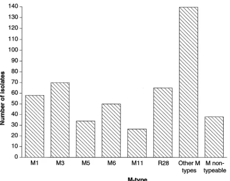
invasive diseases. Although comprehensive M serotyping data were not available, all of those strains within a single M sero-type belonged to a single electropherosero-type (16, 17). However, multilocus enzyme electrophoresis analysis of strains associ-ated with rheumatic fever and acute poststreptococcal glo-meruleronephritis subsequently indicated that single M types contained multiple electropherotypes and vice versa (11). There-fore, systematic studies of the relationship between genotype, M serotype, and disease symptomatology are required to solve apparent anomalies in the literature, to establish a re-producible framework for molecular epidemiological typing, and to quantify the discriminatory power (13) of molecular epidemiological typing. The study described here addresses the question of genetic diversity and its degree of resolution for epidemiological purposes with respect to six of the predomi-nant M serotypes causing invasive disease in the United King-dom in 1993 and 1994 (Fig. 1).
MATERIALS AND METHODS
Bacterial strains and culture conditions.Nine blood culture isolates of S.
pyogenes (obtained in 1993 and 1994 from patients in different geographical
regions of the United Kingdom) belonging to each of the M serotypes M1, M3, M5, M6, M11, and R28 were selected for study and are listed in Table 1. The type strains of each of these six M serotypes were obtained from the National Col-lection of Type Cultures (NCTC; Colindale, United Kingdom). Stock cultures were maintained in 16% blood glycerol broth at2708C.
Minipreparation of genomic DNA from plate cultures.A procedure based on that of Wilson (27) for gram-negative bacteria was developed for the preparation of streptococcal genomic DNA from a single plate culture. Enzyme concentra-tions and incubation times and temperatures were found to be critical for re-producible yield and quality of DNA. Strains were grown overnight on blood agar plates at 378C. The cell growth was scraped off and was resuspended in 1 ml of TE glucose buffer (glucose, 50 mM; Tris, 25 mM [pH 8.0]; EDTA, 10 mM) in a microcentrifuge tube. The cells were spun down (2 min, 13,000 rpm, Jouan M14.11 microcentrifuge), resuspended in 100ml of 40mg of mutanolysin (Sigma) per ml in TE glucose buffer–50ml of 50 mg of lysozyme (Sigma) per ml, and * Corresponding author. Mailing address: Molecular Biology Unit,
Virus Reference Division, Central Public Health Laboratory, 61 Colin-dale Ave., London NW9 5HT, United Kingdom. Phone: 0181 200 4400, extension 3071. Fax: 0181 200 1569.
2850
on May 15, 2020 by guest
http://jcm.asm.org/
incubated at 378C for 1 h. A total of 30ml of 10% sodium dodecyl sulfate (SDS) and 3ml of 20 mg of proteinase K (Sigma) per ml were added, and the combi-nation was mixed and reincubated at 558C for 30 min. Thereafter, 100ml of 5 M NaCl was added with thorough mixing; this was followed by the addition of 80ml of cetyltrimethylammonium bromide (10%) in 0.7 M NaCl. The solution was mixed and incubated for 10 min at 658C, after which lysis was complete. An approximately equal volume of chloroform-isoamyl alcohol (24:1; vol/vol) was mixed in, and the mixture was spun in the microcentrifuge for 5 min. The viscous supernatant above the white interface was transferred to a fresh tube and was extracted with phenol-chloroform-isoamyl alcohol (25:24:1; vol/vol/vol). The aqueous supernatant was again transferred and precipitated by adding an equal volume of isopropanol at room temperature. The final pellet was resuspended in 100ml of TE (Tris, 10 mM; EDTA, 1 mM [pH 7.5]).
Ribotyping with 16S rDNA.A 1,500-bp probe was generated from genomic DNA of the type strain of serotype M1, S. pyogenes NCTC 8198, by PCR with the primers 59-AAGAGTTTGATCCTGGCTCAG-39and 59-GGTTACCTTGTTA CGACTT-39. The PCR products were separated from the primers with Gene-Clean (Bio 101, Inc., La Jolla, Calif.) and were labelled with biotin-16-dUTP by using a random-primed labelling kit (Boehringer Mannheim Biochemica GmbH, Mannheim, Germany). Restriction digests of genomic DNA (5mg per lane) were electrophoresed in 0.7% agarose for 17 h at 55 V (PvuII, EcoRI, and HindIII digests) or 24 h at 60 V (SacI digests). Gels were vacuum blotted (LKB Vacu-Gene apparatus; Pharmacia Biotech Ltd., Milton Keynes, United Kingdom) onto a Hybond N nylon membrane (Amersham Life Science, Bucks, United King-dom). Hybridization was carried out under standard conditions (20) except that the membrane filters were washed twice at 578C for 15 min. Reactions were visualized colorimetrically with BluGene reagent, and molecular weight markers were biotinylated HindIII fragments of phage lambda (Life Technologies, Pais-ley, Scotland, United Kingdom). The numerical index of discriminatory power, or the D value (13), was calculated for each enzyme and for each serotype.
Macrorestriction and PFGE.Streptococci were grown overnight on blood agar plates at 378C, and the cells were resuspended in 2 ml of solution I (1 M NaCl, 10 mM Tris-HCl [pH 7.6]) to a density equivalent to that of a McFarland no. 5 standard. A total of 400ml of this suspension was added to 400ml of molten low-melting-point agarose dissolved in solution I, dispensed into block formers, and cooled to 48C. The solidified blocks were added to 2 ml of solution II (solution I plus 100 mM EDTA, containing 1 mg of lysozyme per ml and 7mg of mutanolysin per ml), and the mixture was incubated overnight at 378C. The solution described above was replaced with 2 ml of solution III (0.5 M EDTA [pH 9.5], 1% Sarkosyl NL-30, 0.5 mg of proteinase K per ml), and the blocks were incubated at 568C for 6 h. After exchange for fresh solution III, the blocks were reincubated overnight at the same temperature. The blocks were washed three times (30 min each time) with 2 ml of TE buffer (10 mM Tris, 10 mM EDTA [pH 7.5]) prior to storage at 48C. Macrorestriction with SmaI was carried out as described previously (1, 18). PFGE was carried out in a CHEF DRII apparatus (Bio-Rad) for 22 h at 200 V. Ramping times were 10 to 35 s (for strains of serotypes M1, M3, M5, and R28) or 1 to 20 s for strains of serotypes M6 and M11). Molecular mass markers were concatamers of phage lambda DNA (size range, 48.5 to 1018.5 kb; New England Biolabs, Hitchin, United Kingdom). D values (13) were calculated for each serotype.
RESULTS
Ribotyping with 16S rDNA.Genomic DNA was prepared as described in Materials and Methods and was digested with the enzymes PvuII, HindIII, SacI, and EcoRI. Southern blots were hybridized with the labelled intragenic 16S rRNA gene probe. The first three of these endonucleases lack a restriction site within the published sequence of the 16S rRNA gene of S.
pyogenes (GenBank accession no. X59029), while EcoRI has
one restriction site at nucleotide 576.
The 60 strains, comprising the type strain and nine clinical isolates of each of the six M serotypes, were all typeable. We found 16S rRNA gene polymorphisms among the 60 strains with each of the four enzymes. In the case of PvuII (coded P) or HindIII (coded H), strains belonging to several different M serotypes sometimes shared a single 16S ribotype. Five to six bands from 4.3 to about 9.5 kbp in size were detected in
HindIII blots. All strains of serotypes M1, M3, M11, and R28
shared a single HindIII ribotype, ribotype H1 (Fig. 2A, lane 1). The type strain of serotype M6 had a unique HindIII ribotype, ribotype H3 (Fig. 2A, lane 3). The nine M6 clinical isolates shared a unique H ribotype, ribotype H4 (Fig. 2A, lane 4), while serotype M5 was composed of two H ribotypes (Fig. 2A, lanes 1 and 2). HindIII failed to discriminate between isolates within four of the six serotypes and yielded low D values for isolates of the other serotypes (0.47 for M5 and 0.2 for M6).
Five to six bands from 2.3 to 9.3 kbp in size were detected in
PvuII blots. All strains of serotypes M1, M3, and M11 shared
ribotype P1 (Fig. 2B, lane 1), as did all but one strain of serotypes M6 and R28. Serotypes M5, M6, and R28 displayed diversity with respect to the PvuII ribotype, but D values were useful only in the first case (serotype M5; D50.65), in which three P types occurred among 10 strains (Fig. 2B, lanes 1 through 3). A unique P type was found for the type strain of M6 (Fig. 2B, lane 4). In EcoRI blots (coded E), from 8 to 11 bands ranging in size from 2 to about 23 kbp were detected. The E ribotypes of the six type strains were different from each other, but they did not differ from those of strains of the same serotype(s) except in the case of M6 (Fig. 2C). Four additional E ribotypes were found (Fig. 2C, lanes 4, 8, 9, and 11). Sero-types M1, M3, and M11 had E riboSero-types which were distinctive and which were shared by all of the strains of those serotypes (Fig. 2C, lanes 1, 2, and 5). Serotype R28 comprised three E ribotypes. There was considerable diversity of the E ribotype (six) within strains of the serotype M5. The E ribotypes of 55 of the 60 strains were serotype specific, while the 5 strains that were exceptions were found in serotype M5. EcoRI failed to discriminate between strains for serotypes M1, M3, and M11, had low D values (0.2 and 0.38) for strains of serotypes M6 and R28, and had a relatively high D value (0.85) only for strains of serotype M5.
[image:2.612.61.295.71.255.2]Four to five bands with large molecular sizes, most greater than 23 kb, were detected in SacI (coded S) blots (data not shown). If serotype M5 is not considered, the S ribotypes of the other serotypes were specific. For example, ribotypes S1, S2, and S3 were associated with serotype M1, ribotypes S4 to S6 were associated with serotype M3, ribotypes S13 to 15 were associated with serotype M6, and ribotypes S16 to S19 were associated with serotype M11. Serotype M5 contained nine different S ribotypes, with four of them overlapping those found in other serotypes (S2, S3, S4, and S10). However, even in serotype M5 there were five specific ribotypes (S7, S8, S9, S11, and S12). The majority of the serotype R28 strains (8 of 10) shared a unique ribotype, ribotype S20, while two serotype R28 strains had ribotypes that overlapped with other serotypes. The D values for SacI within individual serotypes were lowest
FIG. 1. Distribution of M serotypes causing invasive disease in the United Kingdom for the year 1994.
on May 15, 2020 by guest
http://jcm.asm.org/
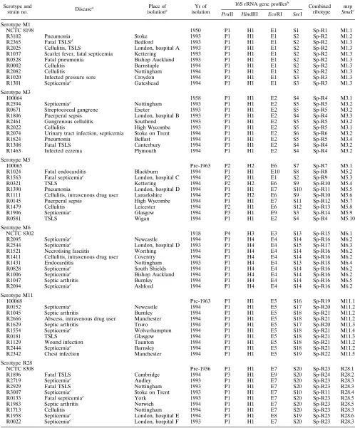
TABLE 1. Epidemiology and genotype markers of S. pyogenes strains
Serotype and
strain no. Disease
a Place of
isolationa isolationYr of
16S rRNA gene profilesb
Combined ribotype
mrp
SmaIc PvuII HindIII EcoRI SacI
Serotype M1
NCTC 8198 1950 P1 H1 E1 S1 Sp-R1 M1.1
R3102 Pneumonia Stoke 1993 P1 H1 E1 S2 Sp-R2 M1.2
R2365 Fatal TSLSd
Bedford 1993 P1 H1 E1 S2 Sp-R2 M1.3
R2025 Cellulitis, TSLS London, hospital A 1993 P1 H1 E1 S2 Sp-R2 M1.3
R1037 Scarlet fever, fatal septicemia Kettering 1993 P1 H1 E1 S2 Sp-R2 M1.3
R0528 Fatal pneumonia Bishop Auckland 1993 P1 H1 E1 S2 Sp-R2 M1.3
R0002 Cellulitis Barnstaple 1994 P1 H1 E1 S2 Sp-R2 M1.3
R2082 Cellulitis Nottingham 1994 P1 H1 E1 S2 Sp-R2 M1.3
R1020 Infected pressure sore Croydon 1994 P1 H1 E1 S3 Sp-R3 M1.3
R1301 Septicemiae Gateshead 1994 P1 H1 E1 S3 Sp-R3 M1.3
Serotype M3
100064 1958 P1 H1 E2 S4 Sp-R4 M3.1
R2594 Septicemiae Nottingham 1993 P1 H1 E2 S5 Sp-R5 M3.2
R0671 Streptococcal gangrene Exeter 1993 P1 H1 E2 S5 Sp-R5 M3.2
R1806 Puerperal sepsis London, hospital B 1993 P1 H1 E2 S4 Sp-R4 M3.3
R2461 Gangrenous cellulitis Southend 1993 P1 H1 E2 S5 Sp-R5 M3.2
R2022 Cellulitis High Wycombe 1993 P1 H1 E2 S5 Sp-R5 M3.1
R2074 Urinary tract infection, septicemia Stoke on Trent 1994 P1 H1 E2 S6 Sp-R6 M3.2
R1824 Pneumonia Belfast 1994 P1 H1 E2 S5 Sp-R5 M3.4
R1308 Fatal TSLS Canterbury 1994 P1 H1 E2 S4 Sp-R4 M3.2
R1463 Infected eczema Plymouth 1994 P1 H1 E2 S4 Sp-R4 M3.2
Serotype M5
100065 Pre-1963 P2 H2 E6 S7 Sp-R7 M5.1
R1024 Fatal endocarditis Blackburn 1994 P1 H1 E10 S8 Sp-R8 M5.2
R1563 Fatal septicemiae London, hospital C 1994 P2 H1 E1 S2 Sp-R9 M5.3
R0321 TSLS Kettering 1994 P2 H2 E6 S9 Sp-R10 M5.4
R1390 Pneumonia London, hospital D 1994 P1 H1 E7 S10 Sp-R11 M5.5
R1111 Cellulitis, intravenous drug user Lanarkshire 1994 P2 H2 E6 S9 Sp-R10 M5.6
R0145 Puerperal sepsis High Wycombe 1994 P1 H1 E7 S11 Sp-R12 M5.7
R1479 Cellulitis Leicester 1994 P2 H1 E6 S12 Sp-R13 M5.8
R1906 Septicemiae Glasgow 1994 P3 H1 E9 S3 Sp-R14 M5.9
R0581 TSLS Wigan 1994 P1 H1 E2 S4 Sp-R4 M5.10
Serotype M6
NCTC 8302 1918 P4 H3 E3 S13 Sp-R15 M6.1
R2095 Septicemiae Newcastle 1994 P1 H4 E4 S14 Sp-R16 M6.2
R2544 Septicemiae London, hospital D 1993 P1 H4 E4 S15 Sp-R17 M6.3
R1521 Necrotising fasciitis Worthing 1994 P1 H4 E4 S14 Sp-R16 M6.2
R1411 Cellulitis, intravenous drug user Coventry 1994 P1 H4 E4 S14 Sp-R16 M6.2
R1431 Endocarditis Nottingham 1993 P1 H4 E4 S13 Sp-R18 M6.4
R0828 Septicemiae South Shields 1994 P1 H4 E4 S14 Sp-R16 M6.2
R1006 Septicemiae Bishop Auckland 1994 P1 H4 E4 S14 Sp-R16 M6.2
R1047 Septic arthritis Burnley 1994 P1 H4 E4 S14 Sp-R16 M6.5
R2094 Septicemiae Ashford 1994 P1 H4 E4 S14 Sp-R16 M6.2
Serotype M11
100068 Pre-1963 P1 H1 E5 S16 Sp-R19 M11.1
R0152 Septicemiae
Newcastle 1994 P1 H1 E5 S17 Sp-R20 M11.2
R1045 Septic arthritis Burnley 1994 P1 H1 E5 S18 Sp-R21 M11.2
R2666 Abscess, intravenous drug user Manchester 1994 P1 H1 E5 S18 Sp-R21 M11.2
R1629 Septic arthritis Truro 1994 P1 H1 E5 S17 Sp-R20 M11.3
R1554 Septicemiae Wolverhampton 1994 P1 H1 E5 S18 Sp-R21 M11.4
R0181 TSLS Glasgow 1994 P1 H1 E5 S18 Sp-R21 M11.3
R1129 Wound infection Taunton 1994 P1 H1 E5 S18 Sp-R21 M11.2
R2444 Septicemiae Barnsley 1994 P1 H1 E5 S18 Sp-R21 M11.2
R2342 Chest infection Manchester 1994 P1 H1 E5 S19 Sp-R22 M11.5
Serotype R28
NCTC 8308 Pre-1936 P1 H1 E7 S20 Sp-R23 R28.1
R1896 Fatal TSLS Cambridge 1994 P3 H1 E9 S20 Sp-R24 R28.2
R2719 Septicemiae
Audley 1993 P1 H1 E7 S20 Sp-R23 R28.3
R2929 Fatal TSLS Nottingham 1993 P1 H1 E7 S20 Sp-R23 R28.3
R3007 Septicemiae Stoke on Trent 1993 P1 H1 E7 S10 Sp-R11 R28.4
R0133 Fatal septicemiae
York 1993 P1 H1 E7 S20 Sp-R23 R28.5
R1983 Septic arthritis Norwich 1994 P1 H1 E7 S20 Sp-R23 R28.5
R1713 Cellulitis Nottingham 1994 P1 H1 E7 S20 Sp-R23 R28.3
R1958 Septicemiae
London, hospital E 1994 P1 H1 E8 S19 Sp-R25 R28.6
R0022 Septicemiae London, hospital F 1993 P1 H1 E7 S20 Sp-R23 R28.3
aAll strains were isolated from blood cultures in the United Kingdom. Data are not applicable to reference strains.
bArabic numbers following the enzyme code (P, PvuII; H, HindIII; E, EcoRI; S, SacI) indicate different patterns of hybridization with the probe. cMacrorestriction profile with SmaI.
dTSLS, streptococcal toxic shock-like syndrome. ePortal of entry unknown.
on May 15, 2020 by guest
http://jcm.asm.org/
for serotype R28 (0.38) and highest for serotype M5 (0.98). The D value for SacI for all six serotypes together was 0.95. Combined ribotypes, designated Sp-R1 to Sp-R25, were de-rived by combining the 16S gene polymorphisms with all four enzymes (Table 1).
Macrorestriction and PFGE.Since the G1C content of S.
pyogenes is low (35 to 39 mol%), the following endonucleases
whose restriction sites are GC-rich were evaluated for macro-restriction: ApaI, BglI, BssHII, EagI, KpnI, NaeI, NgoMI, Ngo AIV, SacII, SalI, SfiI, SmaI, and SstII. Of these, SmaI, which generated 10 to 13 fragments between 40 and 500 kbp in size, yielded macrorestriction patterns (mrps) which were consid-ered to be the most suitable and discriminatory. A protocol was modified for the preparation of DNA from S. pyogenes (see Materials and Methods) and gave 100% typeability for all strains in the study. Differential pulse times were used to improve the resolution of fragments between 40 and 150 kbp in size which were found to be characteristic of serotypes M6 and M11 (Fig. 3B).
The discriminatory power of macrorestriction and PFGE was considerable. The six type strains (other than that of se-rotype M3) had unique mrps (Fig. 3A, lane 2; Fig. 3B, lanes 2 and 8; Fig. 3C, lane 2; Fig. 3D, lane 2). The least discrimination (D50.37) was found within serotype M1, within which 8 of 10 clinical isolates shared mrp M1.3 (Fig. 3A, lane 4). The great-est discrimination was found within serotype M5, in which each isolate had a unique mrp (Fig. 3C, lanes 2 through 11). Sero-types M1, M3, M6, and M11 were each found to contain characteristic sets of mrps, within which only minor band vari-ations were found (Fig. 3A and B). In each case, one of these mrps, e.g., M1.3, M3.2, M6.2, or M11.2, was the mrp for half or more of the strains of that serotype. D values within M6 and M11 were 0.66 and 0.76, respectively. Three of the six R28 mrps (Fig. 3D, lanes 2 through 4) were related and contained 7 of 10 strains, while three unrelated R28 mrps (Fig. 3D, lanes 5 through 7) were found in single strains only, yielding a D value of 0.64 within this serotype. None of the 10 M5 mrps had
any obvious relationship to those of the other M5 strains, yielding the maximal D value of 1.0 within this serotype. These mrps were also unique in the study as a whole.
DISCUSSION
Recent evidence is suggestive of a global upsurge in the number of cases of invasive disease caused by group A strep-tococci, often with fatal outcomes (11, 12, 25). It is imperative for infection control in the community and in hospitals to establish discriminatory subtyping methods which reproducibly subdivide common M serotypes. Such molecular methods should be cross-referenced to classical (phenotypic) M typing, but they should also be capable of standing alone for the comparison of data between laboratories concerning defined epidemiological situations.
Protocols for the preparation of DNA from group A strep-tococci commonly use liquid cultures, whose handling may be hazardous. The method reported here for the rapid extraction of genomic DNA from overnight agar plate cultures gave re-producible cell lysis, with a good yield of high-quality genomic DNA. Such minipreparative methods offer significant advan-tages for molecular epidemiological studies.
[image:4.612.129.484.71.308.2]The discriminatory power of 16S ribotyping was seen to depend on the choice of restriction enzyme used, and three levels of discrimination were evident, with serotype M5 being an exception to this generalization. Although their use was a feature of earlier studies (3, 21), the least discriminatory en-zymes were HindIII or PvuII: these enen-zymes generated poly-morphisms which either overlapped several M serotypes or were at best only moderately discriminatory within certain serotypes (such as M5 or M6). The second level, that of type-specific ribotypes, was found with EcoRI: each M sero-type except serosero-type M5 had a distinct set of E ribosero-types; for the reasons considered below, serotype M5 was exceptional. At the third level, 16S ribotyping could also be used to subtype isolates within an M serotype. Here, SacI was the only enzyme
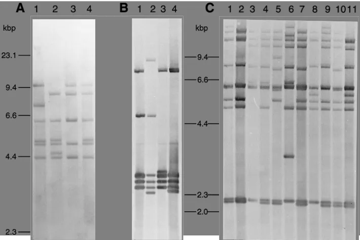
FIG. 2. The 16S ribotypes of S. pyogenes. Genomic Southern blots were probed with an intragenic 16S rRNA gene probe as described in Materials and Methods. (A) HindIII ribotypes. Lanes 1 through 4, ribotypes H1 to H4, respectively. (B) PvuII ribotypes. Lanes 1 through 4, ribotypes P1 through P4, respectively. (C) EcoRI ribotypes. Lanes 1 through 9, ribotypes E1 through E9, respectively; lane 10, ribotype E7; lane 11, ribotype E10.
on May 15, 2020 by guest
http://jcm.asm.org/
to yield intraserotype D values (varying from 0.38 for R28 to 0.98 for M5). Our results parallel those of Bruneau et al. (4), who recently reported the use of this enzyme for ribotyping pharyngeal isolates. We noted that the large molecular sizes of the SacI bands are consistent with the sites concerned being distant from the rRNA operons themselves, and also that their sizes led to difficulty with resolution by standard agarose gel electrophoresis. Intraserotype D values were 0.51 (within se-rotype M1), 0.53 (within sese-rotypes M6 and R28), 0.64 (within serotypes M3 and M11), and 0.98 (within serotype M5). They did not differ between combined ribotyping analysis with all four enzymes or with the two most discriminatory ones. Hence,
EcoRI and SacI provide an adequate basis for 16S ribotyping
of S. pyogenes isolates.
Comparison of M serotypes with 16S ribotypes or with mrps indicated that various degrees of genetic heterogeneity existed within the six serotypes studied. The two methods gave parallel results. Serotype M1 exhibited the least heterogeneity (three combined ribotypes and three mrps). NCTC 8198, the type strain of S. pyogenes and the reference strain for M1, was distinct in both combined ribotype and mrp. Our results thus concur with those of earlier studies (5, 21), which attributed
clonality to recent isolates of this serotype, which has often been associated with invasive disease. Serotypes M3, M6, and M11 exhibited somewhat more heterogeneity than serotype M1 (three to four combined ribotypes or four to five mrps), but the single-band-shift differences in mrps again suggest a com-mon ancestry of isolates within these serotypes. The signifi-cance of minor mrp band shifts is still a matter of interpreta-tion. Maslow et al. (15) consider strains with one or two mrp band shifts consistent with a single genetic event to be clonally related. Nonetheless Baquar et al. (1) showed that outbreak and background strains of Salmonella could be reproducibly distinguished by a single mrp band shift, which was stable over many years. Our interpretation of the large numbers of com-mon bands within a serotype (Fig. 3A and B) is that M3, M6, and M11 resemble M1 in having a clonal basis.
[image:5.612.129.482.72.443.2]Serotype R28, for which four combined ribotypes were found, was distinctive in that 6 of 10 strains appeared to be clonally related by their mrps (Fig. 3D, lanes 2 to 4), while three unrelated mrps were found among the other four strains. Serotype M5 was the most heterogeneous in the study. The 10 strains could be subdivided into nine combined ribotypes or 10 mrps. The latter appeared to be distantly related. These results
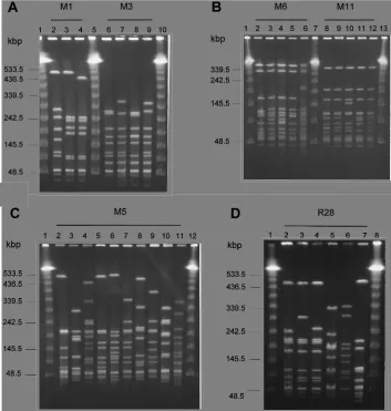
FIG. 3. Macrorestriction profiles of S. pyogenes resolved by PFGE. (A) Lanes 1, 5, and 10, molecular mass markers (concatamers of phage lambda); lanes 2 through 4, mrps M1.1 through M1.3, respectively; lanes 6 through 9, mrps M3.1 through M3.4, respectively. (B) Lanes 1, 7, and 13, molecular mass markers; lanes 2 through 6, mrps M6.1 through M6.5, respectively; lanes 8 through 12, mrps M11.1 through M11.5, respectively. (C) Lanes 1 and 12, molecular mass markers; lanes 2 through 11, mrps M5.1 through M5.10, respectively. (D) Lanes 1 and 8, molecular mass markers; lanes 2 through 7, mrps R28.1 through R28.6, respectively.
on May 15, 2020 by guest
http://jcm.asm.org/
are consistent with polyphyletic origins for serotypes M5 and R28.
The emm gene cluster lies within a chromosomal virulence regulon consisting of one to three tandemly arranged and related genes, which have been categorized into four subfam-ilies according to sequence divergences at their 39ends. The evolution of emm gene diversity is thought to involve intra-genic recombination and gene duplication, but evidence that the horizontal spread of emm genes could lead to homologous recombination within the cluster has also been provided (2). The vehicle for this is unknown, but it might be a plasmid, conjugative transposon, or bacteriophage. The unusual diver-sity of our M5 isolates suggests that the horizontal spread of an
emm gene among genetically diverse clones might account for
the heterogeneity observed within isolates of that serotype. This deserves to be further studied. At least one emm-like gene from an M5 strain has been shown to have a mosaic structure consisting of segments from emm-like genes of different strains (26).
In summary, we have described here methods which can be used for high-resolution genotyping of S. pyogenes isolates and have delineated a scheme for subtyping isolates within the M serotypes. The combination of ribotypes with mrps could dis-tinguish four genotypes within 10 M1 strains, five genotypes within M6 strains, six genotypes within M11 and R28 strains, eight genotypes within M3 strains, and one genotype in each of the 10 M5 strains. Such data provide a framework for molec-ular epidemiological studies as well as tools for investigating the evolutionary genetics of these important pathogenic bacteria.
ACKNOWLEDGMENT
We thank Asha Tanna for assistance with the selection and sero-typing of the strains.
REFERENCES
1. Baquar, N., A. Burnens, and J. Stanley. 1994. Comparative evaluation of molecular typing of strains from a national epidemic due to Salmonella
brandenburg by rRNA gene and IS200 probes and pulsed-field gel
electro-phoresis. J. Clin. Microbiol. 32:1876–1880.
2. Bessen, D. E., and S. K. Hollingshead. 1994. Allelic polymorphism of emm loci provides evidence for horizontal gene spread in group A streptococci. Proc. Natl. Acad. Sci. USA 91:3280–3284.
3. Bingen, E., E. Denamer, N. Lambert-Zechovsky, C. Boissinot, N. Brahimi, Y.
Aujard, P. Blot, and J. Elion.1992. Mother-to-infant vertical transmission and cross colonisation of Streptococcus pyogenes confirmed by DNA restric-tion fragment length polymorphism analysis. J. Infect. Dis. 165:147–150. 4. Bruneau, S., H. de Montclos, E. Dronet, and G. A. Denoyel. 1994. rRNA
gene restriction patterns of Streptococcus pyogenes: epidemiological applica-tions and relation to serotypes. J. Clin. Microbiol. 32:2953–2958. 5. Cleary, P. P., E. L. Kaplan, J. P. Handley, A. Wlazlo, M. H. Kim, A. R.
Hauser, and P. M. Schlievert.1992. Clonal basis for resurgence of serious
Streptococcus pyogenes disease in the 1980s. Lancet 339:518–521.
6. Cleary, P. P., E. L. Kaplan, C. Livdahl, and S. Skjold. 1988. DNA finger-prints of Streptococcus pyogenes are M type specific. J. Infect. Dis. 158:1317– 1323.
7. Colman, G. 1990. Steptococcus & Lactobacillus, p. 120–159. In M. T. Parker and B. I. Duerden (ed.), Topley & Wilson’s principles of bacteriology, 8th ed., vol. 2. Systematic bacteriology. E. Arnold, London.
8. Efstratiou, A., G. Colman, D. Cowley, and D. Pitcher. 1992. Ribotyping of human pyogenic streptococci. New perspectives on streptococci and strep-tococcal infections. Zentrabl. Bakteriol. Parasitenkd. Infektionskr. Hyg. Abt. 1 Orig. Suppl. 22:90–91.
9. Efstratiou, A., and R. C. George. 1994. Invasive group A streptococcal infections. J. Public Health Med. 1995 17:110–115.
10. Efstratiou, A., M. D. Stoyles, A. Tanna, and R. C. George. 1995.
Streptococ-cus pyogenes: serotype distribution and incidence of systemic disease within
the UK 1991–1994, abstr. D-138, p. 273. In Abstracts of the 95th General Meeting of the American Society for Microbiology 1995. American Society for Microbiology, Washington, D.C.
11. Haase, A. M., A. Melder, J. D. Mathews, D. J. Kemp, and M. Adams. 1994. Clonal diversity of Streptococcus pyogenes within some M-types revealed by multilocus enzyme electrophoresis. Epidemiol. Infect. 113:455–462. 12. Hoge, C. W., B. Schwartz, D. F. Talkington, R. F. Breiman, E. M. MacNeill,
and S. J. Englender.1993. The epidemiology of invasive group A strepto-coccal infections and the emergence of streptostrepto-coccal toxic shock-like syn-drome. JAMA 269:384–389.
13. Hunter, P. H., and M. A. Gaston. 1988. Numerical index of the discrimina-tory ability of typing systems: an application of Simpson’s index of diversity. J. Clin. Microbiol. 26:2465–2466.
14. Martin, D. R., and L. A. Single. 1993. Molecular epidemiology of group A
Streptococcus type 1 infections. J. Infect. Dis. 167:1112–1117.
15. Maslow, J. N., M. E. Mulligan, and R. D. Arbeit. 1993. Molecular epidemi-ology: application of contemporary techniques to the typing of microorgan-isms. Clin. Infect. Dis. 17:153–164.
16. Musser, J. M., B. M. Gray, P. M. Schlievert, and M. E. Pichichero. 1992.
Streptococcus pyogenes pharyngitis: characterization of strains by multilocus
enzyme genotype, M and T protein serotype, and pyrogenic exotoxin gene probing. J. Clin. Microbiol. 30:600–603.
17. Musser, J. M., A. R. Hauser, M. H. Kim, P. M. Schlievert, K. Nelson, and
R. K. Selander.1991. Streptococcus pyogenes causing toxic-shock-like syn-drome and other invasive diseases: clonal diversity and pyrogenic exotoxin expression. Proc. Natl. Acad. Sci. USA 88:2668–2672.
18. Owen, R. J., K. Sutherland, C. Fitzgerald, J. Gibson, P. Borman, and J.
Stanley.1995. Molecular subtyping scheme for serotypes HS1 and HS4 of
Campylobacter jejuni. J. Clin. Microbiol. 33:872–877.
19. Ross, P. 1990. Streptococcal diseases, p. 239–262. In G. R. Smith and C. S. F. Easmon (ed.), Topley & Wilson’s principles of bacteriology, 8th ed., vol. 3. Bacterial diseases. E. Arnold, London.
20. Sambrook, J., E. F. Fritsch, and T. Maniatis. 1989. Molecular cloning: a laboratory manual, 2nd ed. Cold Spring Harbor Laboratory Press, Cold Spring Harbor, N.Y.
21. Seppa¨la¨, H., J. Vuopio-Varkila, M. O¨ sterblad, M. Jakkola, M. Rummu-kainen, S. E. Holm, and P. Huovinen.1993. Evaluation of methods for epidemiologic typing of group A streptococci. J. Infect. Dis. 169:519–525. 22. Single, L. A., and D. R. Martin. 1992. Clonal differences within M-types of
the group A Streptococcus revealed by pulsed field gel electrophoresis. FEMS Microbiol. Lett. 91:85–90.
23. Sneath, P. H. A., N. S. Mair, and M. E. Sharpe (ed.). 1986. Bergey’s manual of systematic bacteriology, vol. 2. The Williams & Wilkins Co., Baltimore. 24. Stevens, D. L. 1992. Invasive group A streptococcus infections. Clin. Infect.
Dis. 14:2–11.
25. Stromberg, A., V. Romanus, and L. G. Burman. 1991. Outbreak of group A streptococcal bacteraemia in Sweden; an epidemiologic and clinical study. J. Infect. Dis. 164:595–598.
26. Whatmore, A. M., and M. A. Kehoe. 1994. Horizontal gene transfer in the evolution of group A streptococcal emm-like genes: gene mosaics and vari-ation in Vir regulons. Mol. Microbiol. 11:363–374.
27. Wilson, K. 1987. Preparation of genomic DNA from bacteria, unit 2.4.1. Current protocols in molecular biology. John Wiley & Sons, Inc., New York.