Hypotension and reduced nitric oxide-elicited
vasorelaxation in transgenic mice
overexpressing endothelial nitric oxide
synthase.
Y Ohashi, … , Y Yazaki, M Yokoyama
J Clin Invest.
1998;
102(12)
:2061-2071.
https://doi.org/10.1172/JCI4394
.
Nitric oxide (NO), constitutively produced by endothelial nitric oxide synthase (eNOS), plays
a major role in the regulation of blood pressure and vascular tone. We generated transgenic
mice overexpressing bovine eNOS in the vascular wall using murine preproendothelin-1
promoter. In transgenic lineages with three to eight transgene copies, bovine eNOS-specific
mRNA, protein expression in the particulate fractions, and calcium-dependent NOS activity
were confirmed by RNase protection assay, immunoblotting, and L-arginine/citrulline
conversion. Immunohistochemical studies revealed that eNOS protein was predominantly
localized in the endothelial cells of aorta, heart, and lung. Blood pressure was significantly
lower in eNOS-overexpressing mice than in control littermates. In the transgenic aorta,
basal NO release (estimated by Nomega-nitro-L-arginine-induced facilitation of the
contraction by prostaglandin F2alpha) and basal cGMP levels (measured by enzyme
immunoassay) were significantly increased. In contrast, relaxations of transgenic aorta in
response to acetylcholine and sodium nitroprusside were significantly attenuated, and the
reduced vascular reactivity was associated with reduced response of cGMP elevation to
these agents as compared with control aortas. Thus, our novel mouse model of chronic
eNOS overexpression demonstrates that, in addition to the essential role of eNOS in blood
pressure regulation, tonic NO release by eNOS in the endothelium induces the reduced
vascular reactivity to NO-mediated vasodilators, providing several insights into the
pathogenesis of nitrate tolerance.
Research Article
Find the latest version:
J. Clin. Invest.
© The American Society for Clinical Investigation, Inc. 0021-9738/98/12/2061/11 $2.00
Volume 102, Number 12, December 1998, 2061–2071 http://www.jci.org
Hypotension and Reduced Nitric Oxide–elicited Vasorelaxation in Transgenic
Mice Overexpressing Endothelial Nitric Oxide Synthase
Yoshitaka Ohashi,* Seinosuke Kawashima,* Ken-ichi Hirata,* Tomoya Yamashita,* Tatsuro Ishida,* Nobutaka Inoue,* Tsuyoshi Sakoda,* Hiroki Kurihara,‡ Yoshio Yazaki,‡ and Mitsuhiro Yokoyama*
*The First Department of Internal Medicine, Kobe University School of Medicine, Kobe, Japan; and ‡Department of Cardiovascular Medicine, Graduate School of Medicine, University of Tokyo, Tokyo, Japan
Abstract
Nitric oxide (NO), constitutively produced by endothelial
nitric oxide synthase (eNOS), plays a major role in the
regu-lation of blood pressure and vascular tone. We generated
transgenic mice overexpressing bovine eNOS in the vascular
wall using murine preproendothelin-1 promoter. In
trans-genic lineages with three to eight transgene copies, bovine
eNOS-specific mRNA, protein expression in the particulate
fractions, and calcium-dependent NOS activity were
con-firmed by RNase protection assay, immunoblotting, and
L
-arginine/citrulline conversion. Immunohistochemical
stud-ies revealed that eNOS protein was predominantly localized
in the endothelial cells of aorta, heart, and lung. Blood
pres-sure was significantly lower in eNOS-overexpressing mice
than in control littermates. In the transgenic aorta, basal
NO release (estimated by
N
v-nitro-
L-arginine–induced
facil-itation of the contraction by prostaglandin F
2a) and basal
cGMP levels (measured by enzyme immunoassay) were
sig-nificantly increased. In contrast, relaxations of transgenic
aorta in response to acetylcholine and sodium nitroprusside
were significantly attenuated, and the reduced vascular
re-activity was associated with reduced response of cGMP
ele-vation to these agents as compared with control aortas.
Thus, our novel mouse model of chronic eNOS
overexpres-sion demonstrates that, in addition to the essential role of
eNOS in blood pressure regulation, tonic NO release by eNOS
in the endothelium induces the reduced vascular reactivity
to NO-mediated vasodilators, providing several insights
into the pathogenesis of nitrate tolerance. (
J. Clin. Invest.
1998. 102:2061–2071.) Key words: transgenic animals
•nitric
oxide
•endothelium
•blood pressure
•vascular reactivity
Introduction
Nitric oxide (NO),1 which is produced by endothelial cells,
serves as an endothelium-derived relaxing factor which
medi-ates vascular relaxation in response to vasoactive substances and shear stress (1). Several lines of evidence point to the key role of NO in the regulation of blood pressure and blood flow, and NO acts as an antithrombogenic and antiatherogenic molecule by inhibiting vascular smooth muscle proliferation, platelet aggregation, and leukocyte adhesion (2). Abnormali-ties of endothelial production or metabolism of NO occur in pathological conditions such as atherosclerosis, diabetes, and
hypertension (3). NO is produced from L-arginine and oxygen
by three nitric oxide synthase (NOS) isoforms: two constitu-tive forms, i.e., neuronal NOS (nNOS, NOS1) and endothelial NOS (eNOS, NOS3), and inducible NOS (iNOS, NOS2) (4). NO produced by these NOS isoforms, which are expressed in specific cell types, has been thought to exert distinct actions in various physiological functions and pathological processes. A wide variety of actions of NO has been elucidated in in vitro and in vivo experiments using NOS inhibitors and NO donors (1, 5). However, such pharmacological modulations of NO production affect multiple NOS isoforms, making it difficult to assess the specific roles of each isoform.
Recent advances in molecular biology and genetic engi-neering have enabled us to modulate the expression of a single NOS isoform and to identify the in vivo roles of the specific NOS isoform in complex physiological systems. Experiments involving hypertensive mice lacking the eNOS gene (6, 7) and eNOS gene transfer to normal or injured vessels (8, 9) have demonstrated that NO produced by eNOS plays a pivotal role in the regulation of blood pressure, vascular tone, and vascular homeostasis. In contrast to gene transfer for acute therapeutic benefits, the chronic actions of lifelong overexpression of eNOS in endothelial cells are still unknown. Thus, we gener-ated transgenic mice overexpressing eNOS and determined their phenotypic changes.
Methods
Materials.Nv-nitro-L-arginine methyl ester (L-NAME), Nv-nitro-L -argi-nine (L-NA), NADPH, flavin ade-argi-nine dinucleotide, calmodulin, ni-trate reductase (Aspergillus species), PGF2a, acetylcholine chloride
(ACh), ATPgS, sodium nitroprusside (SNP), forskolin, and N6,29
-O-dibutyryladenosine 39,59-cyclic monophosphate (dibutyryl cAMP) were purchased from Sigma Chemical Co. (St. Louis, MO). Nitro-glycerin (NTG) was obtained from Nippon Kayaku (Tokyo, Japan). EGTA was purchased from Dojindo Laboratories (Kumamoto, Ja-pan). A rabbit polyclonal anti–human eNOS Ab was obtained from Address correspondence to Mitsuhiro Yokoyama, M.D., The First
Department of Internal Medicine, Kobe University School of Medi-cine, 7-5-1 Kusunoki-cho, Chuo-ku, Kobe 650, Japan. Phone: 81-78-341-7451, ext. 5501; FAX: 81-78-341-1390; E-mail: yokoyama@med. kobe-u.ac.jp
Received for publication 24 June 1998 and accepted in revised form 22 October 1998.
Transduction Laboratories (Lexington, KY). This Ab has been proven to cross-react with mouse and bovine eNOS (10) but not to recognize inducible NOS. All other reagents used were of the highest purity commercially available. All animal experiments were con-ducted according to the Guidelines for Animal Experimentation at Kobe University School of Medicine (approval number P950430).
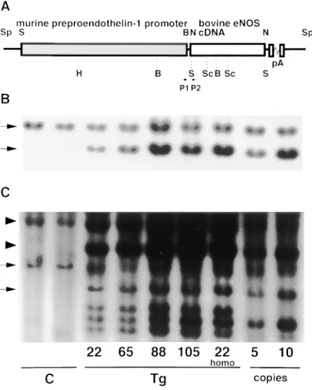
Plasmid construction. To target eNOS gene expression to the vascular wall, we decided to use murine preproendothelin-1 (pre-proET-1) promoter (GenBank accession number U07982) as de-scribed (11). Bovine eNOS cDNA (GenBank accession number M99057, 4.1 kb) (12) was excised from pBluescriptII SK(1) (Strat-agene, La Jolla, CA) plasmids (a generous gift from D.G. Harrison, Emory University, Atlanta, GA) by NotI digestion at the EcoRI linker (EcoRI-NotI-SalI) ligated to both ends of the eNOS cDNA. Plasmids, designated as PEP8, comprised pCDM8 backbone (Invitro-gen, San Diego, CA), the 9.2-kb murine preproET-1 59 flanking pro-moter, single NotI site, and SV40 intron polyA signal (13). Bovine eNOS cDNA was inserted into PEP8 at the NotI site downstream from the preproET-1 promoter. Orientation of the cDNA insert was confirmed by restriction enzyme digestion and the cloning junction was sequenced by the dideoxy termination method (Sequenase Ver-sion II sequencing kit; United States Biochemical, Cleveland, OH). Standard cloning methodologies were used for all DNA manipula-tions. The resultant plasmids, designated as PEP-NOS, were double CsCl-purified and used for in vitro transfection and generation of eNOS transgenic mice.
Generation of transgenic mice. PEP-NOS (16.4 kb) was linear-ized by SpeI digestion, purified by 0.5% agarose gel electrophoresis, and electro-eluted from agarose using Gene Capsule (Geno Technol-ogy, St. Louis, MO). The eluted fragment was further purified and concentrated with an Elutip D column (Schleicher & Schuell, Dassel, Germany) and finally precipitated in ethanol. Purified DNA (2 ng/ ml) free of agarose was finally dissolved in 5 mM Tris/Cl (pH 7.5) and 0.1 mM EDTA before pronuclear microinjection. Fertilized eggs were prepared from superovulated BDF1 (B6D2F1; C57BL/6 3 DBA/2 F1) mice (Charles River Japan, Osaka, Japan). Microinjected embryos were then transferred into the oviducts of pseudopregnant Institute for Cancer Research foster mothers (Clea Japan, Osaka, Ja-pan) and allowed to develop to term. All procedures were performed using standard techniques (14) with microscopes and micromanipula-tors (Leica, Heerbrugg, Switzerland), a micropipette puller (Sutter Instrument, Novata, CA), and a microforge (Technical Products In-ternational, St. Louis, MO). The mice were maintained under con-trolled environmental conditions with respect to temperature (208C) and humidity (64%) on a 12-h light/dark cycle, and provided with standard chow and water ad libitum.
PCR and Southern blot analysis.Founder mice harboring the trans-gene were identified by PCR and subsequent Southern blot analysis of genomic DNA isolated from tail biopsies at 3–4 wk of age. Tail DNA was extracted by proteinase K (1.4 mg/ml; Boehringer Mann-heim, MannMann-heim, Germany) digestion and subsequent phenolization and purification using a Genomix DNA extraction kit (Talent, Tri-este, Italy). PCR detection was performed using transgene-specific oligonucleotide primers, i.e., sense primer at the 39 end of preproET-1 promoter (59 -GAAGTTAGCCGTGATTTCCTCTAGAGCCGG-GTC) and antisense primer at the 59 end of eNOS cDNA (59 -TTG-ATGAAGTCCCTGGCCTGGCTCAGCAG). In genomic Southern blot analysis for eNOS genotyping, tail DNA (15 mg) digested with HindIII and SalI endonucleases was electrophoresed in 0.5% aga-rose, transferred to nitrocellulose filters (Schleicher & Schuell) and hybridized at 428C with a random-primed, 32P-labeled SacI-digested
fragment (1.1 kb) of bovine eNOS cDNA, which contains calmodulin and flavin mononucleotide binding sites (12), or a whole transgene (16.4 kb). After washing, the hybridized filters were analyzed with a bio-imaging analyzer (Fujix BAS2000; Fuji Photo Film, Tokyo, Ja-pan). Transgene copy number was calculated using known amounts of the transgene added to nontransgenic mouse genomic DNA as a control.
Preparation of total RNA and ribonuclease protection assay.Total RNA was extracted from pooled tissues by the acid guanidinium thio-cyanate-phenol/chloroform method (15). To confirm specific expres-sion of bovine eNOS mRNA, we used a ribonuclease (RNase) pro-tection assay that distinguishes the transgene transcript from the endogenous mouse eNOS and other NOS isoform gene transcripts. The region, containing the NH2-terminal myristoylation site of
bo-vine eNOS cDNA (from 215 to 225), amplified with forward primer NP-1 (59-ATAGAATTCACCAGCACCTTTGGGAATGGCGAT) and reverse primer Cp-11 (59 -ATAGAATTCGGATTCACTGTC-TGTGTTGCTGGACTCCTT) and the 124-bp ApaI-AluI fragment of rat GAPDH cDNA, was subcloned, linearized, and in vitro tran-scribed with SP6 and T7 RNA polymerase (Promega, Madison, WI), as described (12, 16). RNase protection assay was performed with a PRAII kit (Ambion, Austin, TX). In brief, an aliquot of total RNA (10 mg) was hybridized with 32P-labeled riboprobes for 14–16 h at
508C in 80% formamide hybridization buffer, followed by digestion with ribonuclease A and ribonuclease T1 at 378C for 30 min. After si-multaneous precipitation of RNA and inactivation of ribonuclease, the protected fragments were separated in 8 M urea/5% acrylamide denaturing gel and the relative signal intensities were determined with the bio-image analyzer.
Immunoblotting of eNOS and Ca-dependent eNOS activity. Crude homogenates of pooled tissues in a homogenizing buffer of 50 mM Tris-HCl, pH 7.4, 1 mM EGTA, 1 mM DTT, 1 mM pepstatin A, 2 mM leupeptin, and 1 mM (p-amidinophenyl) methanesulfonyl fluoride were ultracentrifuged at 100,000 g to collect cytosolic fractions. The pellets were solubilized in the homogenizing buffer containing 10% glycerol and 20 mM 3-[(3-chol-amidopropyl)dimethylammonio]-1-propanesulfonate and ultracentrifuged to extract particulate frac-tions. Protein concentrations were determined by the method of Bradford (Bio-Rad Laboratories, Hercules, CA) with BSA fraction V as a standard protein (17). Immunoblotting was performed as de-scribed (16). In brief, 150 mg of protein samples, unless otherwise in-dicated, from either the cytosolic or particulate fraction was sepa-rated on a 7.5% SDS-polyacrylamide gel under reducing conditions, transferred to a polyvinylidene difluoride membrane (ATTO, Tokyo, Japan), and probed with a rabbit polyclonal (dilution, 1:500) or a mu-rine monoclonal anti–bovine eNOS Ab (clone H32, IgG2a, 1:5,000) (18). Immunoreactive bands were visualized with horseradish peroxi-dase–conjugated anti–rabbit Ig F(ab9)2 fragment or anti–mouse IgG
using an ECL detection kit (Amersham International plc, Bucking-hamshire, UK) and quantified by densitometry. NOS enzymatic ac-tivity was determined by the conversion of L-arginine to L-citrulline with saturating concentrations of substrate and cofactors as described (19). Enzyme activity was expressed as citrulline production in femto-moles per milligram of protein per minute. To test the specificity of calcium-dependent NOS, parallel reactions were performed in a sep-arate buffer omitting calcium and calmodulin or containing 1 mM L-NAME, a competitive NOS inhibitor.
Immunohistochemistry. To determine the tissue distribution of bovine eNOS expression, immunohistochemical staining was per-formed by the labeled streptavidin biotin (LSAB) method using an LSAB kit (DAKOPATTS, Copenhagen, Denmark) as described (20). Frozen sections were fixed in acetone, blocked with 10% BSA, and incubated with 1:50 dilution of the primary rabbit polyclonal anti-eNOS Ab. We used the polyclonal Ab because murine mAb could not be applied to mouse tissues. The sections were then incubated with biotinylated gout anti–rabbit IgG and subsequently with horse-radish peroxidase–labeled streptavidin. Endogenous peroxidase was quenched with H2O2 and the bound primary Ab was detected with
the substrate, diaminobenzidine. Specificity of staining was assessed by substitution of nonimmune serum for primary Ab.
measurement of arterial pressure and heart rate under conscious and unrestrained conditions, the femoral catheter was connected to a sa-line-filled pressure transducer (Bioresearch Center, Nagoya, Japan) using a free-moving cannulation system (Tsumura, Tokyo, Japan) and the signals were amplified and continuously monitored (13). At least 4 h after recovery from anesthesia, physiological parameters (systolic, diastolic, and mean blood pressures and heart rate) were ac-cumulated on a Macintosh computer with MacLab systems (Biore-search Center) every 5 s for 2 h. In another experiment to study the effects of chronic NOS inhibition on systemic blood pressure, L-NAME (1 mg/ml) was given in drinking water for 2 wk to both control litter-mates and eNOS-overexpressing mice.
Basal release of NO from the aorta and vascular reactivity to en-dothelium-dependent and -independent vasodilators.The aortas with intact endothelium from either control or eNOS transgenic mice were dissected and cleaned of adhering tissues. Isometric tension was re-corded as described (21). In brief, 3-mm-wide transverse aortic rings were mounted under 1.5 g resting tension on stainless steel hooks at-tached to force transducers (Nihon Kohden, Tokyo, Japan) in 30-ml organ chambers containing Krebs’ bicarbonate solution (pH 7.4) of the following composition (mM): NaCl 118, KCl 4.0, CaCl2 1.5,
MgSO4 1.2, NaH2PO4 1.2, NaHCO3 25, and glucose 5, and
equili-brated at 378C with a 95% O2/5% CO2 gas mixture. Isometric tension
was displayed using an amplifier system (Nihon Kohden) and a pen recorder (Nippon Densi Kagaku, Kyoto, Japan). A test contraction was induced by 40 mM KCl. To elucidate the tone-related basal re-lease of NO, moderate vascular tone was induced by a low concentra-tion (500 nM) of PGF2a and the rings were subsequently contracted
by cumulative additions of L-NA (1–100 mM). Basal release of NO was indirectly estimated by L-NA–induced endothelium-dependent facilitation of the contraction elicited by PGF2a (22, 23). Next, to
as-sess the vascular reactivity to endothelium-dependent and -indepen-dent vasodilators, aortic rings were submaximally precontracted with 1–3 mM PGF2a to develop z 1.0 g tension. After the contraction
reached a plateau, vasodilatory agents (ACh, ATPgS, SNP, NTG, forskolin, and dibutyryl cAMP) were added in a cumulative manner. Almost all experiments were performed on paired rings from trans-genic mice and control littermates. Relaxations were expressed as the percentage of PGF2a-induced precontraction and a dose–response
curve was obtained for each agent. To address the specificity and re-versibility of NO-mediated changes in vascular reactivity, we mea-sured the isometric tension of aortas from chronic L-NAME–treated mice in the presence of L-NAME (100 mM).
Measurement of cGMP levels in the aorta and plasma nitrite and nitrate levels.Immediately after killing, aortas from either control or transgenic mice were homogenized in 6% TCA and centrifuged at 2,000 g. TCA in the supernatant fraction was extracted with diethyl ether and the samples were then lyophilized. In some experiments, longitudinally opened thoracic aortas were preincubated for 60 min in Krebs’ solution equilibrated at 378C with a 95% O2/5% CO2 gas
mixture and stimulated with ACh and SNP for 1 min, and the in-creases in cGMP levels in response to vasodilators were determined. cGMP was measured using an enzyme immunoassay (EIA) kit (Am-ersham) as described (24). Samples were resuspended, acetylated with triethylamine/acetic anhydride, and subjected to EIA. cGMP levels were expressed as picomoles per milligram of TCA-precipita-ble protein solubilized with 1 N NaOH. At the time of killing, heart, lung, and kidney weights (wet weight) were measured for each animal and blood samples were collected. Nitrite and nitrate levels in hep-arinized plasma were measured as nitrite by using the Griess reaction after enzymatic conversion by nitrate reductase (0.25 U/ml) as de-scribed (25).
Blood biochemistry and analyses of neurohumoral regulatory fac-tors. Blood urea nitrogen and plasma electrolyte concentrations were determined with a multiparameter autoanalyzer. To assess alterations of major neurohumoral factors regulating blood pressure, plasma re-nin activity was determined by radioimmunoassay of generated an-giotensin I. Plasma epinephrine and norepinephrine levels were
mea-sured by HPLC. Plasma ET-1 was extracted with a Seppak C-18 column (Waters, Milford, MA) and measured using a sandwich EIA kit (Wako Pure Chemicals, Osaka, Japan).
Statistics. Student’s t test for unpaired observations was used to determine the significance of differences between transgenic mice and control littermates. Statistical analysis for multiple comparisons was performed using one-way ANOVA with Bonferroni correction. All values are given as means6SE, and statistical significance was set at P , 0.05.
Results
In vitro transfection and generation of eNOS transgenic mice.
[image:4.612.321.549.234.519.2]HeLa cells (American Type Culture Collection, Rockville,
MD), which endogenously express ET-1 and have high trans-fection efficacy, were transfected by using lipofectamine re-agent (GIBCO BRL, Gaithersburg, MD) with the plasmids PEP-NOS. PEP-NOS–transfected cells exhibited a marked in-crease in calcium-dependent NOS activity, as compared with those transfected with control plasmids containing bovine
eNOS cDNA in reverse orientation (110.8614.2 compared
with 23.7619.1 fmol/mg protein/min in three independent
periments, data not shown), indicating that PEP-NOS ex-pressed an enzymatically active eNOS protein under the con-trol of the preproET-1 promoter. SpeI-linearized PEP-NOS was used as a transgene to generate mice overexpressing
bo-vine eNOS (Fig. 1 A). Four potential founder (F0) mice were
independently propagated to obtain offspring. Tail DNA from the transgenic founders (Nos. 22, 65, 88, 105) carrying multiple copies of transgenes was subjected to Southern blot analysis to
detect tandem copies of the transgenes. As shown in Fig. 1 B,
genomic DNA encoding endogenous mouse eNOS allele in tail DNA from nontransgenic control littermates yielded a
band of z 5 kb excised by HindIII and SalI, when probed with
the 32P-labeled SacI fragment of eNOS cDNA (6). In
trans-genic mice, besides the z 5-kb fragment of endogenous eNOS
gene, 4.1 kb of bovine eNOS cDNA excised from transgene by SalI was specifically hybridized with the probe. Transgene copy number calculated by densitometric analysis was three to eight. Southern blot analysis using whole transgene probe showed that, in addition to endogenous and transgene-derived
eNOS signals (z 5 and 4.1 kb, respectively), the 8-kb
frag-ments detected in both transgenic and control DNA repre-sented endogenous preproET-1 promoter excised by HindIII (13) and the 6-kb fragment of preproET-1 promoter excised from the transgene by HindIII and SalI was detected only in
tail DNA from transgenic mice (Fig. 1 C). Transgenic mice did
not differ from control littermates in general appearance. They normally developed and were fertile, and transgenic offspring
were obtained in Mendelian fashion. Since transgenic mice were prepared on a mixed genetic background, founders were subsequently backcrossed for at least three generations with the wild-type C57BL/6J strain of mice (Clea Japan) to ensure a genetically homogeneous background for the use in the studies described below. Offspring were weaned at 3 wk of age and screened by PCR of tail DNA, for which oligonucleotide prim-ers were selected so that the predicted amplification product
was a distinctive fragment (z 750 bp) created by the
juxtaposi-tion of the preproET-1 promoter and the bovine eNOS cDNA
(Fig. 1 A). In established colonies (lines 22, 65, 88, and 105),
mice that were heterozygous at the transgene locus were used in all analyses and nontransgenic littermates served as controls.
Expression of bovine eNOS mRNA and functional pro-tein. RNase protection assay to detect bovine eNOS-specific mRNA revealed the transgene expression in the heart, lung, aorta, and uterus, and albeit to a lesser extent in the brain, liver, kidney, and intestine of four transgenic lineages exam-ined (lines 22, 65, 88, and 105), whereas no transgene
expres-sion was detected in control littermates. Fig. 2 A shows
repre-sentative findings for line 22 and the similar pattern of mRNA expression was observed in other transgenic lineages. In the particulate fractions of heart, lung, and aorta of transgenic mice, increased expression of eNOS protein (135 kD) over en-dogenous mouse eNOS levels of control littermates was
de-tected by immunoblotting with polyclonal Ab (Fig. 2 B).
Im-munoblotting with the murine monoclonal anti–bovine eNOS Ab confirmed the transgene expression in the particulate frac-tions of these organs since this mAb did not cross-react with
mouse eNOS (Fig. 2 C). The calcium-dependent NOS activity
was markedly increased in the particulate fraction of aorta
from transgenic mice (11.066.6 vs. 86.2610.0 fmol citrulline/
mg/min, P , 0.05, for three independent determinations). In
the immunoblotting of the cytosol fraction with the polyclonal Ab, no immunoreactive band was detected. There was no
[image:5.612.56.388.506.745.2]nificant increase in NOS activity in the cytosol fraction and conversion to citrulline was significantly decreased on incuba-tion without calcium and calmodulin or with L-NAME (data not shown).
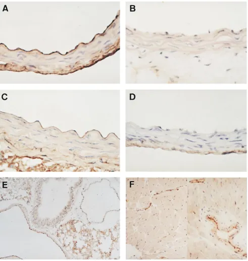
Immunohistochemistry. Immunohistochemical staining for bovine eNOS in transgenic mice (line 22) showed that the
in-creased immunoreactive product for bovine eNOS was pre-dominantly localized in the endothelial cell lining of thoracic
aorta from transgenic mice (Fig. 3 A). Endogenous mouse
[image:6.612.60.551.57.573.2]eNOS in endothelial cells of the aorta from control littermates also cross-reacted with polyclonal anti-eNOS Ab, the same Ab as used for immunoblotting. However, the intensity of eNOS
Figure 3. Immunohistochemical staining for eNOS in the aorta (A–D), lung (E), and heart (F). Frozen sections of thoracic aorta from transgenic mice (A) and control littermates (C) were immunohistochemically stained using a rabbit polyclonal anti-eNOS Ab. Immunoreactive product is predominantly observed in the endothelial cell lining and the immunoreactivity is obviously stronger in transgenic aorta (A) than in control aorta (C). In B and D, sections of transgenic and control aorta were stained with nonimmune serum to assure the specificity of reaction. In the lung (E) and heart (F) of Tg, intense signal is mainly detected in vascular endothelial cells of medium to microsized vessels. A–D, 3400; E and F,
immunoreactivity of transgenic endothelial cells was obviously
stronger than that of control endothelial cells (Fig. 3, A and C).
Sections of transgenic mice and control littermates stained with nonimmune serum were entirely negative for staining
(Fig. 3, B and D). In the lung, a marked increase in eNOS
ex-pression was also observed in the endothelial cell lining of large pulmonary arteries and veins, and weak nonvascular
ex-pression was detected in the airway epithelium (Fig. 3 E). As
depicted in Fig. 3 F, eNOS immunoreactivity was detected in
medium-sized to small coronary arteries, rather than cardiac myocytes in the heart. These findings represent the predomi-nant vascular targeting capacity of the preproET-1 promoter system (11).
Blood pressure and heart rate measurement.There is a gen-der difference in NO level which is possibly affected by estro-gen (21). Therefore, we used male mice to assess the physio-logical consequences of eNOS overexpression. As shown in
Fig. 4 A, systemic arterial pressure was significantly lower in
transgenic mice than in control littermates (systolic, 8964
mmHg in transgenic mice vs. 11163 mmHg in control
litter-mates, P , 0.01; diastolic, 7363 vs. 9263; P , 0.01; mean,
8164 vs. 9963, P , 0.01). L-NAME administration for 2 wk
elevated the mean blood pressure in transgenic mice to a level comparable to that in L-NAME–treated control littermates
(Fig. 4 B) and the blood pressure was lower in independent
transgenic lines (lines 65, 88, and 105) (data not shown). Ac-cordingly, eNOS overexpression was responsible for the hy-potension observed. Plasma nitrite and nitrate levels in trans-genic mice were significantly higher than those in control mice (Table I). There was no significant change in heart rate as compared with control littermates. There was neither differ-ence in body weight, organ weights, nor detectable macro-scopic and micromacro-scopic abnormalities in transgenic mice killed at 2–3 mo of age. Blood urea nitrogen and plasma electrolyte concentrations were not significantly altered in transgenic mice. There were no significant differences in plasma renin ac-tivity and plasma catecholamine and ET-1 levels between con-trol and transgenic mice (Table I).
Basal release of NO and cGMP levels. We next examined the effect of eNOS overexpression on basal tone of the aorta.
L-NA (1–100 mM) dose-dependently contracted the aortic
rings that had been moderately precontracted by PGF2a. Since
L-NA–induced facilitation of PGF2a-induced contraction was
not observed in the aortic rings treated with 100 mM L-NAME
before contraction by PGF2a and was abolished by the
addi-tion of L-arginine (data not shown), the facilitated contraction
[image:7.612.60.417.59.271.2]was ascribed to endothelium-derived basal NO produced by
[image:7.612.315.556.426.636.2]Figure 4. Systemic blood pressure analysis for conscious freely moving mice using a femoral arterial catheter. (A) A summary of systolic, diastolic, and mean blood pres-sures (mmHg) in control littermates (C, n 5 9) and heterozygous eNOS transgenic mice (Tg, line 22, n 5 10) is shown. (B) Ef-fects of chronic NOS inhibition by adminis-tration of L-NAME (1 mg/ml) in drinking water for 2 wk on mean blood pressure. Four animals each are used for C and Tg. Note that a significant reduction in blood pressure is observed in Tg, but is com-pletely reversed by chronic L-NAME treatment. Values are expressed as means6SEM. *P , 0.01, C vs. Tg.
Table I. Basal Characteristics of Physiological Parameters and Biochemical Data
Control (n) Transgenic (n)
Body wt (g) 26.261.1 (13) 26.060.7 (13) Heart wt (mg) 133.265.7 (13) 132.065.6 (13) Lung wt (mg) 224.3612.2 (13) 229.8615.7 (13) Kidney wt (mg) 194.668.2 (13) 191.268.5 (13) Mean blood pressure (mmHg) 9963 (9) 8164 (10)* Heart rate (bpm) 534626 (9) 588619 (10) Plasma nitrite 1 nitrate (mM) 19.463.3 (16) 31.663.1 (18)‡
Blood urea nitrogen (mg/ml) 23.861.8 (7) 24.861.0 (6) Plasma sodium (meq/liter) 148.361.4 (7) 148.261.0 (6) potassium (meq/liter) 5.460.7 (7) 5.760.7 (6) chloride (meq/liter) 113.061.4 (7) 113.561.6 (6) Urine volume (ml/100 g/d) 4.2761.55 (4) 4.2761.42 (4) Plasma renin activity (ng/ml/h) 33.0610.0 (11) 28.669.6 (8) Plasma epinephrine (pg/ml) 52236436 (5) 47086513 (5)
norepinephrine (pg/ml) 49376480 (5) 38746423 (5) Plasma ET-1 (pg/ml) 1.4660.14 (11) 1.8460.18 (9)
eNOS. Fig. 5 A demonstrates that the extent of L-NA–induced endothelium-dependent facilitation of the contraction in
trans-genic mice was twice that in control littermates (1,1366114 vs.
506671 mg, at 100 mM L-NA, respectively). Basal cGMP
lev-els were obviously higher in the aortas from transgenic mice
than in those from control littermates (5.2160.71 vs. 3.2660.20
pmol/mg, respectively, Fig. 5 B). Thus, the basal NO release
estimated in isometric tension recordings was accompanied by increased basal cGMP levels.
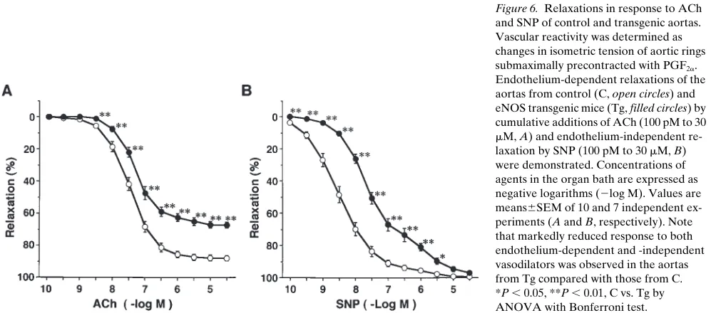
Vascular reactivity to NO- and cAMP-mediated vasodilators and responses of cGMP elevation. An endothelium-dependent vasodilator, ACh, produced concentration-dependent relax-ation of aortas from both control and transgenic mice. This
re-laxation was markedly attenuated by L-NAME treatment (100
mM) (data not shown), suggesting that the relaxation to ACh is
almost exclusively mediated by endothelium-derived NO. Un-expectedly, in eNOS-overexpressing mice, relaxation of the aorta in response to ACh was significantly reduced with an
in-crease in ED50 and a decrease in the maximum response as
compared with control littermates (Fig. 6 A and Table II).
Sim-ilarly, relaxation to ATPgS, another endothelium-dependent
vasodilator, was reduced in these mice (Table II). Relaxation to a NO donor, SNP, was reduced with a significant increase in
ED50 in the transgenic mice (Fig. 6 B and Table II) and
[image:8.612.58.410.117.321.2]relax-ation to NTG was also reduced (Table II), whereas cAMP-ele-vating or mobilizing agents, such as forskolin and dibutyryl
Figure 5. (A) Basal release of endothe-lium-derived NO indirectly estimated by L-NA–induced facilitation of PGF2a
-induced contraction. Moderate vascular tone was induced by 500 nM PGF2a before
cumulative additions of L-NA (1–100 mM). L-NA–induced facilitation of the contrac-tion was measured as milligram changes in developed tension. The magnitude is sig-nificantly greater in aortic rings isolated from eNOS-overexpressing mice (Tg, filled circles, n 5 8) than in those from control littermates (C, open circles, n 5 6). Values are shown as means6SEM. *P , 0.05, **P , 0.01, C vs. Tg. (B) Basal cGMP lev-els in the aorta determined by EIA. Tissue cGMP was extracted and subsequently acetylated for EIA. cGMP levels were cal-culated from the mean OD450 of triplicate
wells in three independent experiments and expressed as picomoles per milligram of protein. The closed bar indicates control littermate (C) value and the shaded bar transgenic mouse (Tg) value. Values are means6SEM of three independent deter-minations. *P , 0.05, C vs. Tg.
Figure 6. Relaxations in response to ACh and SNP of control and transgenic aortas. Vascular reactivity was determined as changes in isometric tension of aortic rings submaximally precontracted with PGF2a.
Endothelium-dependent relaxations of the aortas from control (C, open circles) and eNOS transgenic mice (Tg, filled circles) by cumulative additions of ACh (100 pM to 30
[image:8.612.57.559.519.739.2]cAMP, produced similar concentration–relaxation responses
in the aortas from both control and transgenic mice (Fig. 7, A
and B). In the aortas from transgenic mice treated with
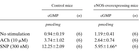
L-NAME for 3 wk, the hyporesponsiveness to SNP was com-pletely recovered in the presence of L-NAME (Table II). Basal cGMP levels were increased, whereas the extents of
cGMP elevation by ACh (10 mM) and SNP (300 nM) were
markedly decreased in transgenic aorta compared with those in control aorta (Table III).
Discussion
NO has been shown to be an endothelium-derived relaxing factor, and NO produced by eNOS diffuses from endothelial cells into underlying smooth muscle cells (1, 4). To study the physiological roles of NO as a local mediator, we generated eNOS transgenic mice with targeted expression to the vascular wall, particularly to endothelial cells in which endogenous
eNOS is constitutively expressed. Endothelium-specific in vivo transgene expression has been reported using promoters of endothelium-specific genes such as the murine Tie2 gene, hu-man vWf gene, and huhu-man intercellular adhesion molecule-2 (ICAM-2) gene (26–28). However, the Tie2 gene promoter was not active in adult mice and vWf gene promoter drove in vivo expression only to the blood vessels in the adult brain. ICAM-2 was also expressed weakly on monocytes, lympho-cytes, platelets, and megakaryolympho-cytes, and use of human pro-moters would potently present species differences in the
ex-pression. In the murine preproET-1 promoter, the 21.4 to
25.9-kb promoter region is suggested to contain endothelial
cell–specific elements and the larger fragments of the murine promoter are expected to exhibit stricter tissue-specific expres-sion (12). We have succeeded in confining constitutive overex-pression of functional eNOS to the vascular tissue in most or-gans using the 9.2-kb murine preproET-1 promoter.
A convincing in vivo feature observed in eNOS-overex-pressing mice was hypotension. Since the hypotensive
pheno-Table II. ED50 Values and Maximal Relaxations of the Aorta from Control and eNOS-overexpressing Mice
Agents
Control mice eNOS-overexpressing mice
ED50 Max. Rel. (n) ED50 Max. Rel. (n)
nM % nM %
ACh 32.763.2 88.262.1 (10) 62.9611.3* 67.562.0‡ (12)
ATPgS 554.1613.3 96.760.8 (6) 780.0676.6* 83.662.2‡ (7)
SNP 3.960.9 99.360.5 (8) 44.064.7‡ 97.361.2 (8)
NTG 21.064.1 89.362.5 (5) 109.8633.9* 62.063.4* (5)
L-NAME–treated
SNP 1.060.4 100.060.0 (5) 0.760.1 100.060.0 (7)
For precise comparison of the sensitivity and maximal response of the aorta of control and transgenic mice, ED50 (concentration which produces 50%
of the maximal relaxation to each agent) and maximal relaxation (percent PGF2a-induced precontraction) were calculated from dose–response curves
[image:9.612.58.557.73.204.2]obtained. L-NAME–treated indicates measurements of isometric tension of aortas from chronic (2–3 wk) L-NAME–treated mice in the presence of L-NAME (100 mM). Each value is a mean6SEM. The numbers of independent experiments are shown as (n). Max. Rel., maximal relaxation. *P, 0.05, ‡P, 0.01 vs. control mice.
Figure 7. Relaxations in response to for-skolin (1 nM to 3 mM, A) and dibutyryl cAMP (1 mM to 300 mM, B) of control (C, open circles) and transgenic aortas (Tg, filled circles). An adenylate cyclase activa-tor and a cAMP analogue were used to as-sess cAMP-mediated vasorelaxation. Con-centrations of agents are expressed as
2log M. Values are means6SEM of seven independent experiments (both A and B). The dose–response curves for both agents were similar between C and Tg, indicating that the cAMP-mediated pathway for vas-cular relaxation is intact in eNOS-overex-pressing mice. Values of ED50 (nM) and
maximal relaxation (%) for forskolin were 156.0645.6 and 99.260.8 in C, and 221.76 38.9 and 98.561.5 in Tg, respectively (not significant). ED50 (mM) and maximal
[image:9.612.55.392.577.739.2]type was also observed in independent transgenic lineages, the functional effects of eNOS overexpression in line 22 were not due to spurious positional effects of transgene insertion. Hy-pertension in mice lacking eNOS has been reported by two groups (6, 7) and our model provides another in vivo valida-tion for that NO produced by eNOS itself regulates blood pressure. The mechanisms of hypotension due to eNOS over-expression have not been determined fully. However, eNOS encoded by the transgene must have contributed to the reduc-tion in blood pressure because chronic administrareduc-tion of L-NAME completely reversed hypotension in transgenic mice without paradoxical reduction in blood pressure observed in L-NA–treated eNOS knockout mice (6). Both cardiac output and peripheral arterial resistance determine blood pressure and depression of cardiac function by NO may lead to de-creased cardiac output. However, in the present model, bovine eNOS was not identified in cardiac myocytes. Of note was that the hypotension in the transgenic mice was associated with both a decrease in basal tone of the aorta and an increase in its basal cGMP levels. Because of technical difficulties, we could not determine total vascular resistance and the vascular tone examined in the aorta might not always reflect that in the resis-tance vessels which regulate blood pressure. But, indeed, the transgene was obviously expressed in large or medium- to
mi-cro-sized vessels in the heart and lung (Fig. 3, E and F).
There-fore, it is likely that enhanced NO release exerted vasorelaxant effects in types of vessels other than the aorta and increases in basal NO release reduced basal vascular tone in resistance ves-sels, thereby produced hypotension. Hypotension has also been observed in transgenic mouse models with overexpres-sion of natriuretic peptides, kallikrein, and bradykinin re-ceptor (29–32). Our model exhibited hypotension without increases in heart rate or urine volume, similar to mice overex-pressing natriuretic peptides that yielded no changes in indica-tors of a volume-depleted state (29, 30). These studies imply that enhanced vasodilatory action of cGMP-mediated pathway by either NO or natriuretic peptides in the vascular wall suffi-ciently reduces blood pressure.
Blood pressure is regulated by integrative neurohumoral
factors. Compensatory mechanisms may operate for the reduc-tion in blood pressure due to chronic activareduc-tion of the NO pathway. Recent reports have demonstrated that NO interacts with the renin-angiotensin system and also the cyclooxygen-ase-prostaglandin system. Elevated plasma renin concentra-tion was observed in eNOS-deficient mice (7). However, there were no significant alterations in plasma renin activity, plasma catecholamine, or ET-1 levels in our transgenic mice (Table I) and the responsiveness of mouse aorta was not modified after preincubation with indomethacin in isometric tension record-ings (data not shown). Collectively, these findrecord-ings further sup-port the notion that the direct vasodilatory action of overpro-duced NO is mainly responsible for hypotension without potential influence of major neurohumoral systems regulating blood pressure.
The most surprising finding observed in eNOS-overex-pressing mice was the reduced vasorelaxant response to NO-mediated vasodilators (Fig. 6). Recent reports of adenovirus-mediated eNOS gene transfer into the endothelium or the adventitia have demonstrated the enhanced endothelium-dependent relaxations in response to ACh and calcium iono-phore (10, 33, 34). We for the first time demonstrated that chronic eNOS overexpression results in attenuation rather than enhancement of NO-mediated vasorelaxation. The differ-ence in vascular reactivity observed between animals with gene transfer and our transgenic model may be possibly due to animal species used, the expression site of eNOS, relative in-tensity of gene expression achieved depending on the pro-moter used in each model, or the duration of gene expression. We could not define the exact mechanisms, but believe that the long duration of eNOS overexpression in the transgenic mice is probably involved. It is likely that, in contrast to the acute effect of eNOS gene transfer, the chronic increase in basal NO release due to eNOS overexpression elicits the re-versible hyporesponsiveness in NO-cGMP pathway. It has been established pharmacologically that chronic in vivo treat-ment of rabbits and rats with organic nitrate results in develop-ment of nitrate tolerance and cross-tolerance to other nitrova-sodilators and endothelium-dependent vanitrova-sodilators (35, 36). Inversely, the sensitivity of isolated rat aorta to NO donors was increased after removal of the endothelium or treatment with a NOS inhibitor (37). Recently, relaxation of the carotid artery in response to low concentrations of SNP was shown to be enhanced in eNOS-deficient mice (38). Our findings are consistent with these reports, which imply that alterations in basal NO release modulate vascular reactivity to NO.
In eNOS-overexpressing mice, cAMP-mediated vasorelax-ation was not altered (Fig. 7), whereas increases in cGMP lev-els in response to ACh and SNP were depressed in spite of the
increased basal cGMP levels (Fig. 5 B and Table III).
[image:10.612.57.298.83.178.2]Concen-tration-dependent increases in cGMP by SNP were signifi-cantly attenuated after chronic NTG injection and were poten-tiated by endothelial denudation or NOS inhibition of the aorta (35, 37). Soluble guanylate cyclase (sGC) purified from NO donor–pretreated rat aorta exhibited desensitization to NO donors (39). sGC mRNA and protein levels and its activity were decreased in cultured smooth muscle cells exposed to NO donors, a cGMP analogue, or an inhibitor of cGMP phos-phodiesterase (40, 41). In view of these reports, desensitization of sGC by continuous overactivation is, at least in part, impli-cated in the reduced vascular reactivity in eNOS-overexpress-ing mice. Furthermore, impairments of signaleNOS-overexpress-ing pathway after
Table III. Increase in cGMP Levels in Response to Endothelium-dependent and -independent Vasodilators
Control mice eNOS-overexpressing mice
cGMP (n) cGMP (n)
pmol/mg pmol/mg
No stimulation 0.9460.19 (6) 1.1960.41 (6) ACh (10 mM) 3.7461.02 (6) 2.6460.74 (6) SNP (300 nM) 12.2562.09 (6) 5.9561.66* (6)
cGMP elevation may occur. Whether it is also the case with our mouse model remains to be clarified.
In conclusion, we generated transgenic mice overexpress-ing eNOS in the vascular wall to study the local effects of NO. eNOS-overexpressing mice displayed marked hypotension which was associated with reduced basal vascular tone and reduced vascular reactivity to NO. Our mouse model provided new in-sights into the in vivo mechanisms of nitrate tolerance in addi-tion to the essential role of eNOS in physiological blood pres-sure regulation and could be a novel useful tool to explore the pathophysiological roles of NO in the cardiovascular system.
Acknowledgments
The authors thank Prof. David G. Harrison (Cardiovascular Division, Emory University School of Medicine) and Dr. Jennifer S. Pollock (Abbott Laboratories, Abbott Park, IL) for bovine aortic eNOS cDNA and an mAb against bovine eNOS (H32). We wish to thank Drs. Akio Inui and Minoru Okita (The Second Department of Inter-nal Medicine, Kobe University School of Medicine) for the technical advice to generate transgenic mice and Dr. Katsuo Kamata (Hoshi University, Tokyo) for his advice and help on isometric tension re-cordings of the mouse aorta. We gratefully appreciate Seiko Tsutsui and Kiyoko Matsui for their support with animal care and secretarial assistance.
This work was supported by grants-in-aid for Scientific Research (No. 08457209) from Ministry of Education, Science and Culture, Ja-pan (1995-97) and grants-in-aid for the research on cardiovascular disease from the Ministry of Health and Welfare, Japan (1995-97), a grant from Japan Cardiovascular Research Foundation, a grant from Research for Molecular Cardiology, and a grant from Kanae Founda-tion of Research for New Medicine.
References
1. Moncada, S., R.M. Palmer, and E.A. Higgs. 1991. Nitric oxide: physiol-ogy, pathophysiolphysiol-ogy, and pharmacology. Pharmacol. Rev. 43:109–142.
2. Dinerman, J.L., C.J. Lowenstein, and S.H. Snyder. 1993. Molecular mechanisms of nitric oxide regulation. Potential relevance to cardiovascular disease. Circ. Res. 73:217–222.
3. Dzau, V.J., and G.H. Gibbons. 1991. Endothelium and growth factors in vascular remodeling of hypertension. Hypertension. 18(Suppl. III):III115– III121.
4. Nathan, C., and Q.-W. Xie. 1994. Nitric oxide synthases: roles, tolls, and controls. Cell. 78:915–918.
5. Moncada, S., and E.A. Higgs. 1993. The L-arginine-nitric oxide pathway.
N. Engl. J. Med. 329:2002–2012.
6. Huang, P.L., Z. Huang, H. Mashimo, K.D. Bloch, M.A. Moskowitz, J.A. Bevan, and M.C. Fishman. 1995. Hypertension in mice lacking the gene for en-dothelial nitric oxide synthase. Nature. 377:239–242.
7. Shesely, E.G., N. Maeda, H.S. Kim, K.M. Desai, J.H. Krege, V.E. Laubach, P.A. Sherman, W.C. Sessa, and O. Smithies. 1996. Elevated blood pressures in mice lacking endothelial nitric oxide synthase. Proc. Natl. Acad. Sci. USA. 93:13176–13181.
8. von der Leyen, H.E., G.H. Gibbons, R. Morishita, N.P. Lewis, L. Zhang, M. Nakajima, Y. Kaneda, J.P. Cooke, and V.J. Dzau. 1995. Gene therapy inhib-iting neointimal vascular lesion: in vivo transfer of endothelial cell nitric oxide synthase gene. Proc. Natl. Acad. Sci. USA. 92:1137–1141.
9. Lin, K.-F., L. Chao, and J. Chao. 1997. Prolonged reduction of high blood pressure with human nitric oxide synthase gene delivery. Hypertension. 30:307– 313.
10. Ooboshi, H., Y. Chu, D. Rios, F.M. Faraci, B.L. Davidson, and D.D. Heistad. 1997. Altered vascular function after adenovirus-mediated overex-pression of endothelial nitric oxide synthase. Am. J. Physiol. 273(Heart Circ.
Physiol. 42):H265–H270.
11. Harats, D., H. Kurihara, P. Belloni, H. Oakley, A. Ziober, D. Ackley, G. Cain, Y. Kurihara, R. Lawn, and E. Sigal. 1995. Targeting gene expression to the vascular wall in transgenic mice using the murine preproendothelin-1 pro-moter. J. Clin. Invest. 95:1335–1344.
12. Nishida, K., D.G. Harrison, J.P. Navas, A.A. Fisher, S.P. Dockery, M. Uematsu, R.M. Nerem, R.W. Alexander, and T.J. Murphy. 1992. Molecular cloning and characterization of the constitutive bovine aortic endothelial cell
nitric oxide synthase. J. Clin. Invest. 90:2092–2096.
13. Maemura, K., H. Kurihara, O. Ueda, Y. Kurihara, T. Kuwaki, H. Morita, T. Kodama, H. Suzuki, M. Kumada, T. Ishikawa, and Y. Yazaki. 1996. Generation and analysis of transgenic mice overexpressing endothelin-1. Circu-lation. 94:I-531.
14. Hogan, B., R. Beddington, F. Costantini, and E. Lacy. 1994. Manipulat-ing the Mouse Embryo: A Laboratory Manual. 2nd ed. Cold SprManipulat-ing Harbor Laboratory, Cold Spring Harbor, NY. 127–188.
15. Chomczynski, P., and N. Sacchi. 1987. Single-step method of RNA isola-tion by acid guanidinium thiocyanate-phenol-chloroform extracisola-tion. Anal. Bio-chem. 162:156–159.
16. Hirata, K., N. Miki, Y. Kuroda, T. Sakoda, S. Kawashima, and M. Yokoyama. 1995. Low concentration of oxidized low-density lipoprotein and lysophosphatidylcholine upregulate constitutive nitric oxide synthase mRNA expression in bovine aortic endothelial cells. Circ. Res. 76:958–962.
17. Bradford, M.M. 1976. A rapid and sensitive method for the quantitation of microgram quantities of protein utilizing the principle of protein-dye bind-ing. Anal. Biochem. 72:248–254.
18. Pollock, J.S., M. Nakane, L.D. Buttery, A. Martinez, D. Springall, J.M. Polak, U. Forstermann, and F. Murad. 1993. Characterization and localization of endothelial nitric oxide synthase using specific monoclonal antibodies. Am. J. Physiol. 265(Cell. Physiol. 34):C1379–C1387.
19. Ohashi, Y., M. Katayama, K. Hirata, M. Suematsu, S. Kawashima, and M. Yokoyama. 1993. Activation of nitric oxide synthase from cultured aortic endothelial cells by phospholipids. Biochem. Biophys. Res. Commun. 195:1314– 1320.
20. Kanazawa, K., S. Kawashima, S. Mikami, Y. Miwa, K. Hirata, M. Sue-matsu, Y. Hayashi, H. Itoh, and M. Yokoyama. 1996. Endothelial constitutive nitric oxide synthase protein and mRNA increased in rabbit atherosclerotic aorta despite impaired endothelium-dependent vascular relaxation. Am. J. Pathol. 148:1949–1956.
21. Kamata, K., M. Sugiura, S. Kojima, and Y. Kasuya. 1996. Preservation of endothelium-dependent relaxation in cholesterol-fed and streptozotocin-induced diabetic mice by the chronic administration of cholestyramine. Br. J.
Pharmacol. 118:385–391.
22. Hayashi, T., J.M. Fukuto, L.J. Ignarro, and G. Chaudhuri. 1992. Basal release of nitric oxide from aortic rings is greater in female rabbits than in male rabbits: implications for atherosclerosis. Proc. Natl. Acad. Sci. USA. 89:11259– 11263.
23. Rubanyi, G.M., A.D. Freay, K. Kauser, D. Sukovich, G. Burton, D.B. Lubahn, J.F. Couse, S.W. Curtis, and K.S. Korach. 1997. Vascular estrogen re-ceptors and endothelium-derived nitric oxide production in the mouse aorta. Gender difference and effect of estrogen receptor gene disruption. J. Clin. In-vest. 99:2429–2437.
24. Janssens, S.P., K.D. Bloch, Z. Nong, R.D. Gerard, P. Zoldhelyi, and D. Collen. 1996. Adenoviral-mediated transfer of the human endothelial nitric ox-ide synthase gene reduces acute hypoxic pulmonary vasoconstriction in rats. J. Clin. Invest. 98:317–324.
25. Moshage, H., B. Kok, J.R. Huizenga, and P.L.M. Jansen. 1995. Nitrite and nitrate determinations in plasma: a critical evaluation. Clin. Chem. 41:892– 896.
26. Schlaeger, T.M., Y. Qin, Y. Fujiwara, J. Magram, and T.N. Sato. 1995. Vascular endothelial cell lineage-specific promoter in transgenic mice.
Devel-opment. 121:1089–1098.
27. Aird, W.C., N. Jahroudi, G.H. Weiler, H.B. Rayburn, and R.D. Rosen-berg. 1995. Human von Willebrand factor gene sequences target expression to a subpopulation of endothelial cells in transgenic mice. Proc. Natl. Acad. Sci. USA. 92:4567–4571.
28. Cowan, P.J., T.A. Shinkel, E.J. Witort, H. Barlow, M.J. Pearse, and A.J. d’Apice. 1996. Targeting gene expression to endothelial cells in transgenic mice using the human intercellular adhesion molecule 2 promoter. Transplantation.
62:155–160.
29. Steinhelper, M.E., K.L. Cochrane, and L.J. Field. 1990. Hypotension in transgenic mice expressing atrial natriuretic factor fusion genes. Hypertension.
16:301–307.
30. Ogawa, Y., H. Itoh, N. Tamura, S. Suga, T. Yoshimasa, M. Uehira, S. Matsuda, S. Shiono, H. Nishimoto, and K. Nakao. 1994. Molecular cloning of the complementary DNA and gene that encode mouse brain natriuretic peptide and generation of transgenic mice that overexpress the brain natriuretic peptide gene. J. Clin. Invest. 93:1911–1921.
31. Wang, J., W. Xiong, Z. Yang, T. Davis, M.J. Dewey, J. Chao, and L. Chao. 1994. Human tissue kallikrein induces hypotension in transgenic mice.
Hypertension. 23:236–243.
32. Wang, D.-Z., L. Chao, and J. Chao. 1997. Hypotension in transgenic mice overexpressing human bradykinin B2 receptor. Hypertension. 29:488–493. 33. Kullo, I.J., G. Mozes, R.S. Schwartz, P. Gloviczki, M. Tsutsui, Z.S. Katu-sic, and T. O’Brien. 1997. Enhanced endothelium-dependent relaxations after gene transfer of recombinant endothelial nitric oxide synthase to rabbit carotid arteries. Hypertension. 30:314–320.
reactivity. Circulation. 96:2254–2261.
35. Molina, C.R., J.W. Andresen, R.M. Rapoport, S. Waldman, and F. Mu-rad. 1987. Effect of in vivo nitroglycerin therapy on endothelium-dependent and independent vascular relaxation and cyclic GMP accumulation in rat aorta.
J. Cardiovasc. Pharmacol. 10:371–378.
36. Münzel, T., H. Seyegh, B.A. Freeman, M.M. Tarpey, and D.G. Harri-son. 1995. Evidence for enhanced vascular superoxide anion production in ni-trate tolerance. A novel mechanism underlying tolerance and cross-tolerance.
J. Clin. Invest. 95:187–194.
37. Moncada, S., D.D. Rees, R. Schulz, and R.M.J. Palmer. 1991. Develop-ment and mechanism of a specific supersensitivity to nitrovasodilators after in-hibition of vascular nitric oxide synthesis in vivo. Proc. Natl. Acad. Sci. USA. 88: 2166–2170.
38. Faraci, F.M., C.D. Sigmund, E.G. Shesely, N. Maeda, and D.D. Heistad. 1998. Responses of carotid artery in mice deficient in expression of the gene for endothelial NO synthase. Am. J. Physiol. 274(Heart Circ. Physiol. 43):H564– H570.
39. Waldman, S.A., R.M. Rapoport, R. Ginsburg, and F. Murad. 1986. De-sensitization to nitroglycerin in vascular smooth muscle from rat and human.
Biochem. Pharmacol. 35:3525–3531.
40. Filippov, G., D.B. Bloch, and K.D. Bloch. 1997. Nitric oxide decreases stability of mRNAs encoding soluble guanylate cyclase subunits in rat pulmo-nary artery smooth muscle cells. J. Clin. Invest. 100:942–948.