Superinduction of cyclooxygenase-2 activity in
human osteoarthritis-affected cartilage.
Influence of nitric oxide.
A R Amin, … , I R Patel, S B Abramson
J Clin Invest.
1997;
99(6)
:1231-1237.
https://doi.org/10.1172/JCI119280
.
Cartilage specimens from osteoarthritis (OA)-affected patients spontaneously released
PGE2 at 48 h in ex vivo culture at levels at least 50-fold higher than in normal cartilage and
18-fold higher than in normal cartilage + cytokines + endotoxin. The superinduction of PGE2
production coincides with the upregulation of cyclooxygenase-2 (COX-2) in OA-affected
cartilage. Production of both nitric oxide (NO) and PGE2 by OA cartilage explants is
regulated at the level of transcription and translation. Dexamethasone inhibited only the
spontaneously released PGE2 production, and not NO, in OA-affected cartilage. The NO
synthase inhibitor HN(G)-monomethyl-L-arginine monoacetate inhibited OA cartilage NO
production by > 90%, but augmented significantly (twofold) the spontaneous production of
PGE2 in the same explants. Similarly, addition of exogenous NO donors to OA cartilage
significantly inhibited PGE2 production. Cytokine + endotoxin stimulation of OA explants
increased PGE2 production above the spontaneous release. Addition of L-NMMA further
augmented cytokine-induced PGE2 production by at least fourfold. Inhibition of PGE2 by
COX-2 inhibitors (dexamethasone or indomethacin) or addition of exogenous PGE2 did not
significantly affect the spontaneous NO production. These data indicate that human
OA-affected cartilage in ex vivo conditions shows (a) superinduction of PGE2 due to
upregulation of COX-2, and (b) spontaneous release of NO that acts as an autacoid to
attenuate the production of the COX-2 products such as PGE2. These studies, […]
Research Article
Find the latest version:
J. Clin. Invest.
© The American Society for Clinical Investigation, Inc. 0021-9738/97/03/1231/07 $2.00
Volume 99, Number 6, March 1997, 1231–1237
Superinduction of Cyclooxygenase-2 Activity in Human
Osteoarthritis-affected Cartilage
Influence of Nitric Oxide
Ashok R. Amin,*§i¶ Mukundan Attur,* Rajesh N. Patel,* Geeta D. Thakker,* Paul J. Marshall,** John Rediske,** Steven A. Stuchin,‡ Indravadan R. Patel,* and Steven B. Abramson*§
*Department of Rheumatology, ‡Department of Orthopedic Surgery, Hospital for Joint Diseases, New York 10003; §Department of
Medicine, iDepartment of Pathology, ¶Kaplan Cancer Research Center, New York University Medical Center, New York 10016; and
**Ciba-Geigy Corporation, Summit, New Jersey 07901
Abstract
Cartilage specimens from osteoarthritis (OA)-affected pa-tients spontaneously released PGE2 at 48 h in ex vivo
cul-ture at levels at least 50-fold higher than in normal cartilage and 18-fold higher than in normal cartilage 1 cytokines 1 endotoxin. The superinduction of PGE2 production
coin-cides with the upregulation of cyclooxygenase-2 (COX-2) in OA-affected cartilage. Production of both nitric oxide (NO) and PGE2 by OA cartilage explants is regulated at the level
of transcription and translation. Dexamethasone inhibited only the spontaneously released PGE2 production, and not
NO, in OA-affected cartilage. The NO synthase inhibitor HNG
-monomethyl-L-arginine monoacetate inhibited OA car-tilage NO production by . 90%, but augmented significantly (twofold) the spontaneous production of PGE2 in the same
explants. Similarly, addition of exogenous NO donors to OA cartilage significantly inhibited PGE2 production. Cytokine 1
endotoxin stimulation of OA explants increased PGE2
pro-duction above the spontaneous release. Addition of L-NMMA further augmented cytokine-induced PGE2 production by at
least fourfold. Inhibition of PGE2 by COX-2 inhibitors
(dexa-methasone or indomethacin) or addition of exogenous PGE2
did not significantly affect the spontaneous NO production. These data indicate that human OA-affected cartilage in ex vivo conditions shows (a) superinduction of PGE2 due to upregulation of COX-2, and (b) spontaneous release of NO that acts as an autacoid to attenuate the production of the COX-2 products such as PGE2. These studies, together with
others, also suggest that PGE2 may be differentially
regu-lated in normal and OA-affected chondrocytes. (J. Clin.
In-vest. 1997. 99:1231–1237.) Key words: nitric oxide •
osteoar-thritis • cyclooxygenase • cytokine • cartilage
Introduction
Osteoarthritis (OA)1 is a disease that affects the weight-bear-ing joints and peripheral and axial articulations. The clinical correlates are pain and restricted motion (1), and although the disease is classified as a noninflammatory arthropathy, several inflammatory components have been implicated in the disease process and have been observed in the synovial fluid. These in-clude inflammatory cytokines, immunoglobulins, and other mediators in the joints (2, 3). Increased production of acute phase reactants such as transferrin, ceruloplasmin, acid glyco-protein, and increased concentrations of substance P have also been reported in OA (4). One of the hallmarks of OA is the progressive degeneration of the cartilage, where the synthetic and degradative forces are operating in tandem. Unchecked, the process leads to an imbalance of these forces and joint de-struction.
Prostaglandins are produced at elevated levels in inflamed tissues including rheumatoid synovium (5, 6). PGE1 and PGE2 contribute to synovial inflammation by increasing local blood flow and potentiating the effects of mediators such as bradyki-nin and IL-1b that induce vasopermeability (7). PGE2 has also been shown to trigger osteoclastic bone resorption (8), sug-gesting that this molecule may contribute to the pathophysiol-ogy of joint erosion in RA.
The human articular chondrocytes, when stimulated with cytokines and/or endotoxin in vitro, release various inflamma-tory mediators, including nitric oxide (NO) and PGE2 (9, 10). We (11) and others (12) have recently observed that OA-affected cartilage in ex vivo conditions spontaneously releases NO in quantities sufficient to cause cartilage damage. The im-portance of these findings is supported by experiments con-ducted in animal models of arthritis, where inhibitors of NO synthase (NOS) (13, 14) or cyclooxygenase (COX)-2 (15) can independently repress joint inflammation, concomitant with the attenuation of PGE2 or NO synthesis. In the present study, we report that OA-affected cartilage (but not normal carti-lage): (a) spontaneously releases PGE2 and NO, which are coordinately regulated; and (b) shows upregulated COX-2 mRNA and COX-2 protein, which spontaneously releases sub-stantial amounts of PGE2 in ex vivo conditions. Furthermore, the NO produced by the OA-affected cartilage inhibits the au-tocrine (or cytokine 1 endotoxin-induced) PGE2 production, thus indicating a negative effect of NO on PGE2 synthesis.
Address correspondence to Ashok R. Amin, Department of Rheu-matology, Room 1600, Hospital for Joint Diseases, 301 E. 17th Street, New York, NY 10003. Phone: 212-598-6537; FAX: 212-598-6168.
Received for publication 7 October 1996 and accepted in revised form 19 December 1996.
1. Abbreviations used in this paper: COX-2, cyclooxygenase-2; L-NMMA, HNG-monomethyl-L-arginine monoacetate; NF, nuclear factor; NO,
Methods
Reagents and cell lines.A macrophage-like cell line, RAW 264.7, was obtained from American Type Culture Collection (Rockville, MD). Monoclonal a-mouse macrophage COX-2 antibody, which also cross-reacts with human COX-2, was obtained from Transduction Laboratories (Lexington, KY). Protease inhibitors, dexamethasone, cycloheximide, indomethacin, pyrrolidine dithiocarbamate (PDTC), aminoguanidine, sodium nitroprusside, and LPS were obtained from Sigma Chemical Co. (St. Louis, MO); human IL-1b and TNF-a were from Fisher Scientific Co. (Pittsburgh, PA); and HNG-monomethyl-L-arginine
monoacetate (L-NMMA) from Cyclo3pss BioChemical Corp. (Salt
Lake City, UT).
Procurement of human cartilage. Cartilage slices were taken from the knees of patients with the diagnosis of advanced OA (age: 50–70 yr) who were undergoing knee replacement surgery, and from nonar-thritic knees (normal controls: age 20–40 yr). The OA patients were free of steroidal/nonsteroidal antiinflammatory drugs for at least 2 wk before surgery. Nonarthritic knee cartilage was obtained from pa-tients with fractures or from accident victims after knee amputation the same day.
Extraction of human cartilage COX-2 and Western blotting.
Slices from articular cartilage were frozen at 2708C, milled to fine particulate in liquid nitrogen, and sequentially extracted (10 ml/g wet wt tissue) with neutral salt buffer (Tris HCl:saline) containing pro-tease inhibitors (11). Samples were run on 10% SDS-PAGE gels un-der nonreducing conditions, transferred to nitrocellulose, and West-ern blotted with a-COX-2 antibody. Bound antibody was detected by a secondary antibody conjugated with horseradish peroxidase, and developed using the enhanced chemiluminescence Western blotting system (Amersham Corp., Arlington Heights, IL) on Kodak Xomatic x-ray film.
Determination of nitrite/PGE2. NO production was measured by
estimating the stable NO metabolite, nitrite, in conditioned medium by modified Griess reaction (16). PGE2 (detection limit: 50 pg/ml)
was determined in the culture supernatant using RIA Kit (Sigma Chemical Co.) according to the manufacturer’s instructions.
RNA extraction from articular chondrocytes. The method we fol-lowed is basically as described by Adams et al. (17). The cartilage was milled into fine powder in liquid nitrogen and extracted with 4 M guanidium thiocyanate, 25 mM sodium citrate, 0.5% sodium dodecyl sarcosine, and 0.1 M 2-mercaptoethanol for 4 h on a rocker. It was then extracted with H2O-saturated phenol followed with
phenol/chlo-roform. The aqueous layer was layered onto cesium trifluoroacetate gradient for ultracentrifugation (24,000 rpm/24 h). The RNA pellet was dissolved in GuSCN and precipitated with alcohol in the pres-ence of acetic acid. The RNA obtained with this method is pure enough for reverse transcriptase (RT)–PCR analysis.
RT-PCR analysis, cloning and sequencing.The presence of mRNA was analyzed by RT of total RNA (1 mg) followed by PCR amplifica-tion of the cDNA. The PCR was carried out in an automated DNA thermal cycle (Perkin-Elmer Cetus Instruments, Norwalk, CT). The cDNA from mRNA was prepared using a SuperScriptTM RNase H
Reverse Transcriptase (Gibco BRL, Gaithersburg, MD) and PCR amplification was performed in 50 ml solution containing 1.5 mM MgCl2, 0.4 mM of each primer, 0.2 mM of each dNTP, 2.0 units of Taq
DNA polymerase (Promega Corp., Madison, WI). The cycle condi-tions were 45 s at 948C, 45 s at 608C, and 2 min at 728C for 20–30 cy-cles for both COX-2 and b-actin. The PCR product was analyzed by electrophoresis on 1.5% agarose gels. Verification that the amplified PCR product originated from RNA rather than DNA was accom-plished by preparing duplicate RNA samples both untreated and treated with reverse transcriptase, followed by PCR amplification. The amplified PCR products were directly cloned into PCRTM II
Prokaryotic TA Cloning System (Invitrogen, San Diego, CA), and then the sequence of clones was determined using M13 primers by the dideoxy chain termination method using an automated laser fluores-cent DNA sequencer (ALF; Pharmacia Diagnostics, Piscataway, NJ).
Statistical analysis. The data represent a minimum of two experi-ments performed from separate normal individuals or OA patients. All experiments using cartilage slices were performed in triplicate or quadruplicate for each parameter studied. The P value was calculated using Student’s t test between the parameters in the same experiment.
Results
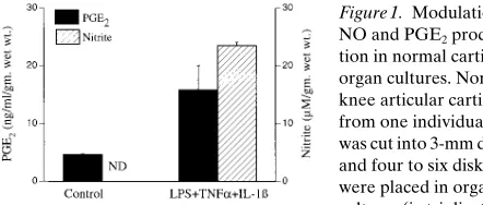
Previous studies have shown that stimulation of articular carti-lage leads to synthesis of (in order of decreasing quantity) PGE2, PGF2a, PGI2, and TXA2 (18). A number of synovial cy-tokines, including IL-1b and TNF-a, or endotoxins (LPS) en-hance PGE2 and NO synthesis in cultured chondrocytes (9, 10). In the present study, we first examined whether cartilage slices from normal human cartilage responded in a similar fashion. Incubation of normal human cartilage slices with IL-1b1 TNF-a1 LPS caused a significant augmentation in PGE2 and induction of NO production within 48 h in culture (Fig. 1). These experiments were consistent with previous ob-servations of basal levels of PGE2 released by normal chondro-cytes (unlike NO production) that could be augmented with LPS or IL-1b (10). These experiments demonstrate that the isolated chondrocytes and those embedded in cartilage in vitro behaved similarly with respect to the simultaneous stimulation of NO and PGE2 production. We next examined whether PGE2 was also spontaneously released by the OA-affected cartilage in ex vivo conditions, as we have previously reported for NO (11). Indeed, all the OA-affected cartilage (samples from 10 different patients) spontaneously released both NO (44–265 mM) and PGE2 (50–670 ng/ml per g tissue) in ex vivo conditions at 48 h (Fig. 2). Each of the samples tested, as ex-pected, had high levels of NO spontaneously released in the culture medium, as previously observed (11). It should be noted that the lowest amount of PGE2 spontaneously released (Fig. 2, patient 2, z 50 ng/ml) was at least 10-fold higher than
[image:3.612.315.536.547.641.2]that produced by normal cartilage (Figs. 1 and 2) or spontane-ously released by equivalent amounts of normal chondrocytes (10). Next, using various inhibitors, we examined whether the spontaneous production of PGE2 by OA-affected cartilage was due to endogenous upregulation of the inducible COX-2. The release of both these mediators (NO and PGE2) was sen-sitive to nuclear-factor (NF)–kB transcription factor inhibitor
Figure 1. Modulation of NO and PGE2
produc-tion in normal cartilage organ cultures. Normal knee articular cartilage from one individual was cut into 3-mm disks, and four to six disks were placed in organ cultures (in triplicate) of 2 ml media in the presence and absence of IL-1b (1 ng/ml) 1 TNF-a
(100 U/ml) 1 LPS (100 mg/ml). The levels of nitrite and PGE2 were
monitored at 48 h as described in Methods. Data are expressed as mi-cromolar of nitrite or nanograms per milliliter of PGE2 per gram (wet
wt) of cartilage. The P values based on the triplicate data (n5 3) be-tween control (unstimulated) and cytokine 1 endotoxin-treated cul-tures were , 0.001 for PGE2 production and , 0.0001 for nitrite
PDTC and translation inhibitor cycloheximide (Fig. 3), indi-cating the inducible nature of these mediators.
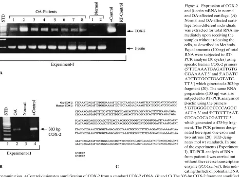
Since recent studies have implicated NF-kB as one of the essential transcription factors for the inducible COX-2 tran-scription (19), we examined the expression of inducible COX-2 mRNA directly from cartilage obtained from normal and OA-affected patients. Fig. 4, A and B, shows that all nine
OA-affected cartilage samples tested showed upregulated COX-2 mRNA that could not be detected in the three normal carti-lage samples tested, where the b-actin signal could be seen with the same mRNA preparations (not shown for
Experi-Figure 2. Spontaneous release of PGE2 from
OA-affected cartilage. Knee articular cartilage from 10 patients under-going knee replace-ment surgery was set up in organ culture in triplicate (n5 3) as de-scribed in Fig. 1, with-out any modulators. Levels of nitrite/PGE2
were monitored after 48 h, as shown in upper panel. The dotted line represents the mean amount of PGE2 released by these 10 patients, which was 285 ng/ml
per g tissue. Data are expressed as micromolar of nitrite or nano-grams per milliliter of PGE2 per gram (wet wt) of cartilage
[image:4.612.55.300.58.190.2]accumu-lated in the same medium at 48 h.
Figure 3. Comparison of NO and PGE2 spontaneously produced by
OA-affected cartilage. OA-affected knee articular cartilage from one patient was cut into 3-mm disks, and four to six disks were placed in organ cultures (in triplicate) in 2 ml of F-12 medium in the presence and absence of PDTC (20 mM) or cycloheximide (2 mg/ml). The lev-els of nitrite and PGE2 were monitored after 48 h. Data are expressed
as micromolar of nitrite or nanograms per milliliter of PGE2 per gram
(wet wt) of cartilage; n5 3. The P values between control (spontane-ous release) versus PDTC-treated (nitrite , 0.0024; PGE2, 0.0024)
or cycloheximide-treated cultures (nitrite , 0.0001; PGE2, 0.0011)
[image:4.612.314.485.58.165.2]were calculated. ND, not detectable (, 0.1 mM nitrite). The data rep-resent one of two similar studies.
Figure 4. Expression of COX-2 and b-actin mRNA in normal and OA-affected cartilage. (A) Normal and OA-affected carti-lage from different individuals was extracted for total RNA im-mediately upon receiving the samples without releasing the cells, as described in Methods. Equal amounts (100 ng) of total RNA were subjected to RT-PCR analysis (30 cycles) using specific human COX-2 primers (59TTCAAATGAGATTGTG GGAAAAT 39 and 59AGATC ATCTCTGCCTGAGTATC-TT 39) which generated a 303-bp fragment (20). The same RNA preparation (100 ng) was also subjected to RT-PCR analysis of
b-actin using the primers 59GTGGGGCGCCCCAGGC ACCA 39 and 59 CTCCTTAAT-GTCACGCACGATTTC 39
[image:4.612.62.555.352.716.2]ment II). The identity of the COX-2 mRNA was confirmed by Southern blot analyses of the 303-bp COX-2 fragments using a full-length COX-2 cDNA (data not shown) and sequencing one of the fragments from COX-2 mRNA amplified from OA-affected cartilage, as shown in Fig. 4, B and C. The partial DNA sequence of the OA-COX-2 obtained from one of the OA patients (Experiment II) showed . 98% homology with the known human COX-2 sequence (20). The expression of COX-2 was confirmed by Western blotting of COX-2 protein from OA-affected cartilage extracts which showed the expres-sion of a 72-kD COX-2 protein as observed in LPS-stimulated macrophages (21) (Fig. 5). These experiments indicate that OA-affected cartilage spontaneously releases an inflammatory compound, PGE2, in levels at least 50-fold higher (on average) than the normal cartilage, and this effect may be due to tran-scriptional upregulation of COX-2, which is sensitive to PDTC and cycloheximide.
We next examined the effect of endogenous NO on PGE2 production in OA-affected cartilage. OA-affected cartilage
slices were incubated in ex vivo conditions and the levels of spontaneously released NO and PGE2 were monitored at 48 h in the presence and absence of the competitive NOS inhibitor
L-NMMA (Table I). Addition of L-NMMA blocked . 90% of spontaneous endogenous NO production in these OA-affected cartilage organ cultures, as previously reported (11). However, the decrease in the NO production caused a significant aug-mentation (approximately twofold) of the PGE2 accumulation in the same experiments. These experiments indicate that the levels of endogenous NO spontaneously released in ex vivo conditions inhibit the spontaneous production of PGE2 in the same OA-affected cartilage slices. The effects of endogenous NO in OA cartilage on PGE2 production could be mimicked by exogenously added NO donors such as sodium nitroprus-side. In one representative experiment where OA-affected cartilage had released 221.7670.2 ng/ml of PGE2 (per g wet wt cartilage) at 72 h, addition of 100 mM of sodium nitroprusside (at 0 h) in the same explants significantly reduced PGE2 pro-duction to 43.3614.7 (P, 0.004; n5 3).
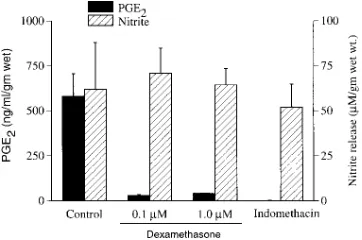
[image:5.612.58.293.61.142.2]We then examined the effects of COX-2 products on NO production in OA explants as shown in Fig. 6. Addition of dexa-methasone or indomethacin, as expected, inhibited (. 90%) the PGE2 production in the OA-affected cartilage but had no significant effect on the levels of NO released by the same car-tilage samples. Furthermore, as expected, addition of a COX-2 inhibitor, CGP28238 (Flosulide; Ciba-Geigy Corp., Summit, NJ) at 5 mM inhibited spontaneous PGE2 production by OA-affected cartilage in ex vivo conditions by . 95% (like in-domethacin), but had no significant effect on NO production (data not shown). We also tested the effects of exogenous PGE2 on the spontaneous accumulation of nitrite in ex vivo conditions in our OA-explant assay. The OA-affected carti-lage on an average released 28 ng/ml of PGE2/100 mg cartilage as shown in Fig. 2. We therefore added 25 or 50 ng/ml of PGE2 to the above OA cartilage explant assay. There was no signifi-cant effect on the accumulation of nitrite in OA-explants sup-plemented with 25 ng/ml PGE2 (5.962.7 mM nitrite at 72 h;
[image:5.612.56.297.474.653.2]Figure 5. Western blot analysis of COX-2 from OA-affected cartilage. RAW 264.7 cells were stimulated with LPS to induce COX-2 as previ-ously described (21). The cell-free extract was loaded on the SDS gel as shown in lane 1. Total cartilage extract from the OA-affected cartilage was made as described in Methods and loaded onto the gel as shown in lane 2. It was then Western blotted and probed with an anti-COX-2 mAb that binds to both murine and human COX-2, giv-ing a signal at 72 kD.
Table I. Regulation of Spontaneously Released PGE2 in OA-affected Chondrocytes by Endogenous NO
OA cartilage (treatment) PGE2 Nitrite
ng/ml per g wt tissue mM/g wet wt tissue
Experiment 1
Control 1 45.468.1 95.062.8
l-NMMA (500 mM) 115.7655.0* 6.063.0*
Experiment 2
Control 1 205.0658.0 155.3647.0
l-NMMA (500 mM) 386.06157.0* 8.263.0* Experiment 3
Control 1 136.9635.5 46.567.6
l-NMMA (500 mM) 187.5642.0‡ 1.060.1*
Experiment 4
Control 1 347.0679.4 207.86105.0
l-NMMA (500 mM) 786.26112.0* 53.5612.2*
Knee articular cartilage from four separate OA-affected patients was
[image:5.612.315.496.491.611.2]in-dividually placed in organ culture in triplicate (n5 3) as described in
Fig. 1. The release of NO/PGE2 in the medium was monitored after 48 h
in the presence and absence of 500 mM of l-NMMA. Data are expressed
as micromolar of nitrite or nanograms per millilter of PGE2 per gram
(wet wt) of cartilage. The P value between the control (spontaneously
released) and l-NMMA–treated cultures was within *, 0.005, ‡, 0.07
for each individual experiment.
Figure 6. Regulation of PGE2 production and NO in OA-affected
cartilage by dexamethasone and indomethacin. Knee articular carti-lage from an OA-affected patient was individually placed in organ culture (in triplicate) as described in Fig. 1. The release of NO/PGE2
in the medium was monitored after 48 h in the presence and absence of dexamethasone (0.1–1 mM) and indomethacin (5 mM). Data are represented as micromolar of nitrite or nanograms per milliliter of PGE2 per gram (wet wt) of cartilage. P values between the control
and dexamethasone-treated cultures were: nitrite , 0.27, PGE2,
0.0001. P values between the control and indomethacin-treated cul-tures were: nitrite , 0.26, PGE2, 0.0001 (n 5 3). The data represent
P, 0.30, n5 3) or 50 ng/ml PGE2 (4.263.3 mM nitrite at 72 h;
P, 0.21, n5 3) as compared to control (2.860.6 mM at 72 h). These experiments demonstrate that in OA-affected cartilage, unlike in rat mesangial cells (22), inhibitors of PGE2 synthesis or addition of exogenous PGE2 had no significant effect on NO production.
We also examined the influence of cytokines and endo-toxin on COX-2 expression and the effect of endogenous NO in OA-affected cartilage. As shown in Table II, production of both NO and PGE2 in OA-affected cartilage is augmented sig-nificantly (1.6- and 2.3-fold, respectively) after exposure to LPS 1 TNFa 1 IL-1b. The inhibition of the NO production with L-NMMA under these conditions caused a dramatic (fourfold) increment in the PGE2 accumulation, which ex-ceeded that observed in the nonstimulated explants (Table II). As expected, PDTC blocked both the spontaneous and the cy-tokine 1 endotoxin-induced release of NO/PGE2. These ex-periments indicate that the decreased levels of endogenous NO (in the presence and absence of cytokines 1 endotoxins 1
L-NMMA) in OA-affected cartilage cause a significant in-crease in PGE2 production, thus again indicating that the en-dogenous NO released by OA-affected cartilage inhibits PGE2 production, and could therefore act as a negative modulator of PGE2 synthesis in OA-affected cartilage.
Discussion
These experiments raise the question: osteoarthritis or os-teoarthrosis? Our data indicate that the spontaneous produc-tion of PGE2 by human OA-affected cartilage is strikingly above that produced by normal cartilage. For example, 1 g of normal human cartilage releases z 5 ng/ml of PGE2 at 48 h in ex vivo conditions described in this study. Addition of cyto-kines 1 endotoxin augments this effect to z 15 ng/ml.
How-ever, equivalent amounts of OA-affected cartilage in ex vivo conditions (and induced in vivo) show superinduction of PGE2 production by releasing an average of z 285 ng/ml/g tissue
(50-fold greater than the spontaneous release and 18-(50-fold greater
than the cytokine 1 endotoxin-induced release). PGE2 is known to exacerbate joint inflammation (18) and induce IL-1b
expression (23). Our experiments suggest that these inflamma-tory mediators (NO and PGE2) may be involved in the disease process of OA. In rheumatoid disease, NO and PGE2 could be expected to exert complex effects on chondrocytes as well as on the variety of cells involved in local and systemic immuno-logical processes. In OA, on the other hand, these inflamma-tory mediators are likely to act in a more limited manner, ex-erting autocrine effects upon the metabolism of articular chondrocytes. For NO, the literature suggests that catabolic ef-fects predominate, including the inhibition of proteoglycan and collagen synthesis as well as activation of metalloproteases (11, 12). For PGE2, the net effects on chondrocytes are less clear. The quantity of PGE2 produced could promote cartilage damage by exacerbating joint inflammation (18) and inducing IL-1b expression (23). However, exogenous PGE2 added to rat chondrocytes stimulates [3H]thymidine incorporation and aggrecan synthesis (24). In human OA-affected cartilage, ex-ogenous PGE2 upregulates glucocorticoid receptors (25). These latter two studies suggest potential beneficial or reparative ef-fects of PGE2 on matrix homeostasis in cartilage.
The elucidation of the role of COX-2 expression in a vari-ety of other tissues may also be relevant to an understanding of its role in OA. Recent studies by Tsujii and DuBois (26) have shown that overexpression of COX-2 in rat intestinal epi-thelial cells causes increased adhesion to extracellular matrix proteins, lack of E-cadherin expression, reduced expression of TGFb2 receptors, and resistance to butyrate-induced apoptosis with increased BCL-2 expression. Furthermore, additional evi-dence that the role of PGE2 goes beyond that of a mere inflam-matory mediator was obtained in experiments with COX-2 knockout mice, which showed high incidence of renal dyspla-sia, cardiac fibrosis, and infertility (in female mice) (27). Therefore, in view of the impact of PGE2 levels (in transgenic and knockout systems) on various cellular functions, a reevalu-ation of its role in the current understanding of OA is war-ranted.
[image:6.612.56.298.93.228.2]One of the most intriguing aspects of this study is that a pleiotropic molecule such as NO regulates another multifunc-tional mediator, PGE2. Several studies in the literature indi-cate that NO (in the presence or absence of cytokines) enhances cyclooxygenase activity and PGE2 production in various cell types including normal human chondrocytes (28–33). Salve-mini et al. (34) have shown that injection of iNOS inhibitors into a rodent air pouch model (carrageenan-induced) blocked both iNOS and COX-2 activity in areas of inflammation. Our stud-ies, in contrast, are similar to the studies by Stadler et al. (35) and Swierkosz et al. (36) that indicate that endogenous NO in-hibits the synthesis of COX products as observed in rodent Kupffer cells and macrophages, respectively. This controversy may be due to the tissue-specific expression of NOS/COX-2 in various cell types, the difference in the pathophysiology of normal and OA-affected chondrocytes, and the influence of various other mediators in the microenvironment of the carti-lage that may influence the production of NO and PGE2. This is well demonstrated by Janabi et al. (37), where endogenous NO activated PGF2a production in human microglial cells but not in astrocytes. Analysis of interactions between PGE2 and NO showed that PGE2 does not induce NO and is not required for NO induction by IL-1b (33). This latter observation is simi-lar to those seen in this study. However, in contrast to studies
Table II. Regulation of COX-2 by Intracellular NO in Cytokine 1 Endotoxin-stimulated OA-affected Cartilage in Ex Vivo Conditions
OA Cartilage (treatment) PGE2 Nitrite
ng/ml per g wet wt tissue mM/g wet wt tissue
Control 205.0658.0* 155.3647.0‡ l-NMMA (500 mM) 386.06157.0* 8.263.0
PDTC (30 mM) 11.068.0 1.461.2 LPS 1 TNF-a1 IL-1b 466.0631.0* 240.7636.9‡
LPS 1 TNF-a1 IL-1b1
l-NMMA 1838.06349.0* 27.364.6
LPS 1 TNF-a1 IL-1b1
PDTC 14.863.2 4.764.2
Cartilage from one OA-affected patient was incubated in organ culture
in triplicate (n5 3) as described in Fig. 1 and treated with cytokines 1
endotoxin, PDTC, or l-NMMA for 48 h. The levels of PGE2 and nitrite
with OA-affected cartilage, exposure of normal chondrocytes to NO donors induced high levels of PGE2, and NOS inhibi-tors (NMA) reduced the IL-1b–induced PGE2 production.
Our studies demonstrate for the first time the negative in-fluence of NO on PGE2 synthesis in human OA-affected carti-lage in ex vivo conditions. Similarities in the induction of these two mediators are of interest. Both NOS and COX-2 are up-regulated in OA cartilage. In vitro, both enzymes are induced in chondrocytes by proinflammatory cytokines such as IL-1 and TNF-a. The production of both NO and PGE2 by OA car-tilage explants can be attenuated by inhibitors of protein synthe-sis (cycloheximide) and NF-kB (PDTC). An interesting diver-gence in the regulation of COX-2 and NOS in chondrocytes, however, is reflected in their sensitivity to glucocorticoids: in OA chondrocytes expression of COX-2, but not NOS, is inhib-ited by prior exposure to dexamethasone (11, 38).
Overall, these observations indicate that PGE2 in chondro-cytes plays a significant physiological role in cartilage homeo-stasis. These studies, together with our recent observation that nonsteroidal antiinflammatory drugs such as aspirin (but not indomethacin or sodium salicylate) can inhibit NOS expres-sion in a COX-2/PGE2-independent fashion (21), indicate that drugs exhibiting selectivity towards both NOS and COX-2 may offer the potential for developing modified/new nonste-roidal antiinflammatory drugs with relatively little mechanism-based toxicity.
Acknowledgments
We thank Dr. Timothy Hla (American Red Cross, Rockville, MD) for generously providing the COX-2 cDNA, National Disease Research Interchange (Philadelphia, PA) for providing some of the tissues used in this study, and Ms. Ann Rupel for preparation of the manuscript.
References
1. Hough, A.J., Jr. 1993. Pathology of osteoarthritis. In Arthritis and Allied
Conditions. D.J. McCarty and W.J. Koopman, editors. Lea & Febiger, Philadel-phia. 1699–1721.
2. Howell, D.S., and J.P. Pelletier. 1993. Etiopathogenesis of osteoarthritis.
In Arthritis and Allied Conditions. D.J. McCarty and W.J. Koopman, editors.
Lea & Febiger, Philadelphia. 1723–1769.
3. Crofford, L.J., R.L. Wilder, A.P. Ristimaki, H. Sano, E.F. Remmers, H.R. Epps, and T. Hla. 1994. Cyclooxygenase-1 and -2 expression in rheumatoid
syn-ovial tissues. Effects of IL-1-b, phorbol ester, and corticosteroids. J. Clin.
In-vest. 93:1095–1101.
4. Denko, C.W. 1989. Osteoarthritis: a metabolic disorder. In New
Devel-opments in Antirheumatic Therapy. K.D. Rainsford and G.P. Velo, editors. Kluwer Academic Publishers, The Netherlands. 29–35.
5. Bombardier, S., P. Cattani, G. Ciabattoni, O. Di Munno, G. Paero, C. Pa-trono, E. Pinca, and F. Pugliese. 1981. The synovial prostaglandin system in chronic inflammatory arthritis: differential effects of steroidal and
non-steroi-dal anti-inflammatory drugs. Br. J. Pharmacol. 73:893–901.
6. Davies, P., P.J. Bailey, M.M. Goldenberg, and A.W. Ford-Hutchinson. 1984. The role of arachidonic acid oxygenation products in pain and
inflamma-tion. Annu. Rev. Immunol. 2:335–357.
7. Davies, P., and D.E. MacIntyre. 1992. Prostaglandins and inflammation.
In Inflammation: Basic Principles and Clinical Correlates. J.I. Gallin, I.M.
Goldstein, and R. Snyderman, editors. Raven Press, New York. 123–137. 8. Robinson, D.R., A.H. Tashjian, and L. Levine. 1975. Prostaglandin-stim-ulated bone resorption by rheumatoid synovia. A possible mechanism for bone
destruction in rheumatoid arthritis. J. Clin. Invest. 56:1181–1188.
9. Palmer, R.M.J., M.S. Hickery, I.G. Charles, S. Moncada, and M.T.
Bay-liss. 1993. Induction of nitric oxide synthase in human chondrocytes. Biochem.
Biophys. Res. Commun. 193:398–405.
10. Geng, Y., F.J. Blanco, M. Cornelisson, and M. Lotz. 1995. Regulation of
cyclooxygenase-2 expression in normal human articular chondrocytes. J.
Immu-nol. 155:796–801.
11. Amin, A.R., P. DiCesare, P. Vyas, M. Attur, E. Tzeng, T.R. Billiar, S.A. Stuchin, and S.B. Abramson. 1995. The expression and regulation of nitric
ox-ide synthase in human osteoarthritis-affected chondrocytes: evox-idence for
upreg-ulated neuronal nitric oxide synthase. J. Exp. Med. 182:2097–2102.
12. Sakurai, H., H. Kohsaka, M.F. Liu, H. Higashiyama, Y. Hirata, K. Kanno, I. Saito, and N. Miyasaka. 1995. Nitric oxide production and inducible
nitric oxide synthase expression in inflammatory arthritides. J. Clin. Invest. 96:
2357–2363.
13. McCartney-Francis, N., J.B. Allen, D.E. Mizel, J.E. Albina, Q.W. Xie, C.F. Nathan, and S.M. Wahl. 1993. Suppression of arthritis by an inhibitor of
ni-tric oxide synthase. J. Exp. Med. 178:749–754.
14. Stefanovic-Racic, M., K. Meyers, C. Meschter, J.W. Coffey, R.A.
Hoff-man, and C.H. Evans. 1994. N-monomethyl arginine, an inhibitor of nitric oxide
synthase, suppresses the development of adjuvant arthritis in rats. Arthritis
Rheum. 37:1062–1069.
15. Anderson, G.C., S.D. Hauser, K.L. McGarity, M.E. Bremer, P.C. Isak-son, and S.A. Gregory. 1996. Selective inhibition of cyclooxygenase (COX)-2 reverses inflammation and expression of COX-2 and interleukin 6 in rat
adju-vant arthritis. J. Clin. Invest. 97:2672–2679.
16. Gilliam, M.B., M.P. Sherman, J.M. Griscavage, and L.J. Ignarro. 1993.
A spectrophotometric assay for nitrate using NADPH oxidation by Aspergillus
nitrate reductase. Anal. Biochem. 212:359–365.
17. Adams, M.E., D.Q. Huang, L.Y. Yao, and L.J. Sandell. 1992. Extraction
and isolation of mRNA from adult articular cartilage. Anal. Biochem. 202:89–
95.
18. Bandara, G., and C.H. Evans. 1992. Intercellular regulation by
synovio-cytes. In Biological Regulation of the Chondrocytes. M. Adolphe, editor. CRC
Press, Boca Raton, FL. 205–222.
19. Wu, K.K. 1995. Inducible cyclooxygenase and nitric oxide synthase.
Adv. Pharmacol. 33:179–207.
20. Ristimaki, A., S. Garfinkel, J. Wessendorf, T. Maciag, and T. Hla. 1994. Induction of cyclooxygenase-2 by interleukin-1 alpha. Evidence for
post-tran-scriptional regulation. J. Biol. Chem. 269:11769–11775.
21. Amin, A.R., P. Vyas, M. Attur, J. Leszczynska-Piziak, I.R. Patel, G. Weissmann, and S.B. Abramson. 1995. The mode of action of aspirin-like
drugs: effect on inducible nitric oxide synthase. Proc. Natl. Acad. Sci. USA. 92:
1926–1930.
22. Tetsuka, T., D. Daphna-Iken, S.K. Srivastava , L.D. Baier, J. DuMaine, and A.R. Morrison. 1994. Cross-talk between cyclooxygenase and nitric oxide
pathways: prostaglandin E2 negatively modulates induction of nitric oxide
syn-thase by interleukin 1. Proc. Natl. Acad. Sci. USA. 91:12168–12172.
23. Lorenz, J.J., P.J. Furdon, J.D. Taylor, M.W. Verghese, G. Chandra, T.A.
Kost, S.A. Haneline, L.A. Roner, and J.G. Gray. 1995. A cyclic adenosine 39,59
-monophosphate signal is required for the induction of IL-1b by TNF-a in
hu-man monocytes. J. Immunol. 155:836–844.
24. Lowe, G.N., Y.H. Fu, S. McDougall, R. Polendo, A. Williams, P.D. Benya, and T.J. Hahn. 1996. Effects of prostaglandins on deoxyribonucleic acid and aggrecan synthesis in the RCJ 3.1C5.18 chondrocyte cell line: role of
sec-ond messengers. Endocrinology. 173:2208–2216.
25. Di Battista, J.A., J. Martel-Pelletier, J.M. Cloutier, and J.P. Pelletier. 1991. Modulation of glucocorticoid receptor expression in human articular
chondrocytes by cAMP and prostaglandins. J. Rheumatol. 27:102–105.
26. Tsujii, M., and R.N. DuBois. 1995. Alterations in cellular adhesion and apoptosis in epithelial cells overexpressing prostaglandin endoperoxide
syn-thase 2. Cell. 83:493–501.
27. Dinchuk, J.E., B.D. Car, R.J. Focht, J.J. Johnston, B.D. Jaffee, M.B. Covington, N.R. Contel, V.M. Eng, R.J. Collins, P.M. Czerniak, et al. 1995. Re-nal abnormalities and an altered inflammatory response in mice lacking
cy-clooxygenase II. Nature (Lond.). 378:406–409.
28. Salvemini, D., T.P. Misko, J.L. Masferrer, K. Seibert, M.G. Currie, and
P. Needleman. 1993. Nitric oxide activates cyclooxygenase enzymes. Proc. Natl.
Acad. Sci. USA. 90:7240–7244.
29. Corbett, J.A., G. Kwon, J. Turk, and M.L. McDaniel. 1993. IL-1b
in-duces the coexpression of both nitric oxide synthase and cyclooxygenase by
is-lets of Langerhans: activation of cyclooxygenase by nitric oxide. Biochemistry.
32:13767–13770.
30. Hajjar, D.P., H.M. Lander, S.F.A. Pearce, R.K. Upmacis, and K.B. Po-merantz. 1995. Nitric oxide enhances prostaglandin-H synthase-1 activity by a
heme-independent mechanism: evidence implicating nitrosothiols. J. Am.
Chem. Soc. 117:3340–3346.
31. Tetsuka, T., D. Daphna-Iken, B.W. Miller, Z. Guan, L.D. Baier, and A.R. Morrison. 1996. Nitric oxide amplifies interleukin 1-induced
cyclooxygen-ase-2 expression in rat mesangial cells. J. Clin. Invest. 97:2051–2056.
32. Salvemini, D., M.G. Currie, and V. Mollace. 1996. Nitric oxide-medi-ated cyclooxygenase activation: a key event in the antiplatelet effects of
nitro-vasodilators. J. Clin. Invest. 97:2562–2568.
33. Blanco, F.J., and M. Lotz. 1995. IL-1-induced nitric oxide inhibits
chon-drocyte proliferation via PGE2. Exp. Cell Res. 218:319–325.
34. Salvemini, D., P.T. Manning, B.S. Zweifel, K. Seibert, J. Connor, M.G. Currie, P. Needleman, and J.L. Masferrer. 1995. Dual inhibition of nitric oxide and prostaglandin production contributes to the antiinflammatory properties of
nitric oxide synthase inhibitors. J. Clin.Invest. 96:301–308.
syn-thesis of cyclooxygenase products and interleukin-6 by rat Kupffer cells. J. Leu-kocyte Biol. 53:165–172.
36. Swierkosz, T.A., J.A. Mitchell, T.D. Warner, R.M. Botting, and J.R. Vane. 1995. Co-induction of nitric oxide synthase and cyclo-oxygenase:
interac-tions between nitric oxide and prostanoids. Br. J. Pharmacol. 114:1335–1342.
37. Janabi, N., S. Chabrier, and M. Tardieu. 1996. Endogenous nitric oxide
activates prostaglandin F2a production in human microglial cells but not in
as-trocytes. A study of interactions between eicosanoids, nitric oxide, and
super-oxide anion (O22) regulatory pathways. J. Immunol. 157:2129–2135.
38. Amin, A.R., M. Attur, P. Vyas, G. Thakker, I. Patel, P. Patel, R. Patel, and S.B. Abramson. 1996. A novel mechanism of action of tetracyclines: effect