BCR/ABL induces multiple abnormalities of
cytoskeletal function.
R Salgia, … , L B Chen, J D Griffin
J Clin Invest.
1997;
100(1)
:46-57.
https://doi.org/10.1172/JCI119520
.
The BCR/ABL oncogene causes human chronic myelogenous leukemia (CML), a
myeloproliferative disease characterized by massive expansion of hematopoietic progenitor
cells and cells of the granulocyte lineage. When transfected into murine hematopoietic cell
lines, BCR/ABL causes cytokine-independence and enhances viability. There is also
growing evidence that p210(BCR/ABL) affects cytoskeletal structure. p210(BCR/ABL) binds
to actin, and several cytoskeletal proteins are tyrosine phosphorylated by this oncoprotein.
Also, at least one aspect of cytoskeletal function is abnormal, in that the affinity of beta1
integrins for fibronectin is altered in CML cells. However, isolated changes in beta1 integrin
function would be unlikely to explain the clinical phenotype of CML. We used time-lapse
video microscopy to study cell motility and cell morphology on extracellular cell matrix
protein-coated surfaces of a series of cell lines before and after transformation by BCR/ABL.
BCR/ABL was associated with a striking increase in spontaneous motility, membrane
ruffling, formation of long actin extensions (filopodia) and accelerated the rate of protrusion
and retraction of pseudopodia on fibronectin-coated surfaces. Also, while untransformed
cells were sessile for long periods, BCR/ABL-transformed cells exhibited persistent motility,
except for brief periods during cell division. Using cell lines transformed by a
temperature-sensitive mutant of BCR/ABL, these kinetic abnormalities of cytoskeletal function were
shown to require BCR/ABL tyrosine kinase activity. Similar abnormalities of cytoskeletal
function on fibronectin-coated surfaces were observed […]
Research Article
Find the latest version:
J. Clin. Invest.
© The American Society for Clinical Investigation, Inc. 0021-9738/97/07/0046/12 $2.00
Volume 100, Number 1, July 1997, 46–57
BCR/ABL
Induces Multiple Abnormalities of Cytoskeletal Function
Ravi Salgia,* Jian-Liang Li,* Darren S. Ewaniuk,* Warren Pear,§ Evan Pisick,* Stephen A. Burky,* Timothy Ernst,* Martin Sattler,* Lan Bo Chen,‡ and James D. Griffin*
*Division of Hematologic Malignancies and ‡Division of Cellular and Molecular Biology, Dana-Farber Cancer Institute and Harvard
Medical School, Boston, Massachusetts 02115; §Department of Pathology, Institute for Medicine and Engineering, University of
Pennsylvania, Stellar Chance Laboratories, Philadelphia, Pennsylvania 19104-6100
Abstract
The BCR/ABL oncogene causes human chronic myeloge-nous leukemia (CML), a myeloproliferative disease charac-terized by massive expansion of hematopoietic progenitor cells and cells of the granulocyte lineage. When transfected into murine hematopoietic cell lines, BCR/ABL causes cyto-kine-independence and enhances viability. There is also growing evidence that p210BCR/ABL affects cytoskeletal
struc-ture. p210BCR/ABL binds to actin, and several cytoskeletal
proteins are tyrosine phosphorylated by this oncoprotein. Also, at least one aspect of cytoskeletal function is abnor-mal, in that the affinity of b1 integrins for fibronectin is al-tered in CML cells. However, isolated changes in b1 integrin function would be unlikely to explain the clinical phenotype of CML. We used time-lapse video microscopy to study cell motility and cell morphology on extracellular cell matrix protein–coated surfaces of a series of cell lines before and after transformation by BCR/ABL. BCR/ABL was associ-ated with a striking increase in spontaneous motility, mem-brane ruffling, formation of long actin extensions (filopodia) and accelerated the rate of protrusion and retraction of pseudopodia on fibronectin-coated surfaces. Also, while un-transformed cells were sessile for long periods, BCR/ABL -transformed cells exhibited persistent motility, except for brief periods during cell division. Using cell lines trans-formed by a temperature-sensitive mutant of BCR/ABL, these kinetic abnormalities of cytoskeletal function were shown to require BCR/ABL tyrosine kinase activity. Similar abnor-malities of cytoskeletal function on fibronectin-coated sur-faces were observed when hematopoietic progenitor cells purified by CD34 selection from patients with CML were compared with CD34 positive cells from normal individuals. Interestingly, a-interferon treatment was found to slowly re-vert the abnormal motility phenotype of BCR/ABL -trans-formed cells towards normal. The increase in spontaneous motility and other defects of cytoskeletal function described here will be useful biological markers of the functional
ef-fects of BCR/ABL in hematopoietic cells. (J. Clin. Invest. 1997. 100:46–57.) Key words: chronic myelogenic leukemia•
cytoskeleton • cell motility • fibronectin • actin
Introduction
Chronic myelogenous leukemia (CML)1 is a clonal
myelopro-liferative disorder that has been extensively studied as a model of neoplastic transformation of a stem cell. It is associated in . 95% of cases with a t(9;22) chromosomal translocation (Philadelphia chromosome) (1). Most patients with CML start with a chronic phase in which there is expansion of all myeloid cells, but this initial phase is eventually followed by blast crisis where there is an accumulation of myeloid or lymphoid blasts in association with new genetic mutations (2).
The biological effects of BCR/ABL have been remarkably difficult to define. CML progenitor cells are actively cycling, possibly more so than normal progenitor cells, but are entirely growth factor dependent for proliferation in vitro. In tissue culture, introduction of BCR/ABL into growth factor-depen-dent cell lines or murine marrow cells typically generates cell lines that evolve from growth factor hypersensitivity (3) to full factor independence, but again, the significance of this promi-nent tissue culture effect is unclear (4). Even modest hypersen-sitivity to growth factors is not a common feature of primary CML progenitor cells, except in more advanced stages of the disease (5). BCR/ABL may also affect the sensitivity of CML cells to apoptosis (6, 7), thereby altering the normal homeosta-sis and potentially contributing to an accumulation of myeloid cells.
There are a number of previous observations that suggest BCR/ABL could affect cytoskeletal function (8–10). First, BCR/ABL localizes in the cytoskeleton by binding to actin through a COOH terminus actin-binding domain, and several of the most prominent tyrosine kinase substrates for BCR/ ABL are cytoskeletal proteins (8–11). This cytoplasmic loca-tion for BCR/ABL is believed to be important for transforma-tion, since BCR/ABL mutants that fail to localize properly have reduced transforming activity (12). Further, there are several observations that suggest BCR/ABL disrupts the nor-mal function of the cytoskeleton. CML cells have a diminished capacity to adhere to stromal layers (13) and to fibronectin or its proteolytic fragments (14), which has been ascribed to al-tered function of beta integrins. Further, CML cells have been reported to have increased adhesion to laminin and collagen type IV (14). These observations are important, as altered ad-hesion to extracellular matrix proteins could lead to premature release of CML cells from the marrow. However, recent stud-ies suggest that the actual situation is more complex, since Address correspondence to Ravi Salgia, M.D., Ph.D., Div. of
Hemato-logic Malignancies, Dana-Farber Cancer Institute and Harvard Medical School, Dana 730B, 44 Binney Street, Boston, MA 02115. Phone: 617-632-4389; FAX: 617-632-4388; E-mail: Ravi_Salgia@dfci.harvard.edu
Received for publication 18 November 1996 and accepted in re-vised form 1 April 1997.
short-term (30 min) adhesion to fibronectin was found to be increased by BCR/ABL, while only longer term adhesion was decreased (15). Finally, studies from our laboratory and others have shown that BCR/ABL activates several signal transduc-tion pathways that can be transiently activated in normal cells by adhesion, binding extracellular matrix proteins, or cross-linking of integrins with monoclonal antibodies. Taken to-gether, these previous studies suggested the extent of BCR/ ABL-induced abnormalities of cytoskeletal function may have been underestimated. In this study, we show that BCR/ABL induces multiple abnormalities of cytoskeletal function associ-ated with motility.
Methods
Cells and cell culture. The murine immature hematopoietic cell line BaF3 was cultured at 378C with 5% CO2 in RPMI 1640 (Mediatech,
Washington, DC) containing 10% (vol/vol) WEHI-3B–conditioned medium as a source of IL-3 and 10% (vol/vol) fetal calf serum (PAA Laboratories Inc., Newport Beach, CA). The BCR/ABL expressing cell lines BaF3.p210BCR/ABL and BaF3.p190BCR/ABL were generated by
transfection with the pGD vector containing the p210BCR/ABL and
p190BCR/ABL cDNAs (16) as previously described (17). BaF3 cells
ex-pressing BCR/ABL were cultured in RPMI 1640 medium with 10% fetal calf serum, but without any source of IL-3. The temperature-sensitive BCR/ABL expressing cell line BaF3 (ts-BaF3.p210) was ob-tained from Dr. L.M. Wiedemann (Institute of Cancer Research, London, UK) (18). At the nonpermissive temperature (398C) ts-BaF3.p210 cells are phenotypically similar to BaF3 cells. At the per-missive temperature (338C), the BCR/ABL tyrosine kinase is acti-vated, the cells become factor independent for viability and adhesion to fibronectin is transiently increased. ts-BaF3.p210 cells were main-tained in RPMI 1640, 10% WEHI conditioned medium, and 10% fe-tal calf serum at 398C with 5% CO2. Murine NIH3T3 cells, NIH3T3
cells expressing p210BCR/ABL, or both Grb2 and p210BCR/ABL,
(gener-ated as described, Pear, W., unpublished observations) were main-tained in DMEM, and 10% (vol/vol) fetal calf serum at 378C with 10% CO2.
[image:3.612.57.558.331.692.2]Hematopoietic progenitor cells from CML patients or normal vol-unteers were enriched by fluorescence-activated cell sorting (FACS; Becton Dickinson Co., Mountain View, CA) using an antibody to CD34 conjugated to phycoerythrin (Caltag Laboratories, Burlin-game, CA). The starting material was bone marrow aspirates ob-tained with informed consent using Dana-Farber Cancer Institute ap-proved protocols. A Coulter Epics Elite ESP was used for sterile cell sorting (Coulter Instruments, Hialeah, FL).
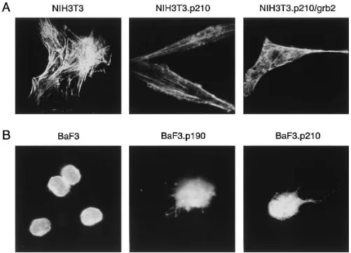
Figure 1. Differences in F-actin staining of untransformed and BCR/ABL transformed cells. Cells were fixed and stained for F-actin using rhodamine-labeled phalloidin. (A) Shown are NIH3T3 cells, NIH3T3 cells transfected by BCR/ABL (NIH3T3.p210), and NIH3T3 cells trans-formed by BCR/ABL and Grb2 (NIH3T3.p210/grb2). (B) Shown are BaF3 cells, p190BCR/ABL transfected BaF3 cells (BaF3.p190), and
Preparation of cell lysates and immunoblotting. Cell lysates were prepared and separated by 7.5% SDS-PAGE under reducing condi-tions, electrophoretically transferred to Immobilon PVDF (Millipore Corp., Bedford, MA) as described (19). The membranes were immuno-blotted with mouse monoclonal antibodies against phosphotyrosine (1:1000, clone 4G10, courtesy of Dr. Brian Druker, Oregon Health Science University, Portland, OR), paxillin (1:5,000, clone 5H11 [20]), talin (1:2,000, clone 8d4; Sigma Chemical Co., St. Louis, MO), focal adhesion kinase (FAK) (1:500; Transduction Labs, Lexington, KY), and vinculin (1:2,000, clone VIN-11c, Sigma) using previously de-scribed methods (9, 19). Signals were detected using an enhanced chemiluminescense technique (Amersham, Arlington Heights, IL) (21).
F-actin staining of cells. F-actin was visualized in fixed cells (1% paraformaldehyde in phosphate-buffered saline) using rhodamine phalloidin (Molecular Probes, Eugene, OR) as previously described (9).
Adhesion assay of BaF3 cells. Adhesion of BaF3, BaF3.p190BCR/ ABL, and BaF3.p210BCR/ABL cells was measured as described (15) on
plastic plates that were uncoated, coated with BSA, or fibronectin coated (Becton Dickinson Labware, Bedford, MA).
Time-lapse video microscopy. Cells were cultured on uncoated plastic tissue culture plates or plates coated with the extracellular ma-trix proteins fibronectin, collagen IV, collagen I, laminin, or vitronec-tinin (35-mm plates; Becton Dickinson Labware) in a temperature controlled chamber in their standard growth media. The cells were examined by video microscopy utilizing a Leitz inverted microscope (Diavert), Omega temperature control device, Hamamatsu C2400 video camera, MTI HR1000 TV, and Panasonic time-lapse S-VHS video recorder. The digital video images were captured and printed with a Kodak DS 8650 digital printer.
a-interferon treatment of cells. BaF3, BaF3.p210, ts-BaF3.p210 cells were plated onto fibronectin-coated plates and treated with mouse
a-interferon (a gift from Dr. Glenn Dranoff, Dana-Farber Cancer In-stitute) using doses of 0–1,000 U/ml for 12 h and observed with
time-lapse video microscopy for 24 h. CD341 CML cells were also treated with 1,000 U/ml human a-interferon (Schering Corporation, Kenil-worth, NJ) and observed with time-lapse video microscopy.
Results
BCR/ABL changes the actin cytoskeleton structure.F-actin was stained with rhodamine–phalloidin and examined using fluo-rescence microscopy (Fig. 1). The F-actin staining of NIH3T3 fibroblasts showed typical actin stress-fiber formation, whereas NIH3T3 cells transformed by BCR/ABL tended to be “spindle” shaped and had long cytoplasmic extensions con-taining actin (“filopodia”). Also, F-actin scon-taining of BCR/ ABL-transfected BaF3 cells (BaF3.p210, BaF3.p190) was brighter and the cells had altered morphology (containing ab-normal extensions with actin) as compared with ab-normal BaF3 cells. These results suggested that BCR/ABL affects the for-mation of actin-containing cell extensions and prompted us to examine the kinetics of formation of cell extensions such as filopodia and pseudopodia, and to look for abnormalities of motility.
Adhesion of BaF3 cells expressing BCR/ABL was mea-sured on fibronectin, and compared to untransformed BaF3 cells. In a 6-h adhesion assay, as shown in Fig. 2, the BaF3.p210 and BaF3.p190 cells have similar adhesion as BaF3 cells on fi-bronectin; however, the BCR/ABL-expressing cells have non-spherical shapes.
[image:4.612.56.554.428.704.2]BCR/ABL induces abnormalities of spontaneous motility on fibronectin-coated surfaces. Several cell lines expressing BCR/ ABL were examined by time-lapse video microscopy, and compared to nontransformed cells. First, the effects of BCR/
Figure 2. Adhesion assay of BaF3 and BCR/ABL expressing BaF3 cells on fibronectin. BaF3 cells, BaF3.p190BCR/ABL, BaF3.p210BCR/ABL cells
Figure 3. BCR/ABL expressing fibroblasts have abnormal cell motility. Shown are time-lapse video microscopy pictures, within the same field, at 30-min intervals. Cells utilized are NIH3T3 cells (left panel), NIH3T3.p210 cells (middle panel), and NIH3T3.p210/grb2 cells (right panel). Ar-rows point to the abnormal extensions containing actin.
[image:5.612.58.556.378.681.2]ABL were examined in NIH3T3 cells transformed by BCR/ ABL (NIH3T3.p210) or NIH3T3 cells made to express both p210BCR/ABL and Grb2 (NIH3T3.p210/grb2) (Fig. 3). Compared
to untransformed NIH3T3 cells, the NIH3T3.p210 cells had
abnormal filopodia extensions in z 10% of the population.
In-terestingly, the NIH3T3.p210/grb2 cells (which have a more transformed phenotype than NIH3T3.p210 cells) have many more filopodia, more lamellipodia and membrane ruffles, and an increased rate of spontaneous motility. The time-lapse photographs revealed that BCR/ABL expressing cells had a strikingly faster spontaneous migration rate than the untrans-formed fibroblasts. These differences were observed on fibro-nectin-coated as well as uncoated surfaces.
Using time-lapse video microscopy, untransformed BaF3 cells on fibronectin coated surfaces were found to be primarily rounded in shape, with little evidence of membrane ruffling or spontaneous motility. At any point in time, , 10% of cells had formed pseudopodia or other types of cell extensions, or were actively moving. In contrast, . 50% of BaF3.p210 cells cul-tured on fibronectin formed multiple pseudopodia or protru-sions, with some of the cells having long extensions similar to filopodia (Fig. 4). Similarly, . 50% of cells were spontane-ously moving and rarely had quiescent periods where they assumed a rounded morphology (with pseudopodia or other protrusions) and stopped moving. The exception was during mitosis, when all cells (BCR/ABL positive or negative) stopped migrating, became rounded and proceeded through cell divi-sion. BaF3.p210 cells also had an increased level of spontane-ous membrane motion interpreted as membrane ruffling com-pared to untransformed cells.
[image:6.612.56.299.58.252.2]The temperature-sensitive ts-BaF3.p210 cell line (18) is a
Figure 5. BaF3 cells ex-pressing the tempera-ture sensitive construct of BCR/ABL demon-strates the inducible ty-rosine phosphorylation of several cellular pro-teins by the BCR/ABL
oncogene. Growth fac-tor-starved BaF3 cells stably transfected with a temperature sensitive
BCR/ABL construct were transferred for the indicated times from the nonpermissive tem-perature (398C) to the permissive tempera-ture (338C). Total cell lysates (2.5 3 105 cells)
were applied to a 7.5% SDS-PAGE gel, transferred to PVDF-mem-brane, and immunoblotted with antiphosphotyrosine (4G10) anti-body. Molecular mass is shown in kD.
[image:6.612.61.557.370.680.2]useful model since at the nonpermissive temperature (398C) these cells have low BCR/ABL tyrosine kinase activity and be-have as untransformed BaF3 cells, and at permissive tempera-ture (338C) the BCR/ABL kinase is activated and phosphory-lates several new cellular proteins (Fig. 5). On fibronectin plates, the ts-BaF3.p210 cells were similar to normal BaF3 cells at 398C (the cells were mostly sessile, had a spherical morphol-ogy with few or no pseudopodia and exhibited slow move-ment), whereas, at 338C they develop the increased motility and abnormal morphology of BaF3.p210 cells (with increased migration even on fibronectin surfaces, more disconcerted movement, and formation of multiple pseudopodia at the same time) (Fig. 6).
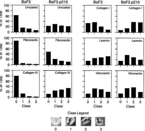
A relative scale was developed to quantitate the degree of cell shape change associated with BCR/ABL-transformation by assigning a grade from 0 (spherical morphology) to 3 (mul-tiple pseudopodia and/or filopodia) (Fig. 7). Single frames were captured from the time-lapse videomicroscopy of cells on fibronectin-coated plates and 300–500 cells were evaluated.
This scale provides a simple way to quantitate the degree of BCR/ABL-induced cell morphology changes and also pro-vides a means of assessing the different degrees of motility ob-served from the video.
[image:7.612.61.556.58.394.2]These studies indicate that BaF3 cells transformed by BCR/ABL have increased spontaneous motility on fibronec-tin-coated surfaces. The morphology and motility of BaF3 and BaF3.p210 cells on fibronectin-coated surfaces was then com-pared to that on uncoated surfaces and surfaces coated with other extracellular matrix proteins (collagen IV, collagen I, laminin, and vitronectin). It is interesting that the degree of spontaneous motility of the BaF3 cells was quite variable on the different surfaces. On collagen I, laminin, vitronectin, and uncoated plastic, BaF3 cells had considerably higher motility than they did on fibronectin or collagen IV, although the de-gree of motility did not reach that seen by BCR/ABL trans-formed BaF3 cells on the same surfaces. In contrast to BaF3 cells, BCR/ABL transformed cells were hyperactive on all of the surfaces (Fig. 8).
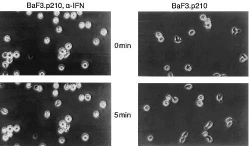
a-interferon reduces cell motility and pseudopod formation of BCR/ABL expressing cells on a fibronectin-coated surface.
a-interferon is useful in the therapy of patients with stable phase CML, although the mechanism of action is unknown. We examined the effects of a-interferon on the cell morphol-ogy and motility of normal and CML cells. a-interferon treat-ment (up to 1,000 U/ml) did not change the spontaneous motil-ity of normal BaF3 cells (data not shown). However, treatment of BaF3.p210 cells with a-interferon (1,000 U/ml for 12 h) re-sulted in reduced spontaneous motility, reduced number of protrusions and pseudopodia per cell, and increased number of cells with spherical morphology (Figs. 7 and 9). Careful mi-croscopic examination of BCR/ABL-transformed cell lines
cultured with a-interferon shows that although they are no longer motile, they still have dramatic ruffling of the cell membrane. Thus, a-interferon normalizes one feature of BCR/ABL-induced cytoskeletal abnormality, but does not affect the increased overall degree of membrane ruffling ac-tivity.
The effects of a-interferon were also examined in the ts-BaF3.p210 cell line model on fibronectin surfaces. When a-interferon was added at the permissive temperature, the cells became less motile and reverted to the morphology and motility rate similar to the cells originally at the nonpermissive temperature (data not shown). When added at the nonpermis-sive temperature, a-interferon had no visible effect, but
[image:8.612.62.531.61.483.2]vented the disordered motility of cells later switched to the permissive temperature.
Cell motility of CD341 progenitor cells from CML patients is increased compared with normal controls on a fibronectin-coated surface. CD341 progenitor cells were enriched by FACS from bone marrow samples from four patients with stable phase CML, and four normal subjects. The spontaneous motility and morphology of the CD341 population from CML patients was different than normal progenitors as viewed by video microscopy on fibronectin-coated plates (Fig. 10). Shown in Fig. 10 are two representative samples from two normal and three CML patients. The CML cells, like the BaF3.p210BCR/ABL
cells, have multiple pseudopodia and protrusions, and sponta-neous motility of the cells was increased. A CML sample was also treated in vitro with a-interferon causing the cells to be-come more adherent to fibronectin and have a spherical mor-phology.
The focal adhesion protein paxillin is degraded in response to BCR/ABL. The results above show that BCR/ABL is asso-ciated with accelerated formation and retraction of pseudopo-dia and filopopseudopo-dia in BaF3 cells, NIH3T3 cells, and primary patient cells. We have previously shown that BCR/ABL phos-phorylates a number of proteins associated with actin or focal adhesions, including tensin, talin, p125FAK, vinculin, and
paxil-lin (9). We asked if the increased rate of cytoskeletal rear-rangement was also associated with degradation of any cyto-skeletal proteins. In NIH3T3 cells transformed by BCR/ABL,
there was accumulation of several lower molecular mass forms of paxillin (46 and 33 kD) which are recognized by an anti-pax-illin monoclonal antibody we have previously generated (Fig. 11) (20). As previously shown, the anti-paxillin antibody (clone 5H11) is specific in recognizing paxillin and its degrada-tion products (20).
We have demonstrated that the p46 and p33 forms are also tyrosine phosphorylated in BCR/ABL-transformed cells (data not shown) by immunoprecipitation studies. No significant ac-cumulation of lower molecular weight forms of vinculin or talin was detected by immunoblotting in BCR/ABL trans-formed cells. Cells were also treated with a-interferon to de-termine if there was any accumulation of the p68 form of paxil-lin; however, there was no difference in the a-interferon treated cells with no treatment (data not shown).
Discussion
[image:9.612.58.556.57.349.2]The Ph chromosomal translocation that causes CML fuses the c-ABL protooncogene from chromosome 9 to part of the BCR gene on chromosome 22. 59BCR exons typically join with the second exon of c-ABL to form BCR/ABL (22). The resulting fusion gene encodes a 210-kD oncoprotein in which the ty-rosine kinase activity of c-ABL is increased by fivefold or more (23). This activation of c-ABL tyrosine kinase is essential for transformation of myeloid and lymphoid target cells (16, 24). An alternative breakpoint in BCR results in the formation
of a smaller fusion protein, p190BCR/ABL, usually associated
with Ph(1) ALL (25). The kinase activity of p190BCR/ABL is
also elevated (26).
The mechanism whereby this oncogene transforms myeloid and lymphoid cells is unknown. Since the tyrosine kinase activ-ity of ABL is required, it is likely that one or more kinase sub-strates exist that transmit signals from p210BCR/ABL. It is
inter-esting that the cellular localization of BCR/ABL is believed to be important for transformation, and at least 70% of the pro-tein is localized to the cytoskeleton (27). Van Etten et al. (10, 11) and McWhirter et al. (8, 27) have identified an actin bind-ing site at the COOH terminus of the ABL protein. In some assays of transformation by BCR/ABL, retention of this actin binding domain is required for full transforming activity.
How-ever, we have used immunofluorescence microscopy to show that p210BCR/ABL has a punctate peripheral staining pattern in
addition to its actin localization, and further that proteins char-acteristic of focal adhesions, such as paxillin and vinculin, tend to colocalize with BCR/ABL in these punctate structures (9). Thus, our previous studies suggest that BCR/ABL accumu-lates in cytoskeletal structures that are likely to be involved in regulation of a variety of important cell functions, including binding to extracellular matrix proteins, cell motility, and cell morphology. The specific attachment sites of p210BCR/ABL
[image:10.612.64.438.52.553.2]within the cytoskeleton, in addition to actin, are currently un-known; although we have previously shown that BCR/ABL can coprecipitate with paxillin (19). In any event, modification of the actin cytoskeleton and cytoskeleton-associated proteins
Figure 10. Cell motility of CD341 progenitor cells from CML cells is abnormal, as com-pared to normal controls. Bone marrow samples from 3 patients with CML stable phase, and 2 normal volunteers, were ob-tained and CD341 progenitor cells were isolated by FACS cell sorting after labeling with anti-CD34-PE antibody. On fibronec-tin-coated plates, the CD341
population from CML stable phase patients was different than normal progenitors as viewed by time-lapse video microscopy. The CML cells, like the BaF3.p210 cells, were more migratory, and had multiple pseudopodia and filopodia. Also one CML CD341
may be an important event in leukemogenesis (12). The results presented here suggest that the localization of BCR/ABL to the cytoskeleton results in more profound abnormalities of cy-toskeletal function than were previously appreciated. Specifi-cally, we observed increased staining for filamentous actin and an enhanced rate of formation and retraction of actin-contain-ing protrusions such as pseudopodia and filopodia. Further, BCR/ABL-containing cells had increased level of spontaneous motility and much shorter periods of quiescence compared with nontransformed cells. These abnormalities required BCR/ABL tyrosine kinase activity and were observed in both
BCR/ABL-transformed cell lines and primary CML progeni-tor cells isolated from patients with active leukemia.
The observed differences between untransformed and
BCR/ABL-transformed cells were dependent on the surface to some degree. Untransformed BaF3 cells exhibited rounded morphology and little spontaneous motility on fibronectin and collagen IV, but higher motility and active pseudopod forma-tion on laminin, collagen I, vitronectin, and uncoated plastic. The surface did not have any significant effect on BCR/ABL-transformed cells, which displayed active motility and pseudo-pod formation on all surfaces. These results can be interpreted in two ways. First, it is possible that BaF3 cells are inherently motile, and attachment to fibronectin and collagen IV send signals that inhibit motility. Second, BaF3 cells are inherently nonmotile, and laminin, collagen I, vitronectin, and uncoated plastic send signals inducing motility. In the first case, BCR/ ABL would inhibit the down-modulating signals of fibronectin and collagen IV receptors. In the second case, BCR/ABL would send signals that lead to hypermotility. The results we have obtained so far do not allow a clear distinction between these possibilities, although the fact that BCR/ABL -trans-formed cells are more motile and morphologically aberrant compared to untransformed cells favors the latter hypothesis.
The significance of finding multiple abnormalities of cy-toskeletal function may underlie some of the unusual clinical aspects of CML. In contrast to most other human leukemia on-cogenes, BCR/ABL does not block differentiation, and does not appear to provide a major mitogenic signal. The major
hallmarks of CML, early release of myeloid cells from the mar-row and accumulation of myeloid cells at all stages of differen-tiation, are difficult to explain (2). One possibility is that the dysregulated motility observed here on certain surfaces may correlate with accelerated exit from the marrow of immature cells in CML patients (13), and it will be of interest to study motility on more complex surfaces such as marrow stromal cells. In the marrow microenvironment, which is likely to have abundant extracellular matrix proteins, the enhanced motility of CML cells could facilitate their early release and subsequent accumulation in other organs such as the spleen. The develop-ment of assays that can be used to study migration from the marrow would be helpful to further evaluate these hypotheses (28). Another possibility is that the hypermotility observed here is simply a manifestation of the abnormal activation of signal transduction pathways in the cytoskeleton by BCR/ABL (9, 19). The biological effects of such signaling activation may be profound. In most normal cells, signals from a variety of cell surface receptors are required to support growth and viability. In many cell types, the mitogenic signal from a growth factor receptor is insufficient, by itself, for normal cell proliferation and viability, and signals from adhesion or costimulatory re-ceptors are also required (29). Such signals might indicate that the cell is located in the “proper” microenvironment, perhaps, or is in contact with other cells that are important for normal regulation of a particular lineage or tissue (28).
One interesting observation reported here is that a biologic therapy, a-interferon, long known to have significant thera-peutic activity in CML (30) reduces some of the hypermotility of these cells. In the early 1980s a-interferon was shown to be beneficial for the therapy for CML. Randomized studies have suggested a survival advantage for patients treated with a-interferon compared with conventional chemotherapy (31). The time of progression to blastic phase is prolonged, and there is a higher rate of cytogenetic remission. Remarkably, although a-interferon is the only therapy other than bone marrow transplantation to induce cytogenetic remission, the mechanism of its action is not understood. At high concen-trations, in vitro, a-interferon inhibits the proliferation of both normal and CML cells, but does not appear to be differ-entially more active on CML cells (30). It has recently been proposed that a-interferon may overcome the defective adher-ence of CML progenitors to stromal cells. a-interferon in-creases the expression of the adhesion molecule LFA-3, which is decreased in CML cells (32). Defects in b1 integrin function have been shown to be corrected by a-interferon (33). In our study, a-interferon treatment of BCR/ABL-expressing cells and CML patient samples reverted the abnormal motility and morphology to a more normal phenotype. It will be of interest to examine motility of cells from patients treated with a -inter-feron to look for in vivo effects of a-interferon that might cor-relate with our in vitro studies. Further, it may be of interest to compare the effects of a-interferon on cells from CML patients who are either sensitive or resistant to a-interferon therapy.
[image:11.612.58.301.59.224.2]It will be important to determine the links between BCR/ ABL and the cytoskeletal abnormalities described here. In preliminary studies, we have found that BCR/ABL induces accumulation of degradation products of the focal adhesion protein paxillin. The antibody for paxillin recognizes paxillin at 68 kD (p68), and also degradation products of p68 at 46 kD (p46) and at 33 kD (p33) (20). In vitro, similar lower molecular
Figure 11. Expression of cytoskeletal pro-teins, and their degrada-tion in BCR/ABL con-taining fibroblasts. Whole cell lysates (50
weight forms of paxillin can be generated by proteolytic cleav-age of paxillin by calpain II (34). These smaller molecular weight forms of paxillin may actually be signaling proteins since we have observed that the adapter protein CRKL ([35], and unpublished observations) and the protooncoprotein p120CBL (36) form a complex with p46 and p33 proteins to a
much greater extent than with p68 paxillin itself. We have also observed the increased tyrosine phosphorylation of the p46 and p33 forms in CML hematopoietic cells (19). In any event, the apparent accumulation of low molecular weight forms of paxillin may be a biochemical marker for the disordered hy-permotility of the BCR/ABL-transformed cell.
In summary, we show here that BCR/ABL globally dis-rupts the function of the cell machinery that interacts with the microenvironment. The outcome may be a cell with disordered motility that is also receiving false “signals” that indicate that certain adhesion receptors (such as integrins) are stimulated even in the absence of actual ligand binding. These cells still need hematopoietic growth factors for growth and viability, but may be able to migrate into capillaries at an immature stage of development, and proliferate in tissues where myeloid cells are not normally found. Overall, we suggest that the sig-nificant abnormalities of spontaneous motility we have found may be a fundamental abnormality of CML cells. If so, then future studies in which abnormalities of signal transduction pathways are analyzed in CML cells should be done with the view to determining their contribution to hypermotility as well as other biological functions ascribed to BCR/ABL.
Acknowledgments
This work was supported by NIH grant CA-01730 (R. Salgia), Jose Carreras International Leukemia Foundation fellowship FIJC-95/ INT (M. Sattler), and NIH grant DK560654 (J.D. Griffin).
References
1. Rowley, J. 1973. A new consistent chromosomal abnormality in chronic myelogenous leukaemia identified by quinacrine fluorescence and Giemsa staining. Nature (Lond.). 243:290–293.
2. Canellos, G.P. 1996. Diagnosis and treatment of chronic granulocytic leu-kemia. In Neoplastic Diseases of the Blood. P.H. Wiernik, G.P. Canellos, J.P. Dutcher, and R.A. Kyle, editors. Churchill Livingstone, Inc., New York. 65–80. 3. Gishizky, J., and O. Witte. 1992. Initiation of deregulated growth of mul-tipotent progenitor cells by BCR-ABL in vitro. Science (Wash. DC). 256:836– 839.
4. Matulonis, U., R. Salgia, K. Okuda, B. Druker, and J. Griffin. 1993. Inter-leukin-3 and p210 BCR/ABL activate both unique and overlapping pathways of signal transduction in a factor-dependent myeloid cell line. Exp. Hematol. 21: 1460–1466.
5. Bedi, A., C.A. Griffin, J.P. Barber, M.S. Vala, A.L. Hawkins, S.J. Sharkis, B.A. Zehnbauer, and R.J. Jones. 1994. Growth factor-mediated termi-nal differentiation of chronic myeloid leukemia. Cancer Res. 54:5535–5538.
6. Bedi, A., B.A. Zehnbauer, J.P. Barber, S.J. Sharkis, and R.J. Jones. 1994. Inhibition of apoptosis by bcr-abl in chronic myeloid leukemia. Blood. 83:2038– 2044.
7. Carlesso, N., J.D. Griffin, and B.J. Druker. 1994. Use of a temperature sensitive mutant to define the biological effects of the p210bcr/abl tyrosine ki-nase on proliferation of a factor-dependent murine myeloid cell line. Oncogene.
9:149–156.
8. McWhirter, J., and J. Wang. 1993. An actin-binding function contributes to transformation by the Bcr-Abl oncoprotein of Philadelphia chromosome-positive human leukemias. EMBO J. 12:1533–1546.
9. Salgia, R., B. Brunkhorst, E. Pisick, J.-L. Li, S.H. Lo, L.B. Chen, and J.D. Griffin. 1995. Increased tyrosine phosphorylation of focal adhesion proteins in myeloid cell lines expressing p210BCR/ABL. Oncogene. 11:1149–1155.
10. Van Etten, R., P. Jackson, D. Baltimore, M. Sanders, P. Matsudaira, and P. Janmey. 1994. The COOH terminus of the c-Abl tyrosine kinase contains distinct F-and G-actin binding domains with bundling activity. J Cell Biol. 124:
325–340.
11. Wetzler, M., M. Talpaz, R. Van Etten, C. Hirsh-Ginsberg, M. Beran, and R. Kurzrock. 1993. Subcellular localization of Bcr, Abl, and Bcr-Abl pro-teins in normal and leukemic cells and correlation of expression with myeloid differentiation. J. Clin. Invest. 92:1925–1939.
12. Renshaw, M.W., J.R. McWhirter, and J. Wang. 1995. The human leuke-mia oncogene bcr-abl abrogates the anchorage requirement but not the growth factor requirement for proliferation. Mol. Cell Biol. 15:1286–1293.
13. Gordon, M.Y., C.R. Dowding, G.P. Riley, J.M. Goldman, and M.F. Greaves. 1987. Altered adhesive interactions with marrow stroma of haemato-poietic progenitor cells in chronic myeloid leukaemia. Nature (Lond.). 328:342– 344.
14. Verfaillie, C.M., J.B. McCarthy, and P.B. McGlave. 1992. Mechanisms underlying abnormal trafficking of malignant progenitors in chronic myeloge-nous leukemia. J. Clin. Invest. 90:1232–1241.
15. Bazzoni, G., N. Carlesso, J.D. Griffin, and M.E. Hemler. 1996. Bcr/abl expression stimulates integrin function in hematopoietic cell lines. J. Clin. In-vest. 98:521–528.
16. Daley, G., and D. Baltimore. 1988. Transformation of an interleu-kin-3-dependent hematopoietic cell line by the chronic myelogenous leukemia-specific p210 BCR/ABL protein. Proc. Natl. Acad. Sci. USA. 85:9312–9316.
17. Sattler, M., R. Salgia, K. Okuda, N. Uemura, M. Durstin, E. Pisick, G. Xu, J.-L. Li, K.V. Prasad, and J.D. Griffin. 1996. The proto-oncogene product p120CBL and the adaptor proteins CRKL and c-CRK link c-ABL, p190BCR/ ABL, and p210BCR/ABL to the phosphatidylinositol-39 kinase pathway. On-cogene. 12:839–846.
18. Kabarowski, J., P.B. Allen, and L.M. Wiedemann. 1994. A temperature sensitive p210 bcr-abl mutant defines the primary consequences of bcr-abl ty-rosine kinase expression in growth factor dependent cells. EMBO J. 13:5887– 5895.
19. Salgia, R., J. Li, S.H. Lo, B. Brunkhorst, G.S. Kansas, E.S. Sobhany, Y.P. Sun, E. Pisick, M. Hallek, T. Ernst, R. Tantravahi, L.B. Chen, and J.D. Griffin. 1995. Molecular cloning of human paxillin, a focal adhesion protein phosphorylated by p210(bcr/abl). J. Biol. Chem. 270:5039–5047.
20. Salgia, R., S. Avraham, E. Pisick, J.-L. Li, S. Raja, E. Greenfield, M. Satt-ler, H. Avraham, and J. Griffin. 1996. The related adhesion focal tyrosine ki-nase forms a complex with paxillin in hematopoietic cells. J. Biol. Chem. 271: 31222–31226.
21. Sattler, M., M.A. Durstin, D.A. Frank, K. Okuda, K. Kaushansky, R. Salgia, and J. Griffin. 1995. The thrombopoietin receptor c-MPL activates Jak2 and TYK2 tyrosine kinases. Exp. Hematol. 23:1040–1048.
22. Chissoe, S.L., A. Bodenteich, Y.F. Wang, Y.P. Wang, D. Burian, S.W. Clifton, J. Crabtree, A. Freeman, K. Iyer, J.A. Li, Y.C. Ma, H.J. Mclaury, H.Q. Pan, O.H. Sarhan, S. Toth, Z.L. Wang, G.Z. Zhang, N. Heisterkamp, J. Grof-fen, and B.A. Roe. 1995. Sequence and analysis of the human abl gene, the bcr gene, and regions involved in the philadelphia chromosomal translocation. Ge-nomics. 27:67–82.
23. Lugo, T., A. Pendergast, A. Muller, and O. Witte. 1990. Tyrosine kinase activity and transformation potency of bcr/abl oncogene products. Science (Wash. DC). 247:1079–1082.
24. Daley, G.Q., J. McLaughlin, O.N. Witte, and D. Baltimore. 1987. The CML-specific P210 bcr/abl protein, unlike v-abl, does not transform NIH/3T3 fibroblasts. Science (Wash. DC). 237:532–535.
25. Fainstein, E., C. Marcelle, A. Rosner, E. Canaani, R.P. Gale, O. Drea-zen, S.D. Smith, and C.M. Croce. 1987. A new fused transcript in Philadelphia chromosome positive acute lymphocytic leukaemia. Nature (Lond.). 330:386– 388.
26. Walker, L.C., T.S. Ganesan, S. Dhut, B. Gibbons, T.A. Lister, J. Roth-bard, and B.D. Young. 1987. Novel chimaeric protein expressed in Philadelphia positive acute lymphoblastic leukaemia. Nature (Lond.). 329:851–853.
27. McWhirter, J., and J. Wang. 1991. Activation of tyrosine kinase and mi-crofilament-binding functions of c-abl by bcr sequences in bcr/abl fusion pro-teins. Mol. Cell Biol. 11:1553–1565.
28. Pedersen, T., K. Yong, J. Pedersen, N. Hansen, K. Dano, and T. Plesner. 1996. Impaired migration in vitro of neutrophils from patients with paroxysmal nocturnal haemoglobinuria. Br. J. Haematol. 95:45–51.
29. Levesque, J.P., D.N. Haylock, and P.J. Simmons. 1996. Cytokine regula-tion of proliferaregula-tion and cell adhesion are correlated events in human CD34 (1) hemopoietic progenitors. Blood. 88:1168–1176.
30. Verma, D., G. Spitzer, J. Gutterman, A. Zander, K. McCradie, and K. Dicke. 1979. Human leukocyte interferon preparation blocks granulopoietic differentiation. Blood. 54:1423–1427.
31. Talpaz, M., H.M. Kantarjian, K. McCredie, J.M. Trujillo, M.J. Keating, and J.U. Gutterman. 1986. Hematologic remission and cytogenetic improve-ment induced by recombinant human interferon alpha A in chronic myeloge-nous leukemia. N. Engl. J. Med. 314:1065–1069.
32. Bhatia, R., E.A. Wayner, P.G. McGlave, and C.M. Verfaillie. 1994. In-terferon-a restores normal adhesion of chronic myelogenous leukemia hemato-poietic progenitors to bone marrow stroma by correcting impaired b1 integrin receptor function. J. Clin. Invest. 94:384–391.
prolifera-tion by the marrow microenvironment in chronic myelogenous leukemia.
Blood. 87:3883–3891.
34. Yamaguchi, R., M. Maki, M. Hatanaka, and H. Sabe. 1994. Unphosphor-ylated and tyrosine-phosphorUnphosphor-ylated forms of a focal adhesion protein, paxillin, are substrates for calpain ii in vitro-implications for the possible involvement of calpain ii in mitosis–specific degradation of paxillin. FEBS Lett. 356:114–116.
35. Salgia, R., N. Uemura, K. Okuda, J.-L. Li, E. Pisick, M. Sattler, R.
de-Jong, B. Druker, N. Heisterkamp, L. Chen, J. Groffen, and J. Griffin. 1995. CRKL links p210BCR/ABL with paxillin in chronic myelogenous leukemia cells. J. Biol. Chem. 270:29145–29150.