Suppression of arthritic bone destruction by
adenovirus-mediated csk gene transfer to
synoviocytes and osteoclasts
Hiroshi Takayanagi, … , Kozo Nakamura, Sakae Tanaka
J Clin Invest.
1999;104(2):137-146. https://doi.org/10.1172/JCI6093.
Rheumatoid arthritis (RA) is characterized by a chronic inflammation of the synovial joints
resulting from hyperplasia of synovial fibroblasts and infiltration of lymphocytes,
macrophages, and plasma cells, all of which manifest signs of activation. Recent studies
have revealed the essential role of osteoclasts in joint destruction in RA. Src family tyrosine
kinases are implicated in various intracellular signaling pathways, including mitogenic
response to growth factors in fibroblasts, activation of lymphocytes, and osteoclastic bone
resorption. Therefore, inhibiting Src activity can be a good therapeutic strategy to prevent
joint inflammation and destruction in RA. We constructed an adenovirus vector carrying the
csk
gene, which negatively regulates Src family tyrosine kinases. Csk overexpression in
cultured rheumatoid synoviocytes remarkably suppressed Src kinase activity and reduced
their proliferation rate and IL-6 production. Bone-resorbing activity of osteoclasts was
strongly inhibited by Csk overexpression. Furthermore, local injection of the virus into rat
ankle joints with adjuvant arthritis not only ameliorated inflammation but suppressed bone
destruction. In conclusion, adenovirus-mediated direct transfer of the
csk
gene is useful in
repressing bone destruction and inflammatory reactions, suggesting the involvement of Src
family tyrosine kinases in arthritic joint breakdown and demonstrating the feasibility of
intervention in the kinases for gene therapy in RA.
J. Clin. Invest
.
104
:137–146 (1999).
Article
Introduction
Rheumatoid arthritis (RA) is a chronic inflammatory dis-ease characterized by invasive synovial hyperplasia lead-ing to progressive joint destruction. Investigations into the pathogenesis of joint destruction in RA have revealed the transformed phenotype of rheumatoid synovial cells (1, 2), although the etiology of RA remains unknown. Rheumatoid synovial cells are not only morphologically characterized by their transformed appearance (3) but also are phenotypically transformed to proliferate abnor-mally (4, 5); they invade bone and cartilage by producing an elevated amount of proinflammatory cytokines (6) and metalloproteinases (7) and by inducing osteoclast formation and activation (8, 9). Their activated pheno-type is marked by an upregulation of proto-oncogenes (10), which are involved in important cellular events such as intracellular signaling and gene transcription. Osteo-clasts, the multinucleated cells exclusively responsible for bone resorption, have been observed to resorb bone actively at the site of invasion of the proliferated synovial membrane into the adjacent bone (11). The cell types
responsible for bone resorption in RA have been charac-terized as authentic osteoclasts (9), and we reported pre-viously that rheumatoid synovial fibroblasts are involved in bone destruction by inducing osteoclastogenesis (8). Thus, rheumatoid bone destruction, attributed to acti-vated synoviocytes and bone-resorbing osteoclasts, can-not be easily controlled by inhibiting only 1 of the factors involved in these multiple pathological processes.
Cytoplasmic tyrosine kinases of the Src family are involved in the regulation of a number of cellular processes, including signal transduction of various growth factor receptors and cytokine receptors (12, 13), immunorecognition of T cells and B cells (13), and acti-vation of osteoclasts (14). Consequently, Src family kinases are implicated in a variety of pathological events in RA, characterized by the proliferation of activated syn-oviocytes, abnormal immune responses, and elevated production of proinflammatory cytokines. Regulation of the activity of Src family kinases can therefore be a good therapeutic target in the treatment of RA.
The kinase activity of Src family kinases is negatively
Suppression of arthritic bone destruction
by adenovirus-mediated csk gene transfer
to synoviocytes and osteoclasts
Hiroshi Takayanagi,
1Takuo Juji,
1Tsuyoshi Miyazaki,
1Hideharu Iizuka,
1Tokiharu Takahashi,
2Masashi Isshiki,
3Masato Okada,
4Yoshiya Tanaka,
5Yasuko Koshihara,
6Hiromi Oda,
1Takahide Kurokawa,
1Kozo Nakamura,
1and Sakae Tanaka
11Department of Orthopaedic Surgery, 2Third Department of Internal Medicine, and
3Fourth Department of Internal Medicine, Faculty of Medicine, the University of Tokyo, Tokyo 113-0033, Japan 4Division of Protein Metabolism, Institute for Protein Research, Osaka University, Osaka 565-0871, Japan
5First Department of Internal Medicine, University of Occupational and Environmental Health, Kitakyushu 807-8555, Japan 6Department of Biosignal Research, Tokyo Metropolitan Institute of Gerontology, Tokyo, 173-0015, Japan
Address correspondence to: Sakae Tanaka, Department of Orthopaedic Surgery, Faculty of Medicine, the University of Tokyo, 7-3-1 Hongo, Bunkyo-ku, Tokyo 113-0033, Japan. Phone: 81-3-3815-5411 ext. 3375; Fax: 81-3-3818-4082; E-mail: TANAKAS-ORT@h.u-tokyo.ac.jp.
Received for publication December 18, 1998, and accepted in revised form June 9, 1999.
Rheumatoid arthritis (RA) is characterized by a chronic inflammation of the synovial joints resulting from hyperplasia of synovial fibroblasts and infiltration of lymphocytes, macrophages, and plasma cells, all of which manifest signs of activation. Recent studies have revealed the essential role of osteoclasts in joint destruction in RA. Src family tyrosine kinases are implicated in various intracellular signaling pathways, including mitogenic response to growth factors in fibroblasts, activation of lymphocytes, and osteoclastic bone resorption. Therefore, inhibiting Src activity can be a good therapeutic strategy to pre-vent joint inflammation and destruction in RA. We constructed an adenovirus vector carrying the csk
gene, which negatively regulates Src family tyrosine kinases. Csk overexpression in cultured rheuma-toid synoviocytes remarkably suppressed Src kinase activity and reduced their proliferation rate and IL-6 production. Bone-resorbing activity of osteoclasts was strongly inhibited by Csk overexpression. Fur-thermore, local injection of the virus into rat ankle joints with adjuvant arthritis not only ameliorated inflammation but suppressed bone destruction. In conclusion, adenovirus-mediated direct transfer of the cskgene is useful in repressing bone destruction and inflammatory reactions, suggesting the involve-ment of Src family tyrosine kinases in arthritic joint breakdown and demonstrating the feasibility of intervention in the kinases for gene therapy in RA.
regulated by another cytoplasmic tyrosine kinase, Csk (C-terminal Src kinase), by the phosphorylation of tyro-sine residue at the carboxyl-terminus (Tyr 527) (15, 16). In the present study, we constructed a replication-defi-cient adenovirus vector carrying the cskgene to investi-gate the effect of introducing the cskgene into synovio-cytes and osteoclasts in vitro and in vivo. Adenovirus-mediated overexpression of Csk, by repress-ing Src kinase activity, dramatically decreased the prolif-eration rate and IL-6 production of human rheumatoid synovial cells in culture; also, the bone-resorbing activi-ty of human osteoclasts formed in coculture of rheuma-toid synoviocytes and PBMCs was remarkably sup-pressed by Csk expression. Importantly, direct injection of Csk virus into rat ankles with adjuvant arthritis not only ameliorated the inflammatory reactions but also suppressed bone destruction.
Methods
Antibodies. Anti-Csk rabbit polyclonal antibody (C-20; Santa Cruz Biotechnology Inc., Santa Cruz, California, USA), anti–v-Src mouse mAb (clone 327; Oncogene Research Products, Cambridge, Massachusetts, USA), anti-Fyn mouse mAb (clone 15; Santa Cruz Biotech-nology Inc.), anti–c-Yes rabbit polyclonal antibody (clone 3; Santa Cruz Biotechnology Inc.), and anti–β -actin polyclonal antibody (Sigma Chemical Co., St. Louis, Missouri, USA) were used. For Western blotting, horseradish peroxidase–linked anti-rabbit IgG and anti-mouse IgG antibodies (Amersham Pharmacia Biotech, Buckinghamshire, United Kingdom) were used as secondary antibodies.
Synovial cell cultures.With the use of enzymatic diges-tion methods described previously (8, 18), primary rheumatoid synovial cells were obtained from synovial tissues dissected from 10 female patients (age range: 49–72 years) who fulfilled the American Rheumatism Association criteria for RA (17). In brief, the minced syn-ovial tissue was digested in 1 mg/mL collagenase (Wako Pure Chemicals Industries, Osaka, Japan), and the cell suspension was purified using Ficoll-Paque (Pharmacia Biotech AB, Uppsala, Sweden). The cells were suspended
in DMEM (GIBCO BRL, Rockville, Maryland, USA) plus 10% FBS (Cell Culture Laboratory, Cleveland, Ohio, USA) and used for experiments after 3–6 passages. Sub-cultured synovial cells were composed of synovial fibrob-lasts that were free of macrophages positively stained for CD11b or nonspecific esterase (data not shown). Informed consent to subsequent experiments was given by each patient.
Adenovirus vector construction and gene transduction in vitro.The replication-deficient recombinant adenovirus vector carrying the rat cskgene (AxCATcsk; Csk virus) under the control of the CAG [cytomegalovirus IE enhancer plus chicken β-actin promoter plus rabbit β -globin poly(A) signal] promoter was constructed using established methods (19). The control virus (Ax1w1; WT virus) and adenovirus vector carrying the β -galac-tosidase (β-gal) gene (AxCASLacZ; LacZ virus) were kindly provided by Izumu Saito (University of Tokyo). The titer of each virus was 1 ×10¹⁰plaque-forming units
(PFU) per milliliter. Infection of adenovirus vectors to cultured synoviocytes and osteoclasts was carried out following the method described previously (20). The cells in culture were incubated with adenovirus (1 ×10¹⁰
PFU/mL) diluted 1:10 in DMEM (the final titer of the virus was 1 ×10⁹PFU/mL) for 1 hour at 37°C, after
which 10 times more medium containing 10% FBS was added. The number of virus particles (measured in PFU) per cell was expressed as moi.
X-gal staining for β-gal expression in vitro. Rheumatoid synovial cells were infected with LacZ virus (moi = 10, 30, 100). After 24 hours, the cells were fixed with 3.7% formaldehyde in PBS for 15 minutes, and incubated overnight at 37°C in X-gal solution [5 mM K4Fe(CN)6,
5 mM K3Fe(CN)6, 2 mM MgCl2, and 2 mM
5-bromo-4-chloro-3-indolyl-β-D-galactoside (X-gal) in PBS] as
described (21).
Immunofluorescence staining and confocal microscopy.WT or Csk virus–infected (moi = 100) synoviocytes plated on glass coverslips were washed with PBS, fixed in 3.7% formaldehyde for 10 minutes, and permeabilized with 0.1% Triton X-100 in PBS for 5 minutes. Cells were sequentially incubated in PBS plus 1% BSA for 30 min-utes, 2.0 µg/mL rabbit anti-Csk polyclonal antibody in PBS for 1 hour, and then with 10 µg/mL goat Cy5-labeled anti-rabbit IgG antibody (FluoroLink; Amer-sham Pharmacia Biotech) in PBS for 1 hour. Microscopy was performed using a confocal microscope (MRC-1000UV; Bio-Rad Laboratories Inc., Hercules, California, USA) as described previously (22).
Protein extraction and Western blot analysis. Twenty-four hours after inoculation with Csk virus (moi = 10, 30, 100) or WT or LacZ virus (moi = 100), synovial cells were lysed in TNE buffer (10 mM Tris-HCl [pH 7.8], 150 mM NaCl, 1 mM EDTA, 1% NP-40, 2 mM Na3VO4,
10 mM NaF, and 10 µg/mL aprotinin). The super-natants were separated by SDS-PAGE under a reducing condition, transferred electrophoretically onto a nitro-cellulose membrane, and probed sequentially with 0.2 µg/mL anti-Csk antibody followed by 0.1 µg/mL horse-radish peroxidase–conjugated secondary antibody. Immunoreactive proteins were viewed by ECL (enhanced chemiluminescence) Western blotting
detec-Figure 1
[image:3.612.73.237.547.674.2]tion reagents (Amersham Pharmacia Biotech), follow-ing the manufacturer’s protocol.
Immunoprecipitation and in vitro kinase assay. Equal amounts of protein (100 µg) extracted from LacZ or Csk virus–infected synoviocytes (moi = 100) were incubated with 1 µg of anti-v-Src, anti-Fyn, or anti-Yes antibody for 1 hour at 4°C, and the immune complexes were recov-ered with protein G-Sepharose (GIBCO BRL). One half of the immunoprecipitate was used for in vitro kinase assay and the other half for Western blotting analysis, to ensure that an equal amount of c-Src protein was recov-ered. In vitro kinase assay was performed as reported pre-viously (23). Briefly, the samples were resuspended in 60 µL of kinase buffer (20 mM HEPES-NaOH [pH 7.4] and 10 mM MgCl2) with 1 µCi (37 kBq) of [γ-32P]ATP
(Amer-sham Pharmacia Biotech) in the presence of 1 µg of acid-treated enolase. They were then incubated for 15 min-utes at 30°C and subjected to 8% SDS-PAGE under a reducing condition followed by autoradiography.
Cell proliferation assay.Rheumatoid synovial cells (5 × 10⁴cells per well) seeded in culture plates were infected
with WT, LacZ, or Csk virus (moi = 100). On days 0, 2, 5, and 8, cells were recovered by trypsin-EDTA treat-ment, and their number was counted. Cell growth rate was also determined by a cell proliferation kit (Amer-sham Pharmacia Biotech) using immunostaining for a thymidine analogue, 5-bromo-2′-deoxyuridine (BrdU), incorporated into replicating DNA according to the manufacturer’s protocol.
ELISA for IL-6. Rheumatoid synovial cells (1 ×10⁵cells
per well) were infected with Csk virus (moi = 10, 30, 100) or WT or LacZ virus (moi = 100). FBS was removed from the medium 24 hours after inoculation. After an addi-tional 24 hours, conditioned medium was recovered, and IL-6 concentration in the medium was determined using a human IL-6 ELISA kit (Fujirebio Inc., Tokyo, Japan).
Northern blot analysis. Total RNA was extracted from rheumatoid synovial cells infected with Csk virus (moi = 10, 30, 100) or WT or LacZ virus (moi = 100) 24 hours after inoculation, using acid-guanidinium isothio-cyanate-phenol-chloroform (Isogen; Nippon Gene, Toya-ma, Japan) following the established method (24). Equal amounts (8 µg) of RNA were denatured in formaldehyde, separated on 1% agarose gel, and transferred to a nitro-cellulose membrane (Hybond-N; Amersham Pharmacia Biotech) followed by ultraviolet cross-linking. ExpressHyb hybridization solution (CLONTECH Labo-ratories Inc., Palo Alto, California, USA) was used accord-ing to the manufacturer’s protocol. The blots were hybridized with a cDNA probe labeled with [α-32P]dCTP
(NEM Life Science Products, Boston, Massachusetts, USA) by a random-primed DNA labeling kit (Boehringer Mannheim Biochemicals Inc., Indianapolis, Indiana, USA). Human IL-6 probe was the PCR product of syn-oviocytes using the primers described previously (25), and human β-actin probe was obtained from Wako Pure Chemicals Industries.
Resorption assay. Pit-forming assay by human osteo-clast-like cells (OCLs) was performed as described pre-viously (8). PBMCs isolated using Ficoll-Paque were cocultured with rheumatoid synovial fibroblasts in α -MEM containing 20% horse serum (GIBCO BRL) in the
presence of 10–7M 1,25(OH)
2 vitamin D3(Wako Pure
Chemicals Industries) and 300 U/mL recombinant human M-CSF (Austral Biologicals, San Ramon, Cali-fornia, USA). After 3 weeks, multinucleated OCLs formed in coculture were isolated by pronase E treat-ment. OCLs (2 ×10⁴cells per well) were seeded on
[image:4.612.307.540.57.432.2]cal-cium phosphate–coated discs (Millenium Biologix Inc., Kingston, Ontario, Canada) (day 0) and infected with WT or LacZ virus (moi = 100) or Csk virus (moi = 10,
Figure 2
30, 100). On day 5, the discs were washed in 6% sodium hypochloride to remove the adherent cells. The resorbed area was measured by scanning densitometry and analyzed by NIH Image software (National Insti-tutes of Health, Bethesda, Maryland, USA).
Animals and induction of adjuvant arthritis.Inbred male Sprague-Dawley rats were purchased from Sankyo Lab-oratory Services (Tokyo, Japan). The animals, 6–7 weeks old, were injected subcutaneously into the right hind paw (day 0) with 100 µL of Freund’s incomplete adjuvant (Difco Laboratories, Detroit, Michigan, USA) containing 10 µg/µL Mycobacterium butyricum (Difco Laboratories). Arthritis of the bilateral ankle joints developed in 100% of animals after day 7. The effect of Csk injection was evaluated in right ankle joints, which showed severe bone destruction, and in left ankle joints, which exhibit-ed significant inflammatory reactions.
β-gal expression in situ and Csk expression in vivo. Fifteen rats with adjuvant arthritis (immunized on day 0) were injected with 30 µL of LacZ virus into the inflamed right ankle joint on day 7. On days 14, 21, 28, 35, and 42 (3 rats each day), the right ankle joint was dissected out en bloc and cut longitudinally. After fixation in 3.7% formaldehyde for 12 hours, the tissues were decal-cified in 10% EDTA and embedded in paraffin to be sectioned. Serial sections were stained for β-gal and tar-trate-resistant acid phosphatase (TRAP) as described previously (21, 26). Counterstaining was performed by
nuclear fast red for β-gal, and by hematoxylin for TRAP. To determine in vivo expression of Csk by the aden-ovirus, 6 rats were injected with 30 µL Csk virus, and 2 rats with 30 µL WT virus, into the right ankle joint (day 0). Csk virus–injected rats were sacrificed on days 7, 21, and 42 (2 rats each day), and WT virus–injected rats (n
= 2) were sacrificed on day 7. The protein was extracted from the synovial and periarticular tissues and applied for Western blot analysis using anti-Csk antibody.
Therapeutic protocol.For virus injection into right ankles, 30 rats were immunized with adjuvant in the right foot (day 0). On day 7, the WT group (n = 10; body weight 151 ± 2.9 g) was intra-articularly injected with 30 µL of WT virus; the LacZ group (n = 10; body weight 150 ± 4.0 g), with 30 µL of LacZ virus; and the Csk group (n = 10; body weight 147 ± 6.7 g), with 30 µL of Csk virus. The viruses (3.0 ×10⁸virus particles per rat) were directly injected
into the inflamed right ankle joint space.
For virus injection into left ankles, 30 rats immunized in the right foot (day 0) were intra-articularly injected with 30 µL of WT virus (n = 10; body weight 148 ± 4.0 g), LacZ virus (n = 10; body weight 150 ± 5.1 g), and Csk virus (n = 10; body weight 150 ± 4.5 g) on day 7.
Assessment of therapeutic effects by Csk virus on adjuvant arthritis. Therapeutic effects of virus injection were exam-ined by arthritis score and paw volume on days 7, 14, 21, 28, and 35 under inhalation anesthesia with diethyl ether. The rats were sacrificed for radiological and
his-Figure 4
Effect of adenovirus-mediated Csk overexpression on IL-6 production. (a) IL-6 concentration in conditioned medium of rheumatoid synovio-cytes inoculated with WT, LacZ, or Csk virus detected by ELISA. IL-6 pro-duction was reduced to about 50% below the normal level in rheuma-toid synoviocytes inoculated with Csk virus at an moi of 10 or greater. Data are expressed as mean ± SD derived from 3 separate experiments (*P < 0.05 vs. LacZ). (b) IL-6 mRNA detected by Northern blot analysis in WT, LacZ, or Csk virus–infected synoviocytes. Significant decrease in IL-6 mRNA signals was observed in synovial cells infected with Csk virus at an moi of 10 or greater. Fold expression of IL-6 mRNA normalized by
[image:5.612.355.534.51.142.2]β-actin is shown below each blot. Data are expressed as the mean derived from 3 separate experiments (*P < 0.05 vs. LacZ).
Figure 3
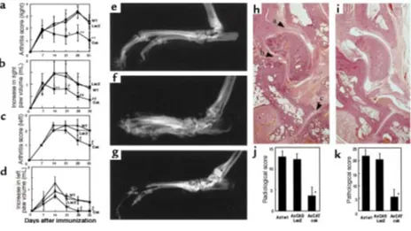
[image:5.612.320.521.503.732.2]tological examinations on day 35. These evaluations were performed by the same observer without knowledge of treatment. (a) Arthritis score. As described previously (27), with modifications, the severity was scored by appearance: 0 = normal, 1 = redness or mild swelling (confined to ankle or toes), 2 = mild swelling (extending from ankle to toes), 3 = moderate swelling, 4 = severe swelling, often with ulceration or ankylosis (maximum 4). (b) Paw volume. The hind-paw volume was measured with an instrument for plethysmography (TK-101; Uni-com, Tokyo, Japan) by vertically immersing the paw to the level of the proximal end of the lateral malleolus in a water-filled tub. (c) Radiological score. Right hind paws were dissected on day 35 and radiologically examined by soft x-ray machine (CMB-2; Softex, Tokyo, Japan). A 0–3 subjective grading system (0 = normal, 1 = mild, 2 = mod-erate, 3 = severe) described previously (28), with modifi-cations, was used to evaluate 5 different parameters including cartilage loss (joint space narrowing), sub-chondral bone erosion, osteoporosis, heterotopic ossifi-cation, and periosteal new bone formation. The radio-logical score refers to the sum of the subjective scores for each of these 5 parameters (maximum 15). (d) Histolog-ical evaluation. After radiography, specimens were placed in 3.7% formaldehyde for 24 hours and decalcified in 10% EDTA for 14 days, after which they were embedded in paraffin and longitudinally sectioned. A 0–3 subjec-tive grading system (0 = normal, 1 = mild, 2 = moderate, 3 = severe) was used to evaluate 3 different parameters including periarticular inflammation (infiltration of mononuclear cells), synovial thickening (pannus forma-tion), and subchondral bone erosion. The pathological score of a joint refers to the sum of the subjective scores for each of these 3 parameters (maximum 9). Each sam-ple was evaluated for the pathological score in the talo-tibial, talo-calcaneal, and calcaneo-navicular joints (see Figure 7h, arrowheads), and the total pathological score equals the sum of scores of these 3 joints (maximum 27).
Imaging system and statistical analysis. The data of West-ern blots, NorthWest-ern blots, and the in vitro kinase assay were quantified by scanning densitometry system (Lumi-nous Imager; Aisin Cosmos Co., Aichi, Japan). All values were expressed as the mean ± SD and were statistically analyzed by Mann-Whitney test.
Results
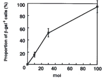
Efficiency of adenovirus-mediated gene transduction into human synoviocytes in vitro.Cultured rheumatoid synovial cells were infected with AxCASLacZ (LacZ virus) to evaluate the efficiency of adenoviral gene transduction into human rheumatoid synovial cells. Histochemical stain-ing with the chromogenic substrate X-gal showed a blue nuclear staining accompanied by a perinuclear cytoplas-mic staining in the cells expressing β-gal. The percentage of synoviocytes positive for β-gal increased in an moi-dependent manner (Figure 1a), and 95% of the cells were positively stained at the moi of 100.
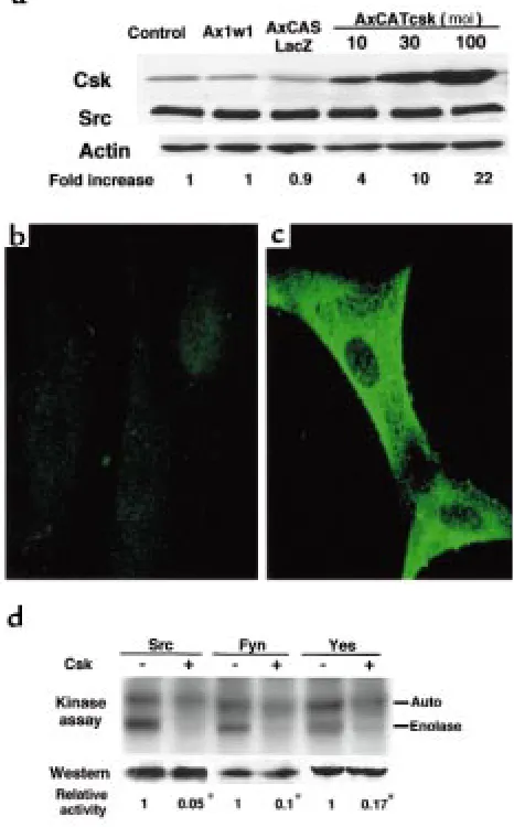
The Src kinase activity was repressed in Csk-overexpressing human synoviocytes.To analyze the effect of Csk overex-pression, human rheumatoid synoviocytes were infect-ed with AxCATcsk (Csk virus). Western blot analysis revealed that the level of expression of Csk protein was
very low in synovial cells infected with Ax1w1 (WT virus) or LacZ virus, but an moi-dependent expression of Csk was observed in Csk virus–infected cells, with no associated change in c-Src or actin expression (Figure 2a). Immunofluorescence staining showed a diffuse cytoplasmic expression of Csk in Csk virus–infected synoviocytes (Figure 2c), whereas only slight perinu-clear staining was observed in WT virus–infected syn-oviocytes (Figure 2b). Although the expression of c-Src was unchanged by Csk expression, the kinase activity of c-Src was remarkably repressed in Csk-overexpressing synoviocytes (Figure 2d). Csk overexpression had repressive effects on the in vitro kinase activity of other members of Src family protein kinases, Fyn and c-Yes.
Adenovirus-mediated Csk overexpression inhibited cell growth of rheumatoid synoviocytes.Effect of adenovirus-mediated Csk on cell proliferation was evaluated by growth curve and by immunostaining for BrdU. Prolif-eration rate of Csk virus–infected synoviocytes was remarkably reduced compared with WT or LacZ virus–infected cells, as determined by the increase in cell number (Figure 3a). After inoculation with Csk virus, the percentage of proliferating (BrdU-positive) cells decreased in an moi-dependent manner (Figure 3b).
Adenovirus-mediated Csk overexpression reduced IL-6 pro-duction at mRNA level.Synovial cells derived from patients with RA spontaneously produce an elevated amount of IL-6 without external stimuli (29). To determine the effect of Csk overexpression on IL-6 production of rheumatoid synovial cells, IL-6 concentration in
condi-Figure 5
[image:6.612.330.521.49.272.2]tioned medium was measured by ELISA, and IL-6 mRNA level was detected by Northern blot analysis in Csk-overexpressing synoviocytes. IL-6 concentration in conditioned medium of Csk virus–infected synoviocytes was reduced to almost 50% of noninfected synoviocytes at an moi of 10 or greater (Figure 4a). Northern blotting showed a dramatic decrease in IL-6 mRNA level in syn-ovial cells infected with Csk virus at an moi of 10 or greater (Figure 4b).
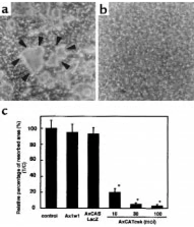
Suppression of bone-resorbing activity of osteoclasts by ade-novirus-mediated Csk.To evaluate the effect of Csk over-expression on bone-resorbing activity of human OCLs, pit-formation assay was performed on calcium phos-phate–coated discs. WT or LacZ virus–infected OCLs formed numerous resorption pits, which were clearly observed after the cells were removed from the disc (Figure 5a, arrowheads). In contrast, pit formation was strongly inhibited by inoculation with Csk virus (moi = 100) (Figure 5b). Pit-forming activity quantified by measuring the resorbed area was drastically sup-pressed by Csk overexpression in an moi-dependent manner (Figure 5c).
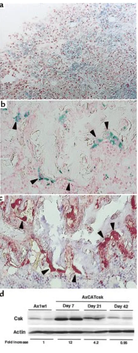
Adenovirus vector–mediated gene transduction in vivo.LacZ virus was injected into rat ankles with adjuvant arthritis to determine the transgene expression in vivo. In situ staining for β-gal showed a diffuse blue staining in the synovial tissues and periarticular tissues of injected ankles. Light microscopy revealed the transgene expres-sion in the synovial-lining and -sublining cells of the inflamed joints (Figure 6a). Immunohistochemical staining showed that both CDpositive and CD 11b-negative synovial cells were stained for LacZ (data not shown). TRAP-positive OCLs (Figure 6c) at the pan-nus/bone interface were preferentially stained for β-gal (Figure 6b). LacZ expression macroscopically decreased in a time-dependent manner and was hardly detectable on day 42. Western blot analysis demonstrated that Csk virus injection led to a 12-fold increase in the expression of Csk protein in synovial tissues on day 7. On day 21, the expression was reduced to about 35% of day 7 levels; on day 42, it returned to the same level as that of the WT virus–injected rat (Figure 6d).
Csk virus injection ameliorated inflammation and suppressed bone destruction in adjuvant arthritis.Csk virus was directly injected into the inflamed ankle joints, and the severity of the disease was evaluated in comparison with WT or LacZ virus–injected rats by arthritis score, paw volume, and radiological and pathohistological examinations. On days 21–35, the arthritis score of Csk virus–injected right ankles was significantly improved compared with that of the WT and LacZ groups (Figure 7a). On days 14–35, the increase in paw volume was also significantly decreased by Csk virus injection into right ankles, compared with the WT and LacZ groups (Figure 7b). Anti-inflammato-ry effects of Csk virus injection into left ankles were shown by arthritis score (Figure 7c) and paw volume (Fig-ure 7d), but the erosive changes in bone were not promi-nent in left ankles. Therefore, we evaluated the effects on bone erosion in the right ankles by radiological and pathological examinations. On day 35, the right ankles of WT or LacZ virus–injected rats showed the radiological findings of severe joint destruction — joint space
nar-Figure 6
[image:7.612.69.294.46.621.2]rowing, erosion, and periarticular osteoporosis (Figure 7f) — compared with a normal rat ankle (Figure 7e), but these destructive changes were improved in Csk virus–injected rats (Figure 7g). Evaluation by radiological scoring confirmed the significant therapeutic effects (Figure 7j). Pathohistological examinations revealed that Csk virus injection obviously suppressed synovial hyper-plasia, characterized by pannus formation and infiltra-tion of inflammatory cells (Figure 7, h and i). Subchon-dral bone resorption and cartilage degeneration were also suppressed by Csk virus injection. Pathological scoring showed significant difference between Csk and WT (or LacZ) groups (Figure 7k).
Discussion
The first gene delivery system used in gene therapy for arthritis was the retrovirus vector (30), but it was
mainly used for ex vivo gene therapy, because retro-virus can deliver genes only into proliferating cells (31) and has a potential risk of insertional mutagenesis when integrated into the host genome. There are sev-eral advantages to the use of adenovirus vectors for gene therapy of arthritis. First, adenovirus vectors can easily be produced and purified at very high titers (>1010PFU/mL) and can be used for in situ gene
trans-fer. Second, the safety of the viral vector has been established from animal experiments; in fact, unat-tenuated adenoviruses have been successfully used for oral vaccine (32). Third, adenovirus vectors have been reported to be the most efficient vector in both in vitro and in vivo gene delivery to synovium (33). Using LacZ virus, we also confirmed that adenovirus vector can effectively transduce synoviocytes both in vitro and in vivo. Finally, adenovirus vectors can transduce
nondi-Figure 7
Therapeutic effects of Csk virus injection on rat adjuvant arthritis. All rats were immunized with adjuvant in the right foot-pad (day 0). Viruses were intra-articularly injected into ankles on day 7. Data are shown as mean ± SD. (a) Effects of Csk virus injection into right ankles, as evalu-ated by arthritis score. The arthritis score of the Csk group (n = 10) was significantly lower than that of the WT (n = 10) and LacZ (n = 10) groups on days 21, 28, and 35 (*P < 0.01 Csk vs. LacZ, Csk vs. WT [day 21]; **P < 0.001 Csk vs. LacZ, Csk vs. WT [days 28 and 35]). (b) Effects of Csk virus injection into right ankles, as evaluated by increase in paw volume. The increase in paw volume of the Csk group was significantly less than that of the WT and LacZ groups on days 14, 21, 28, and 35 (*P < 0.05 Csk vs. WT; #P < 0.01 Csk vs. LacZ [day 21]; **P < 0.01 Csk vs.
WT, Csk vs. LacZ [day 14]; ##P < 0.001 Csk vs. WT, Csk vs. LacZ [days 28 and 35]). (c) Effects of Csk virus injection into left ankles, as
evalu-ated by arthritis score. The arthritis score of the Csk group (n = 10) was significantly lower than that of the WT (n = 10) and LacZ (n = 10) groups on days 28 and 35 (*P < 0.05 Csk vs. LacZ; #P < 0.05 Csk vs. WT). (d) Effects of Csk virus injection into left ankles, as evaluated by increase in
paw volume. The increase in paw volume of the Csk group was significantly less than that of the WT and LacZ groups (*P < 0.05 Csk vs. WT;
#P < 0.05 Csk vs. LacZ). (f) On day 35, the right ankles of WT virus–injected rats showed the radiological findings of severe joint destruction —
[image:8.612.68.532.49.310.2]viding cells as well. This is particularly important because, as we recently reported (20), adenovirus vec-tors can efficiently transduce postmitotic osteoclasts, which play essential roles in bone destruction in RA. We also found, in the present study, that LacZ virus injected in the rat ankle joints can efficiently transduce osteoclasts present at the erosive bone surfaces, as well as synoviocytes (Figure 6, a and b). Several studies have used adenovirus vectors for gene therapy for animal models of arthritis. Transferred genes include inhibitory proteins of proinflammatory cytokines such as IL-1 and TNF-α(34, 35),cytokines such as IL-10 (36) and IL-12 (37), and the apoptosis-inducing molecule FasL (27). These attempts resulted in the suc-cessful amelioration of inflammatory reactions, although no clear effects have been described on inhi-bition of bone destruction.
Src family members of tyrosine kinases are involved in signal-transduction pathways that regulate a variety of biological activities (13, 38). They are implicated in the mitogenic response to several growth factors in fibroblasts; activation of lymphocytes, platelets and osteoclasts; and various cytokine signaling. Therefore, regulating Src family kinase activity can be a good therapeutic approach to RA. Csk is a cytoplasmic tyro-sine kinase that specifically phosphorylates the car-boxylterminus tyrosine residue of Src family tyrosine kinases and, thereby, negatively regulates their kinase activity (16). Csk overexpression reverts transforma-tion of the cells transfected with c-srcand v-crk(39) and negatively regulates antigen receptor–mediated activation of T lymphocytes (40). In addition, Csk overexpression reduces proinflammatory cytokine production in a macrophage cell line (41). These results suggest that the inflammatory reaction observed in RA synovial tissues can be reduced by csk
gene transduction. Here, we demonstrated that the adenovirus vector carrying the cskgene (Csk virus) ameliorates the inflammatory reaction and bone destruction in experimental arthritis.
Csk virus induced an efficient expression of Csk pro-tein in cultured synoviocytes in vitro and in synovial tissues in vivo. Csk overexpression in synoviocytes remarkably repressed the kinase activity of c-Src, with no quantitative changes in the amount of c-Src protein. Cell proliferation rate of rheumatoid synovial cells in vitro was strongly suppressed by overexpression of Csk, suggesting that the signaling mediated by Src family tyrosine kinases plays a crucial role in the proliferation of synoviocytes. Src family tyrosine kinases are involved in the intracellular signal transduction downstream of the receptors of growth factors such as PDGF, EGF, and FGF (12, 13). Therefore, the inhibitory effect of Csk on synoviocyte proliferation can partially be explained by blockade of the signal-transduction path-ways of these growth factors. Because Src family kinas-es are implicated in integrin-mediated signal-trans-duction pathways (42, 43), Csk may modulate the adhesion property of synoviocytes and thereby inhibit anchorage-dependent cell growth.
IL-6 is a pleiotropic cytokine that has been suggest-ed to be involvsuggest-ed in the pathogenesis of RA. First, IL-6
levels are increased in synovial fluids and in the serum in RA, and correlate with the severity of the disease (44, 45). Second, synovial fibroblasts produce a great amount of IL-6 (29), and IL-6 stimulates the prolifer-ation of synoviocytes and formprolifer-ation of osteoclasts in cooperation with soluble IL-6 receptor (46, 47). Third, IL-6 gene transcription is constitutively activated in rheumatoid synovial fibroblasts, owing to the activa-tion of nuclear factor-κB (NF-κB) and C-promoter binding factor 1 (CBF1)(48). Finally, antigen-induced arthritis was poorly developed in IL-6–deficient mice (49), and blockage of IL-6 receptor ameliorated murine collagen-induced arthritis (50). These reports suggest the involvement of IL-6 in autoimmune arthritis. Our results demonstrated that adenovirus vector–mediat-ed Csk overexpression suppressvector–mediat-ed IL-6 production by synoviocytes at the mRNA level, and protein produc-tion of IL-6 was significantly reduced, indicating that Src family protein kinases are also implicated in acti-vated transcription of proinflammatory cytokines by rheumatoid synovial fibroblasts. The detailed mecha-nism by which Csk inhibits IL-6 expression remains elusive, but the inhibition could be explained by the suppression of Src-mediated activation of NF-κB as recently described (51).
Importantly, adenovirus vector–mediated Csk overex-pression strongly inhibited bone-resorbing activity of osteoclasts. Osteoclasts are primary cells responsible for bone resorption, and we recently reported that aden-ovirus vector can efficiently transfer genes into osteo-clasts and modulate osteoclast function (23). Osteoosteo-clasts express a high level of c-Src in the ruffled border (23), and targeted disruption of c-srcresulted in osteopetrosis due to dysfunction of osteoclasts, without any other crit-ical abnormalities (14, 52). This shows that c-srcis essen-tial for osteoclastic function, and its function can be con-trolled by modulating the kinase activity of c-Src without affecting the normal function of other cells. In fact, Csk virus–infected osteoclasts, like src-deficient osteoclasts, were incapable of bone resorption.
transfer had effects on the contralateral knee in a rab-bit model (35).
There are certain drawbacks in using adenovirus tors for gene therapy of arthritis. First, adenovirus vec-tor–mediated gene transduction is transient; therefore, repeated administration will be necessary to obtain fur-ther improvement of arthritis. In fact, as shown in Fig-ure 6d, the expression level of Csk in the synovial cells was reduced on day 21 compared with that on day 7, although significant amounts of Csk protein could still be detected. Second, local injection of adenovirus vector sometimes induced inflammatory reactions because of the persistent expression of adenoviral pro-teins (33, 34). Although no significant difference was observed in the severity of arthritis between control (no virus injection) and WT or LacZ virus–injected groups (data not included) in our experiments, the develop-ment of a less immunogenic and higher-titer vector sys-tem is strongly expected.
In summary, intervention into intracellular signal-transduction pathways of synoviocytes and osteoclasts by adenovirus-mediated gene transfer of Csk resulted in suppression of bone destruction and amelioration of inflammation in rat adjuvant arthritis. These results suggest that signaling mediated by Src family kinases plays critical roles in the inflammation and joint destruction in RA. Adenovirus vector is useful for in vivo gene transfer to intra-articular tissues, and adenovirus-mediated gene transfer of cskgene is a promising means of preventing arthritic bone destruction. There will be no cure for RA until its etiology is elucidated, but suppres-sion of synoviocyte and osteoclast activity by gene trans-fer might lead to a novel therapeutic strategy for pre-venting the joint breakdown associated with RA.
Acknowledgments
We thank I. Saito for the generous gift of the recom-binant adenovirus vector system, and J. Miyazaki (Osaka University, Osaka, Japan) for the kind gift of the CAG promoter. We greatly appreciate the invalu-able technical assistance of R. Yamaguchi and H. Kawahara. This work was supported by a Research Fellowship of the Japan Society for the Promotion of Science for Young Scientists (to H. Takayanagi); Grants-in-Aid from the Ministry of Education, Sci-ence, Sports and Culture of Japan (to S. Tanaka); and Bristol-Myers Squibb/Zimmer Unrestricted Research Grants (to K. Nakamura).
1. Firestein, G.S. 1996. Invasive fibroblast-like synoviocytes in rheumatoid arthritis. Passive responders or transformed aggressors? Arthritis Rheum. 39:1781–1790.
2. Müller-Ladner, U., Gay, R.E., and Gay, S. 1997. Cellular pathways of joint destruction. Curr. Opin. Rheumatol. 9:213–220.
3. Fassbender, H.G. 1983. Histomorphological basis of articular carti-lage destruction in rheumatoid arthritis. Coll. Relat. Res. 3:141–155. 4. Hamilton, J.A. 1983. Hypothesis: in vitro evidence for the invasive and tumor-like properties of the rheumatoid pannus. J. Rheumatol. 10:845–851.
5. Yocum, D.E., Lafyatis, R., Remmers, E.F., Schumacher, H.R., and Wilder, R.L. 1988. Hyperplastic synoviocytes from rats with strepto-coccal cell wall–induced arthritis exhibit a transformed phenotype that is thymic-dependent and retinoid inhibitable. Am. J. Pathol. 132:38–48.
6. Isomaki, P., and Punnonen, J. 1997. Pro- and anti-inflammatory cytokines in rheumatoid arthritis. Ann. Med. 29:499–507.
7. Cawston, T.E. 1995. Proteinases and connective tissue breakdown. In
Mechanisms and models in rheumatoid arthritis. B. Henderson, J.C.W. Edwards, and E.R. Pettipher, editors. Academic Press. London, Unit-ed Kingdom. 333–359.
8. Takayanagi, H., et al. 1997. A new mechanism of bone destruction in rheumatoid arthritis: synovial fibroblasts induce osteoclastogenesis.
Biochem. Biophys. Res. Commun. 240:279–286.
9. Gravallese, E.M., et al. 1998. Identification of cell types responsible for bone resorption in rheumatoid arthritis and juvenile rheumatoid arthritis. Am. J. Pathol. 152:943–951.
10. Müller-Ladner, U., Kriegsmann, J., Gay, R.E., and Gay, S. 1995. Onco-genes in rheumatoid arthritis. Rheum. Dis. Clin. North Am. 21:675–690. 11. Bromley, M., and Woolley, D.E. 1984. Chondroclasts and osteoclasts at subchondral sites of erosion in the rheumatoid joint. Arthritis Rheum. 27:968–975.
12. Parsons, J.T., and Parsons, S.J. 1997. Src family protein tyrosine kinas-es: cooperating with growth factor and adhesion signaling pathways.
Curr. Opin. Cell Biol. 9:187–192.
13. Thomas, S.M., and Brugge, J.S. 1997. Cellular functions regulated by Src family kinases. Annu. Rev. Cell Dev. Biol. 13:513–609.
14. Soriano, P., Montgomery, C., Geske, R., and Bradley, A. 1991. Tar-geted disruption of the c-srcproto-oncogene leads to osteopetrosis in mice. Cell. 64:693–702.
15. Nada, S., Okada, M., MacAuley, A., Cooper, J.A., and Nakagawa, H. 1991. Cloning of a complementary DNA for a protein-tyrosine kinase that specifically phosphorylates a negative regulatory site of p60c-src.
Nature. 351:69–72.
16. Okada, M., Nada, S., Yamanashi, Y., Yamamoto, T., and Nakagawa, H. 1991. CSK: a protein-tyrosine kinase involved in regulation of src family kinases. J. Biol. Chem. 266:24249–24252.
17. Arnett, F.C., et al.1988. The American Rheumatism Association 1987 revised criteria for the classification of rheumatoid arthritis. Arthri-tis Rheum.31:315–324.
18. Burmester, G.R., Dimitriu-Bona, A., Waters, S.J., and Winchester, R.J. 1983. Identification of three major synovial lining cell populations by monoclonal antibodies directed to Ia antigens and antigens asso-ciated with monocytes/macrophages and fibroblasts. Scand. J. Immunol.17:69–82.
19. Miyake, S., et al. 1996. Efficient generation of recombinant aden-oviruses using adenovirus DNA-terminal protein complex and a cos-mid bearing the full-length virus genome. Proc. Natl. Acad. Sci. USA. 93:1320–1324.
20. Tanaka, S., et al. 1998. Modulation of osteoclast function by aden-ovirus vector–induced epidermal growth factor receptor. J. Bone Miner. Res. 13:1714–1720.
21. Sanes, J.R., Rubenstein, J.L., and Nicolas, J.F. 1986. Use of a recombi-nant retrovirus to study post-implantation cell lineage in mouse embryos. EMBO J. 5:3133–3142.
22. Isshiki, M., et al. 1998. Endothelial Ca2+waves preferentially originate
at specific loci in caveolin-rich cell edges. Proc. Natl. Acad. Sci. USA. 95:5009–5014.
23. Tanaka, S., et al. 1992. Osteoclasts express high levels of p60c-src,
pref-erentially on ruffled border membranes. FEBS Lett. 313:85–89. 24. Chomczynski, P., and Sacchi, N. 1987. Single-step method of RNA
isolation by acid guanidinium thiocyanate-phenol-chloroform extraction. Anal. Biochem. 162:156–159.
25. Chandrasekar, B., Melby, P.C., Troyer, D.A., and Freeman, G.L. 1996. Induction of proinflammatory cytokine expression in experimental acute Chagasic cardiomyopathy. Biochem. Biophys. Res. Commun. 223:365–371.
26. Takahashi, N., et al. 1988. Osteoclast-like cell formation and its reg-ulation by osteotropic hormones in mouse bone marrow cultures.
Endocrinology. 122:1373–1382.
27. Zhang, H., et al. 1997. Amelioration of collagen-induced arthritis by CD95 (Apo-1/Fas)-ligand gene transfer. J. Clin. Invest. 100:1951–1957. 28. Ackerman, N.R., et al. 1979. Effects of naproxen on connective tissue changes in the adjuvant arthritic rat. Arthritis Rheum. 22:1365–1374. 29. Guerne, P.A., Zuraw, B.L., Vaughan, J.H., Carson, D.A., and Lotz, M. 1989. Synovium as a source of interleukin 6 in vitro. Contribution to local and systemic manifestations of arthritis. J. Clin. Invest.83:585–592. 30. Bandara, G., et al. 1993. Intraarticular expression of biologically active interleukin 1-receptor–antagonist protein by ex vivo gene transfer. Proc. Natl. Acad. Sci. USA. 90:10764–10768.
31. Miller, D.G., Adam, M.A., and Miller, A.D. 1990. Gene transfer by retrovirus vectors occurs only in cells that are actively replicating at the time of infection. Mol. Cell. Biol. 10:4239–4242.
32. Rubin, B.A., and Rorke, L.B. 1988. Adenovirus vaccines. In Vaccines. S.A. Plotkin and E.A.J. Mortimer, editors. W.B. Saunders. Philadel-phia, PA. 492–512.
Suppres-sion of collagen-induced arthritis through adenovirus-mediated transfer of a modified tumor necrosis factor αreceptor gene. Arthri-tis Rheum. 40:1662–1669.
35. Ghivizzani, S.C., et al. 1998. Direct adenovirus-mediated gene trans-fer of interleukin 1 and tumor necrosis factor αsoluble receptors to rabbit knees with experimental arthritis has local and distal anti-arthritic effects. Proc. Natl. Acad. Sci. USA. 95:4613–4618.
36. Apparailly, F., et al. 1998. Adenovirus-mediated transfer of viral IL-10 gene inhibits murine collagen-induced arthritis. J. Immunol. 160:5213–5220.
37. Parks, E., et al. 1998. Transient gene transfer of IL-12 regulates chemokine expression and disease severity in experimental arthritis.
J. Immunol. 160:4615–4619.
38. Brown, M.T., and Cooper, J.A. 1996. Regulation, substrates and func-tions of src. Biochim. Biophys Acta. 1287:121–149.
39. Sabe, H., Okada, M., Nakagawa, H., and Hanafusa, H. 1992. Activa-tion of c-Src in cells bearing v-Crk and its suppression by Csk. Mol. Cell. Biol. 12:4706–4713.
40. Chow, L.M., Fournel, M., Davidson, D., and Veillette, A. 1993. Nega-tive regulation of T-cell receptor signalling by tyrosine protein kinase p50csk. Nature. 365:156–160.
41. Iwabuchi, K., et al. 1997. Csk overexpression reduces several monokines and nitric oxide productions but enhances prostaglandin E2production in response to lipopolysaccharide in the macrophage
cell line J774A.1. Eur. J. Immunol. 27:742–749.
42. Kaplan, K.B., et al. 1994. Association of the amino-terminal half of c-Src with focal adhesions alters their properties and is regulated by phosphorylation of tyrosine 527. EMBO J. 13:4745–4756.
43. Bergman, M., Joukov, V., Virtanen, I., and Alitalo, K. 1995. Overex-pressed Csk tyrosine kinase is localized in focal adhesions, causes reorganization of αvβ5integrin, and interferes with HeLa cell
spread-ing. Mol. Cell. Biol. 15:711–722.
44. Houssiau, F.A., Devogelaer, J.P., Van Damme, J., de Deuxchaisnes, C.N., and Van Snick, J. 1988. Interleukin-6 in synovial fluid and serum of patients with rheumatoid arthritis and other inflammato-ry arthritides. Arthritis Rheum. 31:784–788.
45. Brozik, M., et al. 1992. Interleukin 6 levels in synovial fluids of patients with different arthritides: correlation with local IgM rheumatoid factor and systemic acute phase protein production. J. Rheumatol. 19:63–68.
46. Mihara, M., Moriya, Y., Kishimoto, T., and Ohsugi, Y. 1995. Inter-leukin-6 (IL-6) induces the proliferation of synovial fibroblastic cells in the presence of soluble IL-6 receptor. Br. J. Rheumatol. 34:321–325. 47. Kotake, S., et al. 1996. Interleukin-6 and soluble interleukin-6 recep-tors in the synovial fluids from rheumatoid arthritis patients are responsible for osteoclast-like cell formation. J. Bone Miner. Res. 11:88–95.
48. Miyazawa, K., Mori, A., Yamamoto, K., and Okudaira, H. 1998. Con-stitutive transcription of the human interleukin-6 gene by rheuma-toid synoviocytes: spontaneous activation of NF-κB and CBF1. Am. J. Pathol. 152:793–803.
49. Ohshima, S., et al. 1998. Interleukin 6 plays a key role in the devel-opment of antigen-induced arthritis. Proc. Natl. Acad. Sci. USA. 95:8222–8226.
50. Takagi, N., et al. 1998. Blockage of interleukin-6 receptor ameliorates joint disease in murine collagen-induced arthritis. Arthritis Rheum. 41:2117–2121.
51. Abu-Amer, Y., et al. 1998. Tumor necrosis factor-αactivation of nuclear transcription factor-κB in marrow macrophages is mediated by c-Src tyrosine phosphorylation of IκBα. J. Biol. Chem. 273:29417–29423.