Echinocandins among Fluconazole-Resistant Bloodstream Isolates of
Candida glabrata
M. A. Pfaller,aM. Castanheira,aS. R. Lockhart,bA. M. Ahlquist,bS. A. Messer,aand R. N. Jonesa,c
JMI Laboratories, North Liberty, Iowa, USAa
; Mycotic Disease Branch, Centers for Disease Control and Prevention, Atlanta, Georgia, USAb
; and Tufts University School of Medicine, Boston, Massachusetts, USAc
The echinocandin class of antifungal agents is considered to be the first-line treatment of bloodstream infections (BSI) due to
Candida glabrata.
Recent reports of BSI due to strains of
C. glabrata
resistant to both fluconazole and the echinocandins are of
concern and prompted us to review the experience of two large surveillance programs, the SENTRY Antimicrobial Surveillance
Program for the years 2006 through 2010 and the Centers for Disease Control and Prevention population-based surveillance
conducted in 2008 to 2010. The
in vitro
susceptibilities of 1,669 BSI isolates of
C. glabrata
to fluconazole, voriconazole,
anidula-fungin, caspoanidula-fungin, and micafungin were determined by CLSI broth microdilution methods. Fluconazole MICs of
>
64
g/ml
were considered resistant. Strains for which anidulafungin and caspofungin MICs were
>
0.5
g/ml and for which micafungin
MICs were
>
0.25
g/ml were considered resistant. A total of 162 isolates (9.7%) were resistant to fluconazole, of which 98.8%
were nonsusceptible to voriconazole (MIC
>
0.5
g/ml) and 9.3%, 9.3%, and 8.0% were resistant to anidulafungin, caspofungin,
and micafungin, respectively. There were 18 fluconazole-resistant isolates that were resistant to one or more of the
echinocan-dins (11.1% of all fluconazole-resistant isolates), all of which contained an acquired mutation in
fks1
or
fks2. By comparison,
there were no echinocandin-resistant strains detected among 110 fluconazole-resistant isolates of
C. glabrata
tested in 2001 to
2004. These data document the broad emergence of coresistance over time to both azoles and echinocandins in clinical isolates of
C. glabrata.
R
esistance to the echinocandin antifungal agents,
anidulafun-gin, caspofunanidulafun-gin, and micafunanidulafun-gin, among the broad
popula-tion of
Candida
spp. causing invasive candidiasis (IC), is distinctly
uncommon (1, 18, 39, 46, 48, 55–58, 62). Among 15,269 clinical
isolates of
Candida
spp. tested at the University of Iowa (Iowa
City), only 25 (0.2%) showed resistance to one or more
echino-candins using the Clinical and Laboratory Standards Institute
(CLSI) broth microdilution (BMD) method and interpretive
cri-teria (39, 44). As such, the echinocandin class of antifungal agents
is recommended as first-line therapy for candidemia and IC,
es-pecially in patients with severe sepsis, those previously exposed to
azoles, and/or those infected with
Candida glabrata
(35).
Ever since the introduction of fluconazole in 1990 for the
treat-ment of candidiasis, including IC, empirical antifungal therapy
has been driven by fear of
C. glabrata
(56, 57). Decreased
suscep-tibility of
C. glabrata
to fluconazole and cross-resistance to other
azoles are well known (21, 22, 32, 35, 41, 43, 45, 56, 57). In the
United States,
C. glabrata
has increased as a cause of IC from 18%
of all bloodstream infection (BSI) isolates in the time period of
1992 to 2001 to 25% in 2001 to 2007, with a concomitant increase
in fluconazole resistance from 9% to 14% (45). Given the distinct
differences between the mechanisms of action and resistance for
the azoles and the echinocandins (37, 38), the documented lack of
cross-resistance between the two classes (27, 31, 42) is not
surpris-ing, and thus the recommendation that the echinocandins be used
for treatment of IC in patients with prior exposure to azoles
and/or infection with
C. glabrata
is well founded (24, 35, 56, 57).
Global surveys have documented the exquisite
in vitro
susceptibil-ity of
C. glabrata
to each of the echinocandins (MIC
90, 0.015
g/ml [micafungin], 0.06
g/ml [caspofungin], or 0.12
g/ml
[anidulafungin]) (39). Furthermore, the clinical experience with
the use of echinocandins to treat IC due to
C. glabrata
is quite
favorable (2, 20, 28, 36, 51, 55).
Although documentation of acquired resistance to the
echino-candins remains sporadic (3, 18, 37, 44, 48, 58, 62), several recent
reports of acquired resistance in clinical isolates of
C. glabrata
focus concern on this species (5, 7, 11–15, 18, 19, 48, 58, 59, 62).
Data from global surveys demonstrate that the frequency of
echi-nocandin resistance among clinical isolates of
C. glabrata
ranges
from 1 to 3% and is higher among isolates from North America
(3%) than among those from Europe (1%), Latin America (0.0%),
or the Asia-Pacific region (0.0%) (39, 47). It is now clear that
clinical isolates of
C. glabrata
with decreased susceptibility to one
or more echinocandins harbor mutations in
fks1
and/or
fks2
and
are also resistant clinically (5, 15, 37, 48, 59, 62).
Chapeland-Leclerc et al. (5) reported a case of IC due to
C. glabrata
in which
the infecting strain acquired resistance to flucytosine, fluconazole,
voriconazole, and caspofungin through successive independent
events following prolonged exposure to each class of antifungal
agent. The recovery of different isolates exhibiting clonality for
microsatellite markers but genetic diversity for antifungal
resis-tance markers (three unique resisresis-tance mechanisms)
demon-strates the high propensity of
C. glabrata
to readily mutate
in vivo
Received14 October 2011Returned for modification7 November 2011 Accepted9 January 2012
Published ahead of print25 January 2012
Address correspondence to M. A. Pfaller, mike-pfaller@jmilabs.com.
Copyright © 2012, American Society for Microbiology. All Rights Reserved.
doi:10.1128/JCM.06112-11
on May 16, 2020 by guest
http://jcm.asm.org/
in a single patient (5). Additional reports from medical centers in
the United States and Denmark provide further documentation of
multidrug-resistant (MDR) (resistant to two or more classes of
antifungal agents) strains of
C. glabrata
(13, 18, 19, 48, 62). One
potential explanation for the emergence of MDR in
C. glabrata
is
that the haploid nature of the organism makes it particularly adept
at acquiring and expressing resistance mutations in response to
drug pressure (5, 37, 48, 62).
The generally excellent wild-type (WT) susceptibility of
C.
glabrata
to the echinocandins coupled with broadening azole
re-sistance has driven the use of echinocandins for treatment of
in-fections due to
C. glabrata
and at the same time has generated
selection pressure for resistant organisms (4, 5, 11, 18, 24, 33–35,
48, 56, 57, 62). The fact that echinocandins are recommended for
use in the setting of prior azole exposure and specifically for the
treatment of IC due to
C. glabrata
raises the concern that acquired
resistance to the echinocandins may emerge independently in
flu-conazole-resistant strains due to mutations in
fks
(5, 24, 48, 62). In
an effort to further examine this issue, we have addressed the
frequency of decreased susceptibility to the echinocandin class of
antifungal agents among fluconazole-resistant strains of
C.
glabrata
BSI isolates from two large antifungal surveys, the
SENTRY Global Surveillance Program for the years 2006 through
2010 and the Centers for Disease Control and Prevention (CDC)
population-based surveillance conducted in the Atlanta, GA, and
Baltimore, MD, metropolitan areas between 2008 and 2010.
Those isolates with decreased susceptibility (either intermediate
[I] or resistant [R]) to one or more of the echinocandins were
further examined for mutations in
fks1
and
fks2.
MATERIALS AND METHODS
Organisms.A total of 1,669 isolates ofC. glabratafrom blood or other normally sterile sites were obtained from diverse medical centers world-wide (SENTRY; 847 isolates) or from those in the Atlanta and Baltimore metropolitan areas (CDC; 822 isolates). All isolates represented incident episodes of IC (first positive blood culture). Isolates were identified by standard methods and stored in water at ambient room temperature (SENTRY) or in glycerol at⫺70°C (CDC) until used in the study. Before testing, each isolate was passaged on Sabouraud dextrose agar (Remel, Lenexa, KS) and CHROMagar (Becton, Dickinson, Sparks, MD) to en-sure purity and viability.
A total of 21 fluconazole-resistant isolates ofC. glabratafor which MICs for one or more echinocandin were I or R were further characterized for the presence or absence of mutations in the hot spot (HS) regions of
fks1andfks2as described previously (3, 62).
Antifungal susceptibility testing.All isolates were tested forin vitro
susceptibilities to anidulafungin, caspofungin, micafungin, fluconazole, and voriconazole using CLSI BMD methods (8, 9). MIC results for all agents were read visually following 24 h of incubation as the lowest con-centration of drug that caused a significant diminution (ⱖ50% inhibi-tion) of growth compared with control levels (8, 9). The recently revised CLSI clinical breakpoints were used to identify strains ofC. glabratathat were either I or R to the echinocandins and R or nonsusceptible (NS) to the azoles (40, 41, 44): anidulafungin and caspofungin MIC values of 0.25 g/ml were considered I, and MIC values ofⱖ0.5g/ml were considered R; micafungin MIC values of 0.12g/ml were considered I, and MIC values ofⱖ0.25g/ml were considered R; fluconazole MIC values ofⱖ64 g/ml were considered R, and voriconazole MIC values of⬎0.5g/ml were considered NS. Quality control was performed by testing the CLSI-recommended strainsCandida kruseiATCC 6258 andCandida parapsilo-sisATCC 22019 (8, 9).
RESULTS AND DISCUSSION
Frequency of resistance to fluconazole and voriconazole.
Among 1,669 BSI isolates of
C. glabrata
collected during the
course of the two surveillance programs, 162 (9.7%) were resistant
to fluconazole, including 62 of 847 (7.3%) isolates from the
SENTRY Program (years 2006 to 2010) and 100 of 822 (12.2%)
isolates from the CDC population-based surveillance (years 2008
to 2010). Among the 162 fluconazole-resistant isolates, 160
(98.8%) were NS to voriconazole (MIC
⬎
0.5
g/ml) (40).
Decreased susceptibility to echinocandins among
flucona-zole-resistant
C. glabrata
isolates.
Among the 162
fluconazole-resistant isolates of
C. glabrata
, there were 21 isolates (13.0%) that
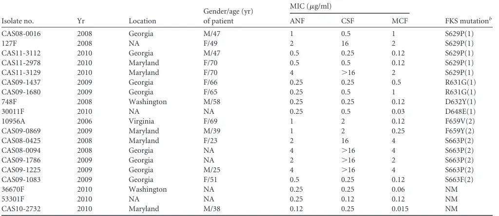
were either I or R to one or more of the echinocandins (Table 1):
3.7% and 9.3% were I or R, respectively, to anidulafungin, 3.7%
and 9.3% to caspofungin, and 4.9% and 8.0% to micafungin.
Mu-tations in either
fks1
or
fks2
were detected in 19 of the 21 (90.5%)
echinocandin I or R isolates (Table 1). There were 18
fluconazole-resistant isolates (11.1% of all fluconazole-fluconazole-resistant isolates) that
were R to one or more of the echinocandins (MIC
ⱖ
0.25
g/ml
[micafungin] or
ⱖ
0.5
g/ml [anidulafungin and caspofungin]),
all (100.0%) of which demonstrated an acquired mutation in
fks1
or
fks2.
Table 2 provides a comparison of the frequency of I or R
among 110 fluconazole-resistant strains of
C. glabrata
collected
from diverse medical centers worldwide between 2001 and 2004
(27, 42) and that of the present collection, representing the time
period 2006 to 2010. Results from the two time periods were
ob-tained with CLSI reference methods in different laboratories;
however, quality control procedures were rigorously performed
during all studies, suggesting that testing conditions did not
influ-ence the differinflu-ences noted.
Whereas there were no echinocandin-resistant strains among
110 fluconazole-resistant isolates of
C. glabrata
tested between
2001 and 2004, a period where only caspofungin was available,
there were 18 echinocandin-resistant strains (11.1%) among the
162 fluconazole-resistant isolates representing the latter time
pe-riod. All of these had mutations in
fks
(Tables 1 and 2).
These data document for the first time the broad emergence of
coresistance over time to both azoles and echinocandins in clinical
isolates of
C. glabrata
. Whereas resistance to azoles in isolates of
C.
glabrata
has been documented to be due to the overexpression of
CDR
efflux pumps (52, 53), there is little or no evidence that efflux
pumps are involved in resistance to the echinocandins (31, 50).
The documentation of
fks
mutations in isolates of
C. glabrata
showing
in vitro
resistance to both azoles and echinocandins
sug-gests the sequential accumulation of acquired resistance
mecha-nisms as demonstrated by Chapeland-Leclerc (5). The lack of
co-resistance in
C. glabrata
in the 2001-to-2004 time period is not
surprising given that among the echinocandins only caspofungin
was available, having been just approved in the United States and
Europe in 2001. Since that time, the overall use of echinocandins
in the United States has increased significantly, from 7.7
⫾
5.3
days of therapy (DOT) per 1,000 patient-days in 2004 to 13.1
⫾
8.6 DOT per 1,000 patient-days in 2008 (mean
⫾
standard
devi-ation [SD]) (
P
⬍
0.001) (33). During the same time period (2004
to 2008), the use of azoles increased as well, from 67.6
⫾
29 to 72
⫾
33 DOT per 1,000 patient-days, but this change was not significant
(
P
⫽
0.1570) (33).
Whereas a shift to non-
albicans
species of
Candida
, particularly
on May 16, 2020 by guest
http://jcm.asm.org/
C. glabrata
, has been a cause for alarm among clinicians caring for
profoundly ill patients in the hospital (23, 26, 61), others have
suggested that this is not a concern in the current “echinocandin
era” (16, 56, 57). Prior to the availability of the echinocandins,
concern over the reduced azole susceptibility of
C. glabrata
was a
major issue for clinicians (10, 22, 29, 32, 54). Both
in vitro
and
clinical studies have documented excellent activities of the
echi-nocandins against
C. glabrata
and other potentially azole-resistant
species, and the echinocandins have been rapidly replacing the
azoles as first-line therapy for IC (4, 17, 18, 24, 30, 33, 35, 55, 58).
Thus, in addition to the continued high use of azoles, the
signifi-cant increase in the use of echinocandins places considerable
se-lection pressure on
C. glabrata
, resulting in the emergence of
MDR strains. It must be noted that thus far,
Candida
isolates
demonstrating reduced echinocandin susceptibility appear
fol-lowing prolonged exposure to the drug, and these infections are
manifested as recurrent episodes of candidemia in severely
immu-nocompromised patients (48, 58).
Exposure to fluconazole is recognized by some (6, 22, 25, 49,
60) but not all (10, 54) investigators as a risk factor for
flucona-zole-resistant
C. glabrata.
Aside from reports of breakthrough
fungemias, some involving
fks
mutations, there is little
compara-ble data for the echinocandins and their influence on species
dis-tribution or resistance patterns in IC (58). Recently Lortholary et
al. (24) examined the effect of exposure to fluconazole (
n
⫽
159)
or caspofungin (
n
⫽
61) on the proportions of the five major
species of
Candida.
For both drugs, preexposure was associated
with a decreased prevalence of
C. albicans
in favor of less
drug-susceptible species (
C. glabrata
and
C. krusei
for the former;
C.
parapilosis
,
C. glabrata
, and
C. krusei
for the latter;
P
⫽
0.001). Not
only did the species distribution change in patients with recent
exposure to fluconazole and caspofungin, but the overall
suscep-tibilities of the isolates to these drugs decreased. Notably, two
C.
glabrata
isolates recovered from patients previously exposed to
caspofungin during incident candidemia had high caspofungin
[image:3.585.39.551.88.310.2]MICs and harbored mutations in
fks1
(24). The authors caution
TABLE 1Fluconazole-resistant bloodstream isolates ofC. glabratawith decreased susceptibility to one or more echinocandin agent as determined by CLSI broth microdilution methodsa
Isolate no. Yr Location
Gender/age (yr) of patient
MIC (g/ml)
FKS mutationb
ANF CSF MCF
CAS08-0016 2008 Georgia M/47 1 0.5 1 S629P(1)
127F 2008 NA F/49 2 16 2 S629P(1)
CAS11-3112 2010 Georgia M/47 0.5 0.25 0.12 S629P(1) CAS11-2978 2010 Maryland F/70 0.5 0.5 0.12 S629P(1) CAS11-3129 2010 Maryland F/70 4 ⬎16 2 S629P(1) CAS09-1437 2009 Georgia F/66 0.25 0.25 0.5 R631G(1) CAS09-1680 2009 Georgia F/65 0.25 0.5 1 R631G(1) 748F 2008 Washington M/58 0.25 0.25 0.12 D632Y(1)
30011F 2010 NA NA 0.25 0.5 0.03 D648E(1)
10956A 2006 Virginia F/69 1 2 0.12 F659V(2)
CAS09-0869 2009 Maryland M/39 1 2 0.25 F659Y(2) CAS08-0425 2008 Maryland F/23 2 16 4 S663P(2)
CAS08-0094 2008 Georgia NA 4 ⬎16 4 S663P(2)
CAS09-1786 2009 Georgia NA 2 ⬎16 2 S663P(2)
CAS09-1225 2009 Georgia M/25 4 ⬎16 4 S663P(2) CAS09-1083 2009 Georgia F/51 0.5 0.25 0.12 S663F(2) 36670F 2010 Washington NA 0.25 0.25 0.06 NM
53301F 2010 NA NA 0.25 0.12 0.12 NM
CAS10-2732 2010 Maryland M/38 0.12 0.25 0.015 NM
aM, male; F, female; ANF, anidulafungin; CSF, caspofungin; MCF, micafungin; NA, data not available.
b
Value in parentheses indicates FKS1 (1) or FKS2 (2); NM, no mutation.
TABLE 2Comparison of the activities of anidulafungin, caspofungin, and micafungin against fluconazole-resistant isolates ofC. glabratafrom two time periods, 2001 to 2004 and 2006 to 2010a
Time period (yr)
Antifungal agent
No. of isolates tested
No. (%) of isolatesb
Reference(s)
I R
2001–2004 Anidulafungin 110 2 (1.8) 0 (0.0) 27, 42 Caspofungin 110 4 (3.6) 0 (0.0)
Micafungin 110 0 (0.0) 0 (0.0)
2006–2010 Anidulafungin 162 6 (3.7) 15 (9.3) Present study Caspofungin 162 6 (3.7) 15 (9.3)
Micafungin 162 8 (4.9) 13 (8.0)
aAll isolates were tested in accordance with CLSI document M27-A3 (8). Fluconazole resistance was defined as an MIC ofⱖ64g/ml.
b
Number of isolates for which the echinocandin MICs were intermediate (I) (anidulafungin and caspofungin MIC of 0.25g/ml; micafungin MIC of 0.12g/ml) or resistant (R) (anidulafungin and caspofungin MIC ofⱖ0.5g/ml; micafungin MIC ofⱖ0.25g/ml).
on May 16, 2020 by guest
http://jcm.asm.org/
[image:3.585.42.545.595.697.2]that a new episode of sepsis after a recent prescription of
antifun-gals may be due to isolates with decreased susceptibility to the
prescribed drugs, including caspofungin (24). In light of this, it
should be noted that among the CDC isolates of
C. glabrata
with
fks
mutations for which epidemiological data were available, all
came from patients with prior exposure to an echinocandin (62).
In summary, we have documented the emergence of
coresis-tance to the echinocandins among BSI isolates of
fluconazole-resistant
C. glabrata.
Among the 18 fluconazole-resistant isolates
with coresistance to the echinocandins, all were from the United
States, consistent with our previous observations concerning the
geographic distribution of echinocandin resistance in
C. glabrata
,
where the highest frequency of resistance was seen in North
Amer-ica (3%) versus Europe (1%), Latin AmerAmer-ica (0.0%), and the
Asia-Pacific region (0.0%) (47). The increased use of both azoles and
echinocandins will most certainly bring selection pressure to bear
against
C. glabrata
, a species that appears to be unique in its ability
to sequentially acquire and express resistance mutations.
Specifi-cally, the targeted use of echinocandins in severely
immunocom-promised individuals with prior exposure to azoles and/or
infec-tion with
C. glabrata
will ensure that this dual drug pressure is
maintained. Although the vast majority of
C. glabrata
isolates
re-main highly susceptible to the echinocandin class of antifungal
agents, the increase in MDR
C. glabrata
strains is a serious concern
and argues for continued close resistance surveillance and the
in-creased application of standardized antifungal susceptibility
test-ing. Heightened awareness, rather than complacency, concerning
the importance of
C. glabrata
should be the watchword for those
caring for patients at risk for IC.
ACKNOWLEDGMENTS
The antifungal global surveillance program which served as the source of data used in the development of the manuscript was supported in part by Pfizer Inc. and by Astellas.
We acknowledge the excellent assistance of S. Benning and P. Clark in the preparation of the manuscript; Tom Chiller, Carol Bolden, and Nau-reen Iqbal for data collection at the CDC; Monica M. Farley and Betsy Stein at Emory University School of Medicine and the Atlanta VA Medical Center and surveillance officers of the Georgia EIP and the hospitals in Georgia Health District 3 for assistance with data collection; Lee H. Har-rison and Rosemary A. Hollick of Johns Hopkins Bloomberg School of Public Health; and the staff of Maryland ABCs and the hospitals in Balti-more city and BaltiBalti-more County, MD. We thank all of the surveillance centers that contributed isolates to both surveillance programs. We also thank G. J. Moet and L. N. Woosley.
The findings and conclusions of this article are ours and do not nec-essarily represent the views of the Centers for Disease Control and Pre-vention.
REFERENCES
1.Arendrup MC, et al.2011. National surveillance of fungemia in Denmark (2004 to 2009). J. Clin. Microbiol.49:325–334.
2.Betts RF, et al.2009. A multicenter, double-blind trial of a high-dose caspofungin treatment regimen versus a standard caspofungin treatment regimen for adult patients with invasive candidiasis. Clin. Infect. Dis.48: 1676 –1684.
3.Castanheira M, et al.2010. Low prevalence offks1hotspot 1 mutations in a worldwide collection ofCandidaspp. Antimicrob. Agents Chemother. 54:2655–2659.
4.Chandwani S, Wentworth C, Burke TA, Patterson TF.2009. Utilization and dosage pattern of echinocandins for treatment of fungal infections in US hospital practice. Curr. Med. Res. Opin.25:385–393.
5.Chapeland-Leclerc F, et al.2010. Acquisition of flucytosine, azole, and caspofungin resistance inCandida glabratabloodstream isolates serially
obtained from a hematopoietic stem cell transplant recipient. Antimicrob. Agents Chemother.54:1360 –1362.
6.Chow JK, et al.2008. Risk factors for albicans and non-albicans candi-demia in the intensive care unit. Crit. Care Med.36:1993–1998. 7.Cleary JD, Garcia-Effron G, Chapman SW, Perlin DS.2008. Reduced
Candida glabratasusceptibility secondary to an FKS1 mutation developed during candidemia treatment. Antimicrob. Agents Chemother.52:2263– 2265.
8.Clinical and Laboratory Standards Institute.2008. M27-A3. Reference method for broth dilution antifungal susceptibility testing of yeasts, 3rd ed. Clinical and Laboratory Standards Institute, Wayne, PA.
9.Clinical and Laboratory Standards Institute.2008. M27–S3. Reference method for broth dilution antifungal susceptibility testing of yeasts, 3rd informational supplement. Clinical and Laboratory Standards Institute, Wayne, PA.
10. Cohen Y, et al.2010. Early prediction ofCandida glabratafungemia in nonneutropenic critically ill patients. Crit. Care Med.38:826 – 830. 11. Costa-de-Oliveira S, et al. 2011. FKS2 mutations associated with
de-creased echinocandin susceptibility ofCandida glabratafollowing anidu-lafungin therapy. Antimicrob. Agents Chemother.55:1312–1314. 12. Daneman N, et al.2006. The emergence of caspofungin resistance during
treatment of recurrentCandida glabratacandidaemia, abstr. P1204. 16th Eur. Congr. Clin. Microbiol. Infect. Dis.,, Nice, France, 1 to 4 April 2006. 13. Dodgson KJ, et al.2005. Caspofungin resistantC. glabrata.Clin.
Micro-biol. Infect.11(Suppl 2):364.
14. Garcia-Effron G, et al.2010. NovelFKSmutations associated with echi-nocandin resistance inCandidaspecies. Antimicrob. Agents Chemother. 54:2225–2227.
15. Garcia-Effron G, Lee S, Park S, Cleary JD, Perlin DS.2009. Effect of
Candida glabrata FKS1andFKS2mutations on echinocandin sensitivity and kinetics of 1,3-beta-D-glucan synthase: implication for the existing susceptibility breakpoint. Antimicrob. Agents Chemother.53:3690 –3699. 16. Hsu DI, Nguyen M, Nguyen L, Law A, Wong-Beringer A. 2010. A multicentre study to evaluate the impact of timing of caspofungin administration on outcomes of invasive candidiasis in non-immunocompromised adult patients. J. Antimicrob. Chemother.65: 1765–1770.
17. Klevay MJ, Horn DL, Neofytos D, Pfaller MA, Diekema DJ.2009. Initial treatment and outcome ofCandida glabrataversusCandida albicans
bloodstream infection. Diagn. Microbiol. Infect. Dis.64:152–157. 18. Kofteridis DP, Lewis RE, Kontoyiannis DP.2010.
Caspofungin-non-susceptibleCandidaisolates in cancer patients. J. Antimicrob. Chemother. 65:293–295.
19. Krogh-Madsen M, Arendrup MC, Heslet L, Knudsen JD.2006. Am-photericin B and caspofungin resistance inCandida glabrataisolates re-covered from a critically ill patient. Clin. Infect. Dis.42:938 –944. 20. Kuse ER, et al.2007. Micafungin versus liposomal amphotericin B for
candidaemia and invasive candidosis: a phase III randomised double-blind trial. Lancet369:1519 –1527.
21. Lee I, et al.2010. Clinical and economic outcomes of decreased flucona-zole susceptibility in patients withCandida glabratabloodstream infec-tions. Am. J. Infect. Control38:740 –745.
22. Lee I, et al.2010. Risk factors for fluconazole resistance in patients with
Candida glabratabloodstream infection: potential impact of control group selection on characterizing the association between previous flu-conazole use and fluflu-conazole resistance. Am. J. Infect. Control38:456 – 460.
23. Lipsett PA.2006. Surgical critical care: fungal infections in surgical pa-tients. Crit. Care Med.34:S215–S224.
24. Lortholary O, et al.2011. Recent exposure to caspofungin or fluconazole influences the epidemiology of candidemia: a prospective multicenter study involving 2,441 patients. Antimicrob. Agents Chemother.55:532– 538.
25. Marr KA, Seidel K, White TC, Bowden RA.2000. Candidemia in allo-geneic blood and marrow transplant recipients: evolution of risk factors after the adoption of prophylactic fluconazole. J. Infect. Dis.181:309 –316. 26. Mean M, Marchetti O, Calandra T. 2008. Bench-to-bedside review:
Candidainfections in the intensive care unit. Crit. Care12:204 –212. 27. Messer SA, et al. 2006. Activities of micafungin against 315 invasive
clinical isolates of fluconazole-resistantCandidaspp. J. Clin. Microbiol. 44:324 –326.
28. Mora-Duarte J, et al.2002. Comparison of caspofungin and amphoteri-cin B for invasive candidiasis. N. Engl. J. Med.347:2020 –2029.
on May 16, 2020 by guest
http://jcm.asm.org/
29. Moran C, Grussemeyer CA, Spalding JR, Benjamin DK, Jr, Reed SD. 2010. Comparison of costs, length of stay, and mortality associated with
Candida glabrataandCandida albicansbloodstream infections. Am. J. Infect. Control38:78 – 80.
30. Neoh CF, et al.2011. Cost-effectiveness analysis of anidulafungin versus fluconazole for the treatment of invasive candidiasis. J. Antimicrob. Che-mother.66:1906 –1915.
31. Niimi K, et al.2006. Overexpression ofCandida albicans CDR1,CDR2, or
MDR1does not produce significant changes in echinocandin susceptibil-ity. Antimicrob. Agents Chemother.50:1148 –1155.
32. Oxman DA, et al.2010. Candidaemia associated with decreased in vitro fluconazole susceptibility: isCandidaspeciation predictive of the suscep-tibility pattern? J. Antimicrob. Chemother.65:1460 –1465.
33. Pakyz AL, Gurgle HE, Oinonen MJ.2011. Antifungal use in hospitalized adults in U.S. academic health centers. Am. J. Health Syst. Pharm.68:415– 418.
34. Pappas PG.2011. Candidemia in the intensive care unit: miles to go before we sleep. Crit. Care Med.39:884 – 885.
35. Pappas PG, et al.2009. Clinical practice guidelines for the management of candidiasis: 2009 update by the Infectious Diseases Society of America. Clin. Infect. Dis.48:503–535.
36. Pappas PG, et al.2007. Micafungin versus caspofungin for treatment of candidemia and other forms of invasive candidiasis. Clin. Infect. Dis.45: 883– 893.
37. Perlin DS. 2011. Echinocandin-resistantCandida: molecular methods and phenotypes. Curr. Fungal Infect. Rep.5:113–119.
38. Perlin DS.2007. Resistance to echinocandin-class antifungal drugs. Drug Resist. Updat.10:121–130.
39. Pfaller M, et al.2011. Use of epidemiological cutoff values to examine 9-year trends in susceptibility of Candida species to anidulafungin, caspo-fungin, and micafungin. J. Clin. Microbiol.49:624 – 629.
40. Pfaller MA, et al.2011. Clinical breakpoints for voriconazole and Can-didaspp. revisited: review of microbiologic, molecular, pharmacody-namic, and clinical data as they pertain to the development of species-specific interpretive criteria. Diagn. Microbiol. Infect. Dis.70:330 –343. 41. Pfaller MA, et al.2010. Wild-type MIC distributions, epidemiological
cutoff values and species-specific clinical breakpoints for fluconazole and
Candida: time for harmonization of CLSI and EUCAST broth microdilu-tion methods. Drug Resist. Updat.13:180 –195.
42. Pfaller MA, et al.2005. In vitro activities of anidulafungin against more than 2,500 clinical isolates ofCandidaspp., including 315 isolates resistant to fluconazole. J. Clin. Microbiol.43:5425–5427.
43. Pfaller MA, Diekema DJ.2007. Azole antifungal drug cross-resistance: mechanisms, epidemiology, and clinical significance. J. Invasive Fungal Infect.1:74 –92.
44. Pfaller MA, et al.2011. Clinical breakpoints for the echinocandins and Candida revisited: integration of molecular, clinical, and microbiological data to arrive at species-specific interpretive criteria. Drug Resist. Updat. 14:164 –176.
45. Pfaller MA, et al.2009. Variation in susceptibility of bloodstream isolates ofCandida glabratato fluconazole according to patient age and geo-graphic location in the United States in 2001 to 2007. J. Clin. Microbiol. 47:3185–3190.
46. Pfaller MA, Messer SA, Moet GJ, Jones RN, Castanheira M. 2011.
Candidabloodstream infections: comparison of species distribution and resistance to echinocandin and azole antifungal agents in intensive care unit (ICU) and non-ICU settings in the SENTRY Antimicrobial Surveil-lance Program (2008 –2009). Int. J. Antimicrob. Agents38:65– 69. 47. Pfaller MA, Moet GJ, Messer SA, Jones RN, Castanheira M. 2011.
Geographic variations in species distribution and echinocandin and azole antifungal resistance rates amongCandidabloodstream infection isolates: report from the SENTRY Antimicrobial Surveillance Program (2008 to 2009). J. Clin. Microbiol.49:396 –399.
48. Pfeiffer CD, et al.2010. Breakthrough invasive candidiasis in patients on micafungin. J. Clin. Microbiol.48:2373–2380.
49. Playford EG, et al.2008. Candidemia in nonneutropenic critically ill patients: risk factors for non-albicans Candidaspp. Crit. Care Med.36: 2034 –2039.
50. Posteraro B, et al.2006. Caspofungin activity against clinical isolates of azole cross-resistantCandida glabrataoverexpressing efflux pump genes. J. Antimicrob. Chemother.58:458 – 461.
51. Reboli AC, et al.2007. Anidulafungin versus fluconazole for invasive candidiasis. N. Engl. J. Med.356:2472–2482.
52. Sanglard D, Ischer F, Calabrese D, Majcherczyk PA, Bille J.1999. The ATP binding cassette transporter geneCgCDR1fromCandida glabratais involved in the resistance of clinical isolates to azole antifungal agents. Antimicrob. Agents Chemother.43:2753–2765.
53. Sanguinetti M, et al.2005. Mechanisms of azole resistance in clinical isolates ofCandida glabratacollected during a hospital survey of antifun-gal resistance. Antimicrob. Agents Chemother.49:668 – 679.
54. Shorr AF, et al.2007. Do clinical features allow for accurate prediction of fungal pathogenesis in bloodstream infections? Potential implications of the increasing prevalence of non-albicans candidemia. Crit. Care Med. 35:1077–1083.
55. Shorr AF, Wu C, Kothari S.2011. Outcomes with micafungin in patients with candidaemia or invasive candidiasis due toCandida glabrataand
Candida krusei.J. Antimicrob. Chemother.66:375–380.
56. Sobel JD.2008. Changing epidemiology of invasive candidiasis in inten-sive care units—much ado about nothing? Crit. Care Med.36:2188 –2189. 57. Sobel JD.2010. Changing trends in the epidemiology ofCandidablood
stream infections: a matter for concern? Crit. Care Med.38:990 –992. 58. Sun HY, Singh N.2010. Characterization of breakthrough invasive
my-coses in echinocandin recipients: an evidence-based review. Int. J. Anti-microb. Agents35:211–218.
59. Thompson GR, et al.2008. Development of caspofungin resistance fol-lowing prolonged therapy for invasive candidiasis secondary toCandida glabratainfection. Antimicrob. Agents Chemother.52:3783–3785. 60. Tumbarello M, et al.2008. Fungaemia caused byCandida glabratawith
reduced susceptibility to fluconazole due to altered gene expression: risk factors, antifungal treatment and outcome. J. Antimicrob. Chemother. 62:1379 –1385.
61. Zilberberg MD, Shorr AF.2009. Fungal infections in the ICU. Infect. Dis. Clin. North Am.23:625– 642.
62. Zimbeck AJ, et al.2010.FKSmutations and elevated echinocandin MIC values amongCandida glabrataisolates from U.S. population-based sur-veillance. Antimicrob. Agents Chemother.54:5042–5047.