RESEARCH
Enhancing full-length antibody
production by signal peptide engineering
Yizhou Zhou
1, Peter Liu
2, Yutian Gan
2, Wendy Sandoval
2, Anand Kumar Katakam
3, Mike Reichelt
3,
Linda Rangell
3and Dorothea Reilly
1*Abstract
Background: Protein secretion to the periplasm of Escherichia coli offers an attractive route for producing heterolo-gous proteins including antibodies. In this approach, a signal peptide is fused to the N-terminus of the heteroloheterolo-gous protein. The signal peptide mediates translocation of the heterologous protein from the cytoplasm to the periplasm and is cleaved during the translocation process. It was previously shown that optimization of the translation initiation region (TIR) which overlaps with the nucleotide sequence of the signal sequence improves the production of heter-ologous proteins. Despite the progress, there is still room to improve yields using secretion as a means to produce protein complexes such as full-length monoclonal antibodies (mAbs).
Results: In this study we identified the inefficient secretion of heavy chain as the limitation for full-length mAb accu-mulation in the periplasm. To improve heavy chain secretion we investigated the effects of various signal peptides at controlled TIR strengths. The signal peptide of disulfide oxidoreductase (DsbA) mediated more efficient secretion of heavy chain than the other signal peptides tested. Mutagenesis studies demonstrated that at controlled trans-lational levels, hydrophobicity of the hydrophobic core (H-region) of the signal peptide is a critical factor for heavy chain secretion and full-length mAb accumulation in the periplasm. Increasing the hydrophobicity of a signal peptide enhanced heavy chain secretion and periplasmic levels of assembled full-length mAbs, while decreasing the hydro-phobicity had the opposite effect.
Conclusions: This study demonstrates that under similar translational strengths, the hydrophobicity of the signal peptide plays an important role in heavy chain secretion. Increasing the hydrophobicity of the H-region and control-ling TIR strengths can serve as an approach to improve heavy chain secretion and full-length mAb production in E. coli.
Keywords: Monoclonal antibody, Signal peptide, Escherichia coli, Secretion, Protein production
© 2016 Zhou et al. This article is distributed under the terms of the Creative Commons Attribution 4.0 International License (http://creativecommons.org/licenses/by/4.0/), which permits unrestricted use, distribution, and reproduction in any medium, provided you give appropriate credit to the original author(s) and the source, provide a link to the Creative Commons license, and indicate if changes were made. The Creative Commons Public Domain Dedication waiver (http://creativecommons.org/ publicdomain/zero/1.0/) applies to the data made available in this article, unless otherwise stated.
Background
Protein secretion to the periplasm of Escherichia coli offers an attractive route to produce heterologous pro-teins that contain disulfide bonds [1–4]. In this approach, the N-terminus of the heterologous protein is fused to a signal peptide that mediates translocation of the protein from the cytoplasm to the periplasm. The signal peptide is cleaved during the translocation process. Compared to cytoplasmic accumulation, secretory production of
heterologous proteins has several advantages. First, the native N-terminal amino acid of the heterologous protein is maintained after the signal peptide is cleaved. Second, the oxidizing environment and enzymes in the periplasm facilitate correct disulfide bond formation [4]. Moreover, low concentrations of endogenous proteins in the peri-plasm make it easier to isolate the heterologous protein from host protein contaminants at laboratory scale [1, 3,
5–7].
Despite the advantages, it is still in general challeng-ing to use secretion as a means to produce some heter-ologous proteins, especially protein complexes such as full-length mAbs [3, 8]. Limitations include inefficient
Open Access
*Correspondence: reilly.dorothea@gene.com
1 Department of Early Stage Cell Culture, Genentech Inc., 1 DNA way, South San Francisco, CA 94080, USA
translocation of heterologous proteins from the cyto-plasm to the pericyto-plasm and incomplete processing of the signal peptide [3, 7, 9, 10]. Unprocessed precursors tend to aggregate and form inclusion bodies in the cytoplasm [9, 11]. As a result, the yields of heterologous proteins in the periplasm are often reported to be low [1, 3].
To improve protein accumulation in the periplasm, extensive studies have focused on the primary structures of signal peptides. Signal peptides are commonly com-posed of three distinct regions: a charged N-terminal region, a hydrophobic core region often referred to as the H-region, and a C-terminal region recognized by the signal peptidase [12, 13]. A large body of literature using E. coli proteins or fusion proteins as cargo proteins sug-gests that increasing the hydrophobicity of the H-region promotes protein translocation [14–24]. However, a few mutagenesis studies of heterologous protein production showed that increasing the signal peptide hydrophobicity did not improve the yields [25–29].
The above studies did not account for the translational strengths of signal peptides. The nucleotide sequence of the signal peptide overlaps with the translation initia-tion region (TIR) which starts immediately upstream of the Shine-Dalgarno sequence and extends to around 20 nucleotides downstream of the initiation codon [30]. Changes in the TIR sequence can greatly affect secre-tion and periplasmic levels of heterologous proteins [31]. Changes to amino acid residues in the N-terminal portion of the signal sequence can alter the translation strength and make it challenging to evaluate the underly-ing cause of observed effects.
To address this problem, we controlled the translational strength of various signal peptides by silent mutagen-esis and analyzed their effects on the production of full-length mAbs. Our results demonstrated that under conditions of similar translational strength, the hydro-phobicity of the signal peptide is critical for heavy chain secretion to the periplasm. Increasing hydrophobicity of the signal peptide enhanced the periplasmic levels of heavy chain and further increased the yield of assembled full-length mAbs.
Results
Production of full‑length hu5D5 in the periplasm of E. coli
and challenges in antibody chain secretion
We started with a humanized MET hu5D5.v2 anti-body [32] (referred to as hu5D5 in this study) as a model IgG1 to study the production of full-length mAb in the periplasm of E. coli. A two-cistron expression vector [33] was used, in which hu5D5 light chain and heavy chain sequences were each inserted downstream of the phoA promoter, trp Shine-Dalgarno sequence, and the signal sequence of Heat-Stable Enterotoxin II (ssSTII). Simmons
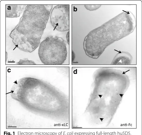
et al. previously constructed ssSTII variants with various TIRs [31] and showed that lower TIR strengths resulted in increased full-length mAb production [33]. Based on this study, we used an ssSTII variant with a relative TIR strength of 0.3 (ssSTII0.3) for the light chain and an ssS-TII variant with a relative TIR strength of 1 (ssSssS-TII1) for the heavy chain. The DNA sequences and relative TIR strengths of the signal sequences are listed in Additional file 1: Table S1. The corresponding amino acid sequences are listed in Table 1. The expression vector was trans-formed into a periplasmic protease-deficient host strain 64B4 (Additional file 2: Table S2) and antibody chain expression was induced upon phosphate depletion in the shake flask culture. E. coli cells expressing hu5D5 formed large inclusion bodies in the cytoplasm (Fig. 1b). E. coli expressing the empty vector did not form inclusion bod-ies (Fig. 1a). This observation suggested inefficient secre-tion of hu5D5 light and/or heavy chain to the periplasm. Immunogold electron microscopy showed that hu5D5 light chain predominantly localized in the periplasm whereas heavy chain localized in the cytoplasm (Fig. 1c, d), indicating hu5D5 heavy chain may be trapped in the cytoplasm.
To confirm that the secretion of hu5D5 heavy chain was limited, we examined the appearance of the pre-cursor and mature antibody chains. The non-secreted precursor form of an antibody chain maintains the sig-nal peptide at the N-terminus. During protein trans-location, the signal peptide is cleaved and therefore the secreted mature chain no longer contains the signal pep-tide sequence [34, 35]. For hu5D5 light chain, the precur-sor and mature forms can be distinguished by apparent molecular weight differences on SDS-PAGE. No precur-sor of light chain was detected using SDS-PAGE followed
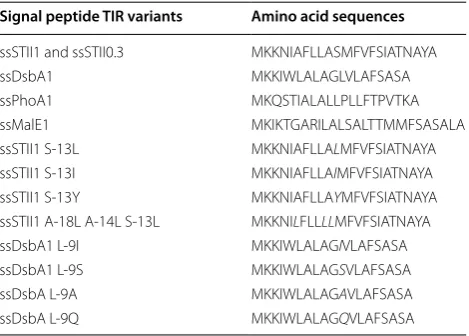
Table 1 The amino acid sequences of the signal peptide TIR variants
Amino acid substitutions are marked in italics
Signal peptide TIR variants Amino acid sequences
ssSTII1 and ssSTII0.3 MKKNIAFLLASMFVFSIATNAYA
ssDsbA1 MKKIWLALAGLVLAFSASA
ssPhoA1 MKQSTIALALLPLLFTPVTKA
ssMalE1 MKIKTGARILALSALTTMMFSASALA
ssSTII1 S-13L MKKNIAFLLALMFVFSIATNAYA
ssSTII1 S-13I MKKNIAFLLAIMFVFSIATNAYA
ssSTII1 S-13Y MKKNIAFLLAYMFVFSIATNAYA
ssSTII1 A-18L A-14L S-13L MKKNILFLLLLMFVFSIATNAYA
ssDsbA1 L-9I MKKIWLALAGIVLAFSASA
ssDsbA1 L-9S MKKIWLALAGSVLAFSASA
ssDsbA L-9A MKKIWLALAGAVLAFSASA
by Western blot analysis probing for light chain. N-termi-nal Edman sequencing showed that the sigN-termi-nal sequence was partially detected, however, the yield of the amino acids was too low to be quantified (data not shown). For the hu5D5 heavy chain, N-terminal Edman sequenc-ing was used to detect the precursor and mature forms because the small difference in molecular weight made it challenging to separate the two forms via SDS-PAGE. Interpretation of the sequencing data showed that less than half of the total amount of heavy chain was secreted (Table 2). To eliminate the possibility that light chain co-expression might interfere with heavy chain secretion, we expressed heavy chain with the STII signal sequence in the absence of light chain, and did not observe an increase in heavy chain secretion efficiency (Table 2). In contrast, no precursor was detected when hu5D5 light chain was expressed alone (Additional file 3: Figure S1). Taken together, these results demonstrated that the secretion of hu5D5 heavy chain, but not the light chain, was limiting.
The effect of various signal peptides on hu5D5 heavy chain secretion to the periplasm
In order to improve the secretion of hu5D5 heavy chain to the periplasm, we tested signal peptides representing
two major bacterial secretion pathways: the signal sequence of DsbA (ssDsbA), which mediates co-transla-tional secretion and is dependent upon the signal recog-nition particle (SRP) [36]; and the signal sequences of the maltose binding protein MalE and alkaline phosphatase PhoA (ssMalE, ssPhoA), which mediate sec-dependent post-translational secretion [37–40]. To control the translational strengths, we utilized silent mutations to generate signal peptide variants with various TIRs (see “Methods” section). ssSTII, ssDsbA, ssPhoA, and ssMalE TIR variants with relative translational strengths of ~1 (Additional file 1: Table S1, named as ssSTII1, ssDsbA1, ssPhoA1, ssMalE1) were each separately fused to hu5D5 heavy chain and tested for their ability to mediate hu5D5 secretion to the periplasm in the absence of light chain.
N-terminal Edman sequencing revealed that ssS-TII1, ssMalE1, and ssPhoA1 resulted in more precursor than mature heavy chain, whereas ssDsbA1 led to more mature heavy chain than precursor (Table 2), whether light chain was co-expressed or not. Collectively, with a TIR strength of ~1, ssDsbA showed increased secretion efficiency for hu5D5 heavy chain compared to ssSTII, ssPhoA, and ssMalE.
The effect of signal peptide hydrophobicity on hu5D5 heavy chain secretion at TIR strength 1
We sought to understand the molecular mechanism of why ssDsbA mediated more efficient secretion of hu5D5 heavy chain compared to the other signal peptides tested. Most E. coli signal peptides are composed of three dis-tinct regions: an N-terminal region which contains one or two positively charged amino acid residues, a hydro-phobic core region often referred to as the H-region, and a C-terminal region recognized by the signal pepti-dase [12, 13]. A comparison between ssSTII and ssDsbA showed that they have the same N-terminal residues and small residues Ala at the -1 and -3 positions of the cleav-age site, suggesting that the N- and C-termini may not be
c
an-κLC
d
an-Fc
a
b
Fig. 1 Electron microscopy of E. coli expressing full-length hu5D5.
a–b Transmission electron microscopy (TEM) images of 64B4 host strain expressing the empty vector pBR322 (a) or pBR-ssSTII0.3-ssSTII1-hu5D5 (b). White arrowheads point to inclusion bodies and black arrows point to the periplasm. c–d Immunostaining EM probing for light chain using anti-LC antibody (c) or heavy chain using anti-Fc antibody (d). Black arrows point to the periplasm. Black arrowheads point to gold particles indicating the cellular localization of light chain or heavy chain. Scale bars equal to 200 nm. The complete plasmid sequences were confirmed by DNA sequencing
Table 2 The processing of hu5D5 heavy chain mediated by ssSTII, ssDsbA, ssPhoA, and ssMalE at a relative TIR strength of one
Signal peptide Mature HC %
LC and HC co-expressed ssSTII1 45
ssDsbA1 86
ssPhoA1 6
ssMalE1 35
HC only ssSTII1 29
ssDsbA1 84
ssPhoA1 4
the key factors that cause different secretion efficiency. Hydrophobicity calculation of the four signal peptides revealed that the H-region of ssDsbA is more hydro-phobic than that of ssSTII, ssMalE, and ssPhoA (Fig. 2). Moreover, studies of multiple signal peptides suggest the hydrophobicity of the H-region is important for protein secretion, although the TIR strengths of the signal pep-tides were not controlled in these studies [14, 16, 21,
41, 42]. Therefore, we hypothesized that modulating the hydrophobicity of the H-region and controlling the TIR strength could improve hu5D5 heavy chain secretion.
To test this hypothesis, we introduced single amino acid changes into the H-region of ssSTII and ssDsbA (Table 3). The H-region of ssSTII contains two polar amino acid residues, Ser-13 and Ser-8. To increase the H-region hydrophobicity, Ser-13 was mutated to the resi-due Leu or Ile; as a control Ser-13 was also mutated to the non-charged polar residue Tyr, which has hydrophobicity similar to Ser (Table 3). Ser-8 was not changed due to the concern that it might affect cleavage by the signal pepti-dase. The TIR strengths of ssSTII hydrophobicity variants were normalized to ~1 by wobble-codon silent muta-tions (Additional file 1: Table S1). In the absence of light chain co-expression, both S-13L and S-13I substitutions in ssSTII increased hu5D5 heavy chain secretion to the periplasm (Table 3). The control S-13Y did not increase
the secretion (Table 3). We also engineered a highly hydrophobic ssSTII TIR1 variant by introducing three leucines into the H-region. ssSTII A-18L, A-14L, S-13L did not further improve the secretion of hu5D5 heavy chain above the results obtained for S-13L (Table 3), sug-gesting the effects of H-region hydrophobicity reached a plateau. Similar plateau effects were observed in a study that modulated the hydrophobicity of ssPhoA [20]. For ssDsbA, we generated variants with decreased hydropho-bicity by mutating Leu-9 to Ala, Ser, or Gln (Table 3). All the variants have a TIR strength of one. In the absence of light chain, both L-9S and L-9Q resulted in a decrease in secreted hu5D5 heavy chain. L-9A appeared to have a more modest impact on the secretion of hu5D5 heavy chain (Table 3). As a control, replacing Leu-9 with the hydrophobic residue Ile did not affect hu5D5 heavy chain secretion compared to the native signal peptide sequence (Table 3). We observed similar effects of signal peptide hydrophobicity on hu5D5 heavy chain secretion when light chain was co-expressed (Table 3). Collectively, at a TIR strength of 1, the hydrophobicity of the H-region is a critical factor for the efficient secretion of hu5D5 heavy chain.
Improving assembled full‑length hu5D5 levels by modulating signal peptide hydrophobicity at TIR strength 1
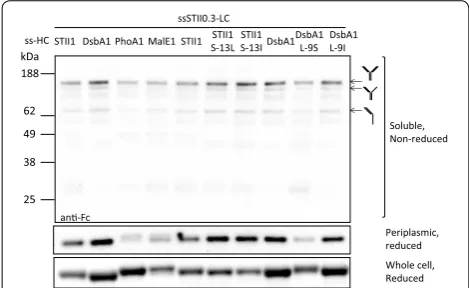
We next asked if the hydrophobicity of the H-region also affects assembled full-length hu5D5 accumulation in the periplasm. To test this idea, the hu5D5 light chain was fused to ssSTII0.3 and hu5D5 heavy chain was fused to ssSTII1, ssDsbA1, ssPhoA1, ssMalE1, or the H-region hydrophobicity variants of ssSTII1 and ssDsbA1. Levels of soluble hu5D5 heavy chain and full-length hu5D5 in the periplasm were examined by Western blot analyses (Fig. 3). ssDsbA1 resulted in more soluble hu5D5 heavy chain and assembled full-length hu5D5 accumulation in the periplasm than ssSTII1, ssPhoA1, or ssMalE1. Decreasing the hydrophobicity of the ssDsbA1 H-region 0.92 11.04
0.73 10.89 0.41 6.51 0.75 9.00
N H-region C Ave Hydro. H-region Sum Hydro. H-region
ssDsbA: ssStII: ssMalE: ssPhoA:
MKK MKK MKIK MKQ
IWLALAGLVLAF NIAFLLASMFVFSIA TGARILALSALTTMMF STIALALLPLLF
SASA TNAYA SASALA TPVTKA
Fig. 2 The amino acid sequences of signal peptides and the hydrophobicity of the H-region. The N-terminus, H-region, and the C-terminus were assigned following previous literature [35, 42, 53, 60]. The average and sum hydrophobicity of the H-region were calculated using the Eisenberg scale [61]
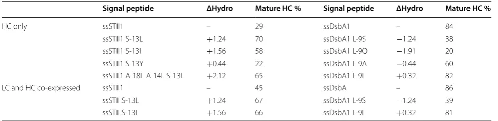
Table 3 The effects of signal peptide hydrophobicity on hu5D5 heavy chain processing
ΔHydro represents the change in total hydrophobicity resulting from the amino acid variants
Signal peptide ΔHydro Mature HC % Signal peptide ΔHydro Mature HC %
HC only ssSTII1 – 29 ssDsbA1 – 84
ssSTII1 S-13L +1.24 70 ssDsbA1 L-9S −1.24 38
ssSTII1 S-13I +1.56 58 ssDsbA1 L-9Q −1.91 20
ssSTII1 S-13Y +0.44 22 ssDsbA1 L-9A −0.44 60
ssSTII1 A-18L A-14L S-13L +2.12 65 ssDsbA1 L-9I +0.32 82
LC and HC co-expressed ssSTII1 – 45 ssDsbA – 86
ssSTII S-13L +1.24 67 ssDsbA1 L-9S −1.24 39
by using L-9S resulted in decreased levels of soluble heavy chain and assembled full-length hu5D5 in the peri-plasm (Fig. 3). As a control, ssDsbA L-9I did not affect the periplasmic accumulation of soluble heavy chain and assembled full-length hu5D5. For ssSTII, increas-ing the hydrophobicity of the native STII signal peptide by using S-13L or S-13I increased the accumulation of heavy chain and assembled full-length hu5D5. We did not observe any correlation between H-region hydropho-bicity and total levels of hu5D5 heavy chain in the cells. However, the hu5D5 heavy chain migrated at a higher apparent molecular weight for ssMalE1, ssPhoA1, and ssDsbA L-9S. N-terminal sequencing confirmed that the major components of these bands were heavy chain pre-cursors (Tables 2, 3). Collectively, the H-region hydro-phobicity of the signal peptide plays an important role in full-length hu5D5 production, probably by increasing the amount of heavy chain secreted to the periplasm.
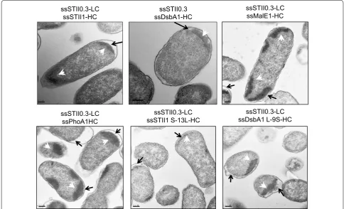
H-region hydrophobicity also influenced the cellular localization of inclusion bodies (Fig. 4). Signal peptides with a less hydrophobic H-region fused to hu5D5 heavy chain (ssSTII1, ssPhoA1, ssMalE1, or ssDsbA1 L-9SS) resulted in inclusion body formation in the cytoplasm, suggesting that proteins were trapped in the cytoplasm and aggregated. In contrast, signal peptides with a more hydrophobic H-region (ssDsbA1 and ssSTII S-13L) resulted in more inclusion bodies in the periplasm, indi-cating that proteins were secreted to the periplasm and
aggregated. Strategies to optimize antibody folding and prevent aggregation may further improve full-length hu5D5 production.
The effects of signal peptide hydrophobicity on the production of other full‑length mAbs
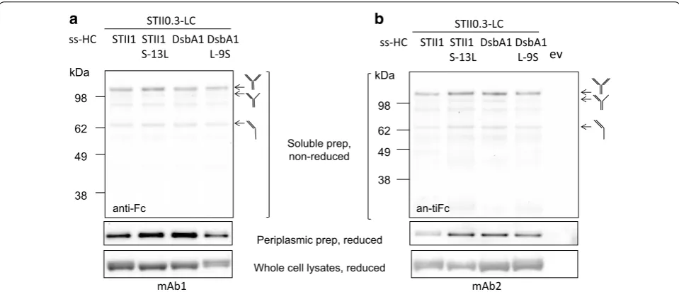
To test if the effects of signal peptide hydrophobicity apply to other full-length mAbs, two additional IgG1s (designated as mAb1 and mAb2) were included in this study. We first examined the secretion of mAb1 heavy chain and mAb2 heavy chain in the absence of their corresponding light chain. ssDsbA1 was found to medi-ate more efficient secretion of both mAb1 and mAb2 heavy chain compared to ssSTII1. Increasing the hydro-phobicity of ssSTII1 with S-13L increased the secretion efficiency of both heavy chains, whereas decreasing the hydrophobicity of DsbA1 by L-9S had the opposite effect (Table 4). However, the impact of L-9S on the secretion of mAb1 heavy chain was greater than for mAb2 heavy chain (Table 4).
To examine the effects of signal peptide hydrophobic-ity on the production of full-length mAb1 and mAb2, ssSTII0.3 was used for light chain and ssSTII1, ssDsbA1, ssSTII1 S-13L, or ssDsbA1 L11S for heavy chain. We observed for mAb1 that although ssDsbA1 led to more soluble heavy chain accumulation than ssSTII1, it did not lead to increased amounts of assembled full-length mAb (Fig. 5a). The use of ssSTII1 S-13L increased lev-els of both soluble heavy chain and assembled full-length mAb1 in the periplasm, and use of ssDsbA L-9S decreased the levels of soluble heavy chain and assem-bled full-length mAb1 (Fig. 5a). However, the differences in mAb1 levels were not as dramatic as those seen for hu5D5. For mAb2, the more hydrophobic signal peptides ssDsbA1 and ssSTII1 S-13L resulted in higher levels of soluble heavy chain and assembled full-length mAb in the periplasm than the less hydrophobic signal peptides ssSTII1 and ssDsbA1 L-9S (Fig. 5b). These results sug-gest that modulation of the signal peptide hydrophobicity impacts the accumulation of heavy chain and can impact the assembly of full-length mAb1 and mAb2. It is impor-tant to note that other factors such as antibody assembly efficiency and light chain-heavy chain interactions may limit the levels of full-length mAbs in the periplasm.
Discussion
Production of aglycosylated full-length mAbs in E. coli has been previously demonstrated by secreting the light and heavy chain to the periplasm where the antibody fragments are assembled [33, 43, 44]. However, the secre-tory production of mAbs often faces challenges such as protein aggregation, proteolytic degradation, and ineffi-cient secretion [3]. In this study we found that inefficient STII1 DsbA1 PhoA1 MalE1
ssSTII0.3-LC ss-HC
Soluble, Non-reduced
Periplasmic, reduced Whole cell, Reduced
188 62 49 38 25
kDa STII1 STII1 S-13L
STII1
S-13I DsbA1 DsbA1 L-9S DsbA1 L-9I
an-Fc
secretion of heavy chain can potentially limit the assem-bled full-length mAb level in the periplasm. We were able to improve heavy chain secretion and mAb accumulation by using a signal peptide with hydrophobic H-region at a controlled translational strength. The signal peptide engineering technique presented here may also apply to improve secretory production of other heterologous pro-teins in bacteria and to expand the range of propro-teins that can be efficiently presented by phage display [45].
Two aspects were considered for signal peptide engi-neering. First, we introduced amino acid substitutions to increase the hydrophobicity of the H-region, an impor-tant factor for protein secretion. Second, because amino acid changes in signal sequences alter the TIR sequence and potentially change the translational strength, and because high translational strength can result in ineffi-cient secretion of the target protein [31], we used silent mutations that change the wobble base pairs in the sig-nal sequence to control the translatiosig-nal strengths. Many previous studies on signal peptide mutagenesis showed that increasing signal peptide hydrophobicity improved protein translocation [14–24], whereas a few studies showed the opposite results [25–29]. The different obser-vations may be due to the use of different signal peptides and/or cargo proteins in these studies. However, it is also important to note that these studies generally did not control the translational strength.
In E. coli, the majority of secreted proteins are tar-geted to the SecYEG translocon through two pathways: the SRP-dependent co-translational pathway and the SRP-independent post-translational pathway. In the co-translational pathway, SRP binds co-co-translationally to
ssSTII0.3-LC
ssSTII1-HC ssSTII0.3-LC ssMalE1-HC
ssSTII0.3-LC ssPhoA1HC
ssSTII0.3 ssDsbA1-HC
ssSTII0.3-LC ssDsbA1 L-9S-HC ssSTII0.3-LC
ssSTII1 S-13L-HC
Fig. 4 The effects of signal peptide hydrophobicity on the cellular localization of inclusion bodies. TEM of E. coli expressing ssSTII0.3-ssSTII1-hu5D5, ssSTII0.3-ssDsbA1-ssSTII0.3-ssSTII1-hu5D5, ssSTII0.3-ssMalE1-ssSTII0.3-ssSTII1-hu5D5, ssSTII0.3-ssPhoA1-ssSTII0.3-ssSTII1-hu5D5, ssSTII0.3-ssSTII1 S-13L-ssSTII0.3-ssSTII1-hu5D5, or pBR-ssSTII0.3-ssDsbA1 L-9S-hu5D5. Black arrows point to the periplasmic space, and the white arrowheads point to the inclusion bodies
Table 4 The effects of signal peptide hydrophobicity on the secretion of mAb1 and mAb2 heavy chain
Signal peptide Cargo protein Mature HC %
ssSTII1 mAb1HC 35
ssSTII1 S-13L mAb1HC 79
ssDsbA1 mAb1HC 86
ssDsbA1 L-9S mAb1HC 13
ssSTII1 mAb2HC 17
ssSTII1 S-13L mAb2HC 78
ssDsbA1 mAb2HC 89
the newly synthesized signal peptide at the ribosome and directs the ribosome-nascent chain (RNC) complex to the SRP receptor FtsY. The RNCs are then targeted to the SecYEG translocon where translocation occurs simulta-neously with translation. In the post-translational path-way, the polypeptide is released from the ribosome as the precursor form prior to translocation. The precursor is then targeted to the membrane-bound SecA and SecYEG in the inner membrane (reviewed in [1]). Although post-translational signal peptides have been extensively used for recombinant protein production, they may not be a good choice for recombinant proteins that fold or aggre-gate rapidly in the cytoplasm [46]. Such issues could potentially be prevented by using SRP-dependent sig-nal sequences. With the co-translation pathway protein translation, translocation, and folding are coordinated. Among the signal peptides tested, ssDsbA mediates co-translational secretion [36], while ssPhoA and ssMalE have been shown to mediate post-translational secretion [37, 40]. Indeed, we observed that ssDsbA led to more efficient secretion of heavy chain than the two post-trans-lational signal peptides (Table 2). Lee et al., also recently reported that using the DsbA signal sequence to replace the PelB signal sequence enhanced the production of full-length IgG [43]. It is unclear if ssSTII mediates co-trans-lational or post-translation secretion. The observation of a large percentage of precursor heavy chains suggests that ssSTII preferentially uses the post-translational
pathway, although it does not rule out the possibil-ity that ssSTII mediates heavy chain secretion through both the post- and co-translational pathways. It has been shown that some proteins use both pathways for secre-tion [34, 47]. Moreover, recombinant protein expression and secretion can result in exceeding the capacity of one secretion pathway. In such a scenario it is possible that protein secretion utilizes both pathways.
How does signal peptide hydrophobicity affect heavy chain secretion? One possible explanation is that a less hydrophobic signal peptide such as ssSTII preferentially uses the post-translational pathway. The heavy chain pre-cursors are likely to fold or aggregate in the cytoplasm and become secretion incompetent. Increasing the H-region hydrophobicity could re-route heavy chain to the co-trans-lational secretion pathway and prevent heavy chain folding or aggregation in the cytoplasm. This hypothesis is sup-ported by studies showing that hydrophobic signal pep-tides interact with SPR more efficiently [48–50] and often mediate co-translational secretion [24, 51]. Alternatively, it is possible that increasing the signal peptide hydrophobic-ity results in more efficient interaction between the heavy chain precursor and SecA [48] or cytoplasmic chaperones.
In addition to signal peptide hydrophobicity and TIR strengths, other factors can also impact antibody chain secretion. For instance, a positively charged amino acid at the N-terminus of the signal sequence has been shown to promote the interaction with SRP [52]. The alpha-helix
Soluble prep, non-reduced
Whole cell lysates, reduced Periplasmic prep, reduced
a
kDa
98
62
49
38
STII1 STII1
S-13L DsbA1 DsbA1 L-9S STII0.3-LC ss-HC
mAb1 anti-Fc
b
62 49
38 kDa
98
STII1 STII1
S-13L DsbA1 DsbA1 L-9S STII0.3-LC ss-HC
mAb2 an-tiFc
ev
content in the signal sequence is also important for effi-cient secretion [53, 54]. The amino acid composition of C-terminus is critical for the cleavage of signal peptide [35]. Moreover, several studies have shown that signal peptides can modulate the folding, thermodynamic sta-bility, and aggregation propensities of cargo proteins [55,
56]. With the post-translational secretion pathway, inter-actions between the signal peptide and heavy chain in the cytoplasm may affect secretion efficiency.
Using signal peptide engineering, we increased the yields of hu5D5 in the shake flask cultures approximately 2.5–3 fold. Many factors remain to be tested to further optimize full-length mAb accumulation in the periplasm. For example, co-expression of key components of the secretion machinery may further increase antibody chain secretion. It’s been shown that over-expression of SRP (Ffh) increased the yields of full-length IgG [43], presum-ably due to increased secretion efficiency. Also, based on our preliminary results we suspect that heavy chain and light chain may compete for the limited secretion capac-ity. Therefore, the ratio of light chain to heavy chain may need to be finely tuned. Moreover, secreted light chain and heavy chain can form dimeric species or aggregates in the periplasm (Figs. 3, 5). Co-expression of periplasmic chaperones provides a feasible strategy to prevent protein aggregation and increase yields of mAbs [8, 43, 44].
Conclusions
Escherichia coli is an attractive vehicle for the production of therapeutic proteins. However, it remains challenging to achieve high yields for complex, multi-subunit pro-teins including full-length mAbs. In this study we dem-onstrated that one of the bottlenecks for the production of assembled full-length mAbs is the secretion of heavy chain to the periplasm. Signal peptide engineering by controlling the TIR strength and increasing H-region hydrophobicity improved the efficiency of heavy chain secretion to the periplasm and further improved the accumulation of several full-length mAbs. The technol-ogy described here offers an effective approach for pro-duction of full-length mAbs in E. coli.
Methods
Strains and plasmids
Strains and plasmids used in this study are listed in Addi-tional file 2: Table S2. To construct expression vectors, DNA fragments of heavy chain or light chain containing the phoA promoter and signal sequence were cloned into a pBR322-derived expression vector [33]. To construct phoA reporter plasmids for translational strength meas-urement, the fragments containing the signal peptide sequences were cloned upstream of the phoA reporter gene in the pPhoA86 vector [31]. Additional site-specific
mutations in signal peptide sequences were introduced by the QuickChange Site-Directed Mutagenesis Kit (Stratagene). The sequence of each plasmid was con-firmed by DNA sequencing (Genentech Inc.).
Construction of signal peptide TIR libraries
Libraries of signal sequences with a range of translational strengths were generated as described previously [31, 57]. Briefly, degenerate oligos were used to introduce silent mutations in the first seven codons of signal sequences from thestII, dsbA, malE, and phoA. Restriction sites of XbaI, BssHII, or MluI were inserted upstream of the start codon to diversify the TIR region. The TIR variant sequences were inserted upstream of the mature phoA gene on the pPhoA81 plasmid. The constructs were transformed into the host strain 27C7. The transformants were plated on LB agar plates containing carbenicillin and 100 µg/mL of the substrate 5-bromo-4-chloro-3-in-dolyl phosphate (BCIP, Sigma) and grown at 37 °C over-night. Colonies with light blue or dark blue color were selected for plasmid extraction and sequencing (Genen-tech, Inc.).
Translational strength measurement
The translational strengths of the signal peptide variants were determined by the alkaline phosphatase assay using para-nitrophenylphosphate as the substrate. [31, 57].
Induction of antibody expression
Bacterial cultures were grown in Luria–Bertani (LB) or complete C.R.A.P. phosphate-limiting media [33] con-taining antibiotics at 37 °C or at 30 °C as indicated. The following concentrations of antibiotics were used: car-benicillin 50 µg/mL, and tetracycline 20 µg/mL. For protein expression, the host strain 64B4 harboring the antibody expression vector was inoculated into 5 mL of LB supplemented with 20 µL/mL tetracycline and 5 mM sodium phosphate, pH 7 in a polypropylene culture tube and incubated at 30 °C with shaking overnight. OD550 of
the overnight culture was measured to ensure the strain did not have growth defects. 0.5 mL of the overnight culture was inoculated into 25 mL of complete C.R.A.P. supplemented with 20 µL/mL tetracycline in a 125 mL baffled shake flask and cells were grown at 30 °C with shaking for 24 h. The optical density of the culture at 7 and 24 h was measured at 550 nm.
Protein extraction and Western blotting analysis
End point samples from shake flask cultures were col-lected for Western blot analysis. To measure total heavy chain levels, 1 mL of 1 OD550 of bacteria was spun down,
To extract soluble proteins, a previously described sonication approach [57] was used because it resulted in more reproducible results and higher enrichment of anti-body chains compared to the conventional osmotic shock method. Briefly, whole cell broth was diluted into chilled lysis buffer (10 mM Tris, pH 6.8, 5 mM EDTA, 0.2 mg/ mL Lysozyme, and 5 mM iodoacetic acid) to a final OD550
of 3.0. 600 µL of samples were then sonicated using two rounds of 10 × 1 s pulses and centrifuged for 15 min at 16,000 ×g at 4 °C. Supernatant was collected for SDS-PAGE and Western blot analysis. For periplasmic pro-tein extraction, 10 OD550 of bacteria were pelleted and
resuspended in TBS buffer (200 mM Tris, pH8.0, 0.5 mM sucrose, 1 mM EDTA) with 1 tablet of protease inhibitor cocktail (Roche) as described [58]. Soluble protein sam-ples or periplasmic extracts were mixed 1:1 (v/v) with tricine SDS buffer with or without 0.2 M DTT and then analyzed by SDS-PAGE and Western blotting. Lane inten-sities on coomassie stained gels were used as a loading control. Heavy chain-containing species were probed with HRP-conjugated goat anti-human Fc antibody (Pierce). Light chain-containing species were probed with HRP-conjugated goat-anti-human kLC antibody (Bethyl Labo-ratories). Target proteins on immunoblots were detected by enhanced chemiluminescent (GE Healthcare).
Edman sequencing of the N‑terminus of heavy chain End point samples from shake flask cultures were nor-malized to 4 OD550 and harvested by centrifugation at
16,000×g for 3 min. Whole cell lysates resuspended in 200 µL tricine SDS buffer containing 0.2 M DTT were loaded onto 10 % Bis–Tris SDS-PAGE. After electropho-retic separation, the proteins were transferred to PVDF membranes using wet transfer (Biorad) in CAPS buffer. The heavy chain band at ~50 kDa on the membrane was excised and subjected to Edman sequencing analysis using the Applied Biosystems Procise Sequencer Model 494HT. Picomole values of each amino acid were calcu-lated by the sequence analysis program SEQX normal-ized against the uncorrected phenylthiohydantoin amino acid standards [59]. An average of 10 cycles was used to produce the repetitive yield plot to calculate the lin-ear regression, and the initial yields of major and minor sequences were defined as the y-intercepts of the plot-ted lines. The heavy chain processing efficiency was cal-culated as the percentage of mature heavy chain, which equals Initial yieldmature/(Initial yieldmature + Initial yieldprecursor).
Transmission electron microscopy (TEM)
End point samples from shake flask cultures were imme-diately collected, pelleted to 50–100 µL pellets, and resuspended in 1 mL of in the modified Karnovsky’s
fixative (2 % paraformaldehyde and 2.5 % glutaraldehyde in 0.1 M sodium cacodylate buffer, pH7.2) and then post-fixed in 1 % aqueous osmium tetroxide (EM Sciences, Hatfield, PA) for 1 h followed by overnight incubation in 0.5 % uranyl acetate at 4 °C. The samples were then dehy-drated through a series of increasing ethanol concentra-tions (50, 70, 90, 100 %), followed by propylene oxide (each step was for 15 min) and embedded in Eponate 12 (Ted Pella, Redding, CA). Ultrathin sections (80 nm) were cut with an Ultracut microtome (Leica), stained with 0.2 % lead citrate and examined using a JEOL JEM-1400 transmission electron microscope (TEM) at 120 kV. Digital images were captured with a GATAN Ultrascan 1000 CCD camera.
Immunogold electron microscopy (immunoEM)
For the immunogold EM experiments, samples were first prepared for cryosectioning. The cells were fixed in 4 % paraformaldehyde with 0.1 % glutaraldehyde in phosphate buffer (0.1 M, pH 7.2), washed several times in PBS, embedded in 12 % gelatin and infiltrated in 2.3 M sucrose overnight at 4 °C. Samples were then mounted on pins for cryo-ultramicrotomy frozen in a cryosectioning chamber (supplied with liquid nitrogen). Ultrathin cryosections (100 nm) were prepared with a diamond knife (Diatome) at −80 °C using an ultrami-crotome (Ultracut; Leica) equipped with a cryosection-ing chamber. Thawed cryosections were transferred to Formvar- and carbon-coated EM grids (Nickel) with a drop of 2.3 M sucrose, immunolabeled (see below) and then counterstained for EM with 0.5 % uranyl acetate in 2 % methylcellulose for 1 min at room temperature. For immunogold labeling, the thawed cryosections on grids were blocked in blocking agent (Aurion Inc) for 30 min and incubated with an anti-light chain (LC) or anti-heavy chain (Fc) HRP-conjugated goat antibody for 45 min at room temperature, followed by incuba-tion with an anti-HRP gold conjugated goat antibody (Jackson ImmmunoResearch) for 30 min. Sections were then counterstained as described above. Immunogold-labeled sections were visualized and examined in a JEOL JEM-1400 transmission electron microscope (TEM) at 120 kV. Digital images were captured with a GATAN Ultrascan 1000 CCD camera.
Additional files
Additional file 1: Table S1. The DNA sequences of the signal peptide TIR variants and the relative TIR strengths.
Authors’ contributions
YZ designed the experiments and analyzed the data. DR supervised the research. YZ and DR wrote the manuscript. YG, PL, and WS performed the Edman sequencing and analyzed the sequencing data. AKK, MR, and LR performed the TEM and data analysis. All authors read and approved the final manuscript.
Author details
1 Department of Early Stage Cell Culture, Genentech Inc., 1 DNA way, South San Francisco, CA 94080, USA. 2 Department of Protein Chemistry, Genentech Inc., 1 DNA way, South San Francisco, CA 94080, USA. 3 Department of Pathol-ogy, Genentech Inc., 1 DNA way, South San Francisco, CA 94080, USA.
Acknowledgements
We thank the DNA plasmid preparation lab and the Media Prep lab at Genentech for providing the research reagents. We acknowledge the DNA sequencing lab at Genentech for sequencing the constructs. We acknowl-edge Matthew Marrichi for construction of the signal peptide TIR library. We acknowledge Laura Simmons, Christoph Spiess, Scott Chamberlain, Thomas Patapoff, and the Early Stage Cell Culture E. coli group for helpful discussions.
Competing interests
The authors declare that they have no competing interests.
Received: 5 December 2015 Accepted: 21 February 2016
References
1. Mergulhao FJ, Summers DK, Monteiro GA. Recombinant protein secretion in Escherichia coli. Biotechnol Adv. 2005;23:177–202.
2. Andersen DC, Reilly DE. Production technologies for monoclonal anti-bodies and their fragments. Curr Opin Biotechnol. 2004;15:456–62. 3. Choi JH, Lee SY. Secretory and extracellular production of
recom-binant proteins using Escherichia coli. Appl Microbiol Biotechnol. 2004;64:625–35.
4. de Marco A. Strategies for successful recombinant expression of disulfide bond-dependent proteins in Escherichia coli. Microb Cell Fact. 2009;8:26. 5. Nossal NG, Heppel LA. The release of enzymes by osmotic shock from
Escherichia coli in exponential phase. J Biol Chem. 1966;241:3055–62. 6. Shokri A, Sanden AM, Larsson G. Cell and process design for targeting of
recombinant protein into the culture medium of Escherichia coli. Appl Microbiol Biotechnol. 2003;60:654–64.
7. Rosenberg HF. Isolation of recombinant secretory proteins by limited induction and quantitative harvest. Biotechniques. 1998;24(188–190):192. 8. Reilly DE, Yansura DG. Production of monoclonal antibodies in E. coli.
Springer: Berlin; 2010.
9. Jeong KJ, Lee SY. Secretory production of human leptin in Escherichia coli. Biotechnol Bioeng. 2000;67:398–407.
10. Pritchard MP, Ossetian R, Li DN, Henderson CJ, Burchell B, Wolf CR, Friedberg T. A general strategy for the expression of recombinant human cytochrome P450s in Escherichia coli using bacterial signal peptides: expression of CYP3A4, CYP2A6, and CYP2E1. Arch Biochem Biophys. 1997;345:342–54.
11. Schlegel S, Rujas E, Ytterberg AJ, Zubarev RA, Luirink J, de Gier JW. Optimizing heterologous protein production in the periplasm of E. coli by regulating gene expression levels. Microb Cell Fact. 2013;12:24. 12. Perlman D, Halvorson HO. A putative signal peptidase recognition site
and sequence in eukaryotic and prokaryotic signal peptides. J Mol Biol. 1983;167:391–409.
13. von Heijne G, Abrahmsen L. Species-specific variation in signal peptide design. Implications for protein secretion in foreign hosts. FEBS Lett. 1989;244:439–46.
14. Izard JW, Rusch SL, Kendall DA. The amino-terminal charge and core region hydrophobicity interdependently contribute to the function of signal sequences. J Biol Chem. 1996;271:21579–82.
15. Rusch SL, Mascolo CL, Kebir MO, Kendall DA. Juxtaposition of signal-peptide charge and core region hydrophobicity is critical for functional signal peptides. Arch Microbiol. 2002;178:306–10.
16. Rusch SLCH, Izard JW, Kendall DA. Signal peptide hydrophobicity is finely tailored for function. J Cell Biochem. 1994;55:209–17.
17. Kendall DA, Bock SC, Kaiser ET. Idealization of the hydrophobic segment of the alkaline phosphatase signal peptide. Nature. 1986;321:706–8. 18. Kendall DA, Doud SK, Kaiser ET. A comparative analysis of single- and
multiple-residue substitutions in the alkaline phosphatase signal peptide. Biopolymers. 1990;29:139–47.
19. Kendall DA, Kaiser ET. A functional decaisoleucine-containing sig-nal sequence. Construction by cassette mutagenesis. J Biol Chem. 1988;263:7261–5.
20. Doud SK, Chou MM, Kendall DA. Titration of protein transport activity by incremental changes in signal peptide hydrophobicity. Biochemistry. 1993;32:1251–6.
21. Rusch SL, Kendall DA. Transport of an export-defective protein by a highly hydrophobic signal peptide. J Biol Chem. 1994;269:1243–8.
22. Goldstein J, Lehnhardt S, Inouye M. Enhancement of protein transloca-tion across the membrane by specific mutatransloca-tions in the hydrophobic region of the signal peptide. J Bacteriol. 1990;172:1225–31. 23. Hikita C, Mizushima S. Effects of total hydrophobicity and length of
the hydrophobic domain of a signal peptide on in vitro translocation efficiency. J Biol Chem. 1992;267:4882–8.
24. Bowers CW, Lau F, Silhavy TJ. Secretion of LamB-LacZ by the Sig-nal recognition particle pathway of Escherichia coli. J Bacteriol. 2003;185:5697–705.
25. Morioka-Fujimoto K, Marumoto R, Fukuda T. Modified enterotoxin signal sequences increase secretion level of the recombinant human epidermal growth factor in Escherichia coli. J Biol Chem. 1991;266:1728–32. 26. Jonet MA, Mahadi NM, Murad AM, Rabu A, Bakar FD, Rahim RA, Low KO,
Illias RM. Optimization of a heterologous signal peptide by site-directed mutagenesis for improved secretion of recombinant proteins in Escheri-chia coli. J Mol Microbiol Biotechnol. 2012;22:48–58.
27. Ravn PAJ, Madsen SM, Vrang A, Israelsen H. Optimization of signal peptide SP310 for heterologous protein production in Lactococcus lactis. Microbiology. 2003;149:2193–201.
28. Nakamura K, Fujita Y, Itoh Y, Yamane K. Modification of length, hydropho-bic properties and electric charge of Bacillus subtilis alpha-amylase signal peptide and their different effects on the production of secretory pro-teins in B. subtilis and Escherichia coli cells. Mol Gen Genet. 1989;216:1–9. 29. Zanen G, Houben EN, Meima R, Tjalsma H, Jongbloed JD, Westers H,
Oudega B, Luirink J, van Dijl JM, Quax WJ. Signal peptide hydrophobic-ity is critical for early stages in protein export by Bacillus subtilis. FEBS J. 2005;272:4617–30.
30. McCarthy JE, Gualerzi C. Translational control of prokaryotic gene expres-sion. Trends Genet. 1990;6:78–85.
31. Simmons LC, Yansura DG. Translational level is a critical factor for the secretion of heterologous proteins in Escherichia coli. Nat Biotechnol. 1996;14:629–34.
32. Merchant M, Ma X, Maun HR, Zheng Z, Peng J, Romero M, Huang A, Yang NY, Nishimura M, Greve J, et al. Monovalent antibody design and mecha-nism of action of onartuzumab, a MET antagonist with anti-tumor activity as a therapeutic agent. Proc Natl Acad Sci USA. 2013;110:E2987–96. 33. Simmons LC, Reilly D, Klimowski L, Raju TS, Meng G, Sims P, Hong K,
Shields RL, Damico LA, Rancatore P, Yansura DG. Expression of full-length immunoglobulins in Escherichia coli: rapid and efficient production of aglycosylated antibodies. J Immunol Methods. 2002;263:133–47. 34. Josefsson LG, Randall LL. Different exported proteins in E. coli show
differ-ences in the temporal mode of processing in vivo. Cell. 1981;25:151–7. 35. Auclair SM, Bhanu MK, Kendall DA. Signal peptidase I: cleaving the way to
mature proteins. Protein Sci. 2012;21:13–25.
36. Schierle CF, Berkmen M, Huber D, Kumamoto C, Boyd D, Beckwith J. The DsbA signal sequence directs efficient, cotranslational export of passen-ger proteins to the Escherichia coli periplasm via the signal recognition particle pathway. J Bacteriol. 2003;185:5706–13.
37. Collier DN, Bankaitis VA, Weiss JB, Bassford PJ Jr. The antifolding activity of SecB promotes the export of the E. coli maltose-binding protein. Cell. 1988;53:273–83.
38. Weiss JB, Ray PH, Bassford PJ Jr. Purified secB protein of Escherichia coli retards folding and promotes membrane translocation of the maltose-binding protein in vitro. Proc Natl Acad Sci USA. 1988;85:8978–82. 39. Kumamoto CA, Beckwith J. Evidence for specificity at an early step in
• We accept pre-submission inquiries
• Our selector tool helps you to find the most relevant journal • We provide round the clock customer support
• Convenient online submission • Thorough peer review
• Inclusion in PubMed and all major indexing services • Maximum visibility for your research
Submit your manuscript at www.biomedcentral.com/submit
Submit your next manuscript to BioMed Central
and we will help you at every step:
40. Chen L, Rhoads D, Tai PC. Alkaline phosphatase and OmpA protein can be translocated posttranslationally into membrane vesicles of Escherichia coli. J Bacteriol. 1985;161:973–80.
41. Chou MM, Kendall DA. Polymeric sequences reveal a functional inter-relationship between hydrophobicity and length of signal peptides. J Biol Chem. 1990;165:2873–80.
42. Izard JW, Kendall DA. Signal peptides: exquisitely designed transport promoters. Mol Microbiol. 1994;13:765–73.
43. Lee YJ, Kim HS, Ryu AJ, Jeong KJ. Enhanced production of full-length immunoglobulin G via the signal recognition particle (SRP)-dependent pathway in Escherichia coli. J Biotechnol. 2013;165:102–8.
44. Lee YJ, Lee DH, Jeong KJ. Enhanced production of human full-length immunoglobulin G1 in the periplasm of Escherichia coli. Appl Microbiol Biotechnol. 2014;98(3):1237–46.
45. Steiner D, Forrer P, Stumpp MT, Pluckthun A. Signal sequences directing cotranslational translocation expand the range of proteins amenable to phage display. Nat Biotechnol. 2006;24:823–31.
46. Huber D, Cha MI, Debarbieux L, Planson AG, Cruz N, Lopez G, Tasayco ML, Chaffotte A, Beckwith J. A selection for mutants that interfere with folding of Escherichia coli thioredoxin-1 in vivo. Proc Natl Acad Sci USA. 2005;102:18872–7.
47. Froderberg L, Houben E, Samuelson JC, Chen M, Park SK, Phillips GJ, Dalbey R, Luirink J, De Gier JW. Versatility of inner membrane protein biogenesis in Escherichia coli. Mol Microbiol. 2003;47:1015–27. 48. Valent QA, Scotti PA, High S, de Gier JW, von Heijne G, Lentzen G,
Winter-meyer W, Oudega B, Luirink J. The Escherichia coli SRP and SecB targeting pathways converge at the translocon. EMBO J. 1998;17:2504–12. 49. Lee HC, Bernstein HD. The targeting pathway of Escherichia coli
presecretory and integral membrane proteins is specified by the hydrophobicity of the targeting signal. Proc Natl Acad Sci USA. 2001;98:3471–6.
50. Zhang X, Rashid R, Wang K, Shan SO. Sequential checkpoints govern substrate selection during cotranslational protein targeting. Science. 2010;328:757–60.
51. Huber D, Boyd D, Xia Y, Olma MH, Gerstein M, Beckwith J. Use of thioredoxin as a reporter to identify a subset of Escherichia coli signal sequences that promote signal recognition particle-dependent translo-cation. J Bacteriol. 2005;187:2983–91.
52. Peterson JH, Woolhead CA, Bernstein HD. Basic amino acids in a distinct subset of signal peptides promote interaction with the signal recognition particle. J Biol Chem. 2003;278:46155–62.
53. Izard JW, Doughty MB, Kendall DA. Physical and conformational proper-ties of synthetic idealized signal sequences parallel their biological func-tion. Biochemistry. 1995;34:9904–12.
54. Adams H, Scotti PA, de Cock H, Luirink J, Tommassen J. The presence of a helix breaker in the hydrophobic core of signal sequences of secre-tory proteins prevents recognition by the signal-recognition particle in
Escherichia coli. Eur J Biochem. 2002;269:5564–71.
55. Park S, Liu G, Topping TB, Cover WH, Randall LL. Modulation of fold-ing pathways of exported proteins by the leader sequence. Science. 1988;239:1033–5.
56. Singh P, Sharma L, Kulothungan SR, Adkar BV, Prajapati RS, Ali PS, Krishnan B, Varadarajan R. Effect of signal peptide on stability and folding of
Escherichia coli thioredoxin. PLoS One. 2013;8:e63442.
57. Marrichi M, Reilly DE. Methods and composition for secretion of heterolo-gous polypeptides. US Patent 8,361,744. 2013.
58. Quan S, Hiniker A, Collet JF, Bardwell JC. Isolation of bacteria envelope proteins. Methods Mol Biol. 2013;966:359–66.
59. Henzel WJ, Rodriguez H, Watanabe C. Computer analysis of automated Edman degradation and amino acid analysis data. J Chromatogr. 1987;404:41–52. 60. Collier DN. Escherichia coli signal peptides direct inefficient secretion of
an outer membrane protein (OmpA) and periplasmic proteins (maltose-binding protein, ribose-(maltose-binding protein, and alkaline phosphatase) in
Bacillus subtilis. J Bacteriol. 1994;176:3013–20.