R E S E A R C H A R T I C L E
Open Access
Does all single infarction have lower risk of
stroke recurrence than multiple infarctions
in minor stroke?
Guangyao Wang
1,2,3,4†, Jing Jing
1,2,3,4†, Yuesong Pan
1,2,3,4, Xia Meng
1,2,3,4, Xingquan Zhao
1,2,3,4, Liping Liu
1,2,3,4,
Hao Li
1,2,3,4, David Wang
5, Yongjun Wang
1,2,3,4, Yilong Wang
1,2,3,4*and On behalf of the CHANCE Investigatores
Abstract
Background:
Single acute infarction (SAI) usually had lower risk of stroke recurrence than multiple acute infarctions
(MAIs) in minor stroke. To evaluate whether all SAI had lower risk of stroke recurrence than MAIs in minor stroke.
Methods:
We derived data from the imaging subgroup of the Clopidogrel in High-risk Patients with Acute
Nondisabling Cerebrovascular Events (CHANCE) trial. Minor stroke were categorized into SAI and MAIs by infarction
numbers in diffusion weighted imaging. SAI were classified as lacunar infarction and non-lacunar infarction. The
outcome was stroke recurrence within one-year follow-up. We assessed the associations between infarction
patterns and stroke recurrence using multivariable Cox regression models.
Results:
Overall, 834 patients with minor stroke were included in this subgroup, 553 SAI (381 lacunar infarction, 172
non-lacunar infarction) and 281 MAIs. The rate of stroke recurrence was 7.6%, 15.1% and 15.3% in lacunar infarction
of SAI, non-lacunar infarction of SAI and MAIs at one year, respectively. Compared with MAIs, lacunar infarction of
SAI had lower risk of stroke recurrence (hazard ratio [HR] 0.41, 95% confidence interval [CI] 0.21
–
0.80,
P
= 0.009), but
not in non-lacunar infarction of SAI (HR 1.01, 95% CI 0.60
–
1.69,
P
= 0.98).
Conclusions:
Lacunar infarction of SAI have lower risk of stroke recurrence than MAIs, while non-lacunar infarction
of SAI might have similar risk as MAIs. Except for the number of infarctions, size and location should also be
considered to stratify risk of stroke recurrence in minor stroke.
Trial registration:
http://www.clinicaltrials.gov Unique identifier:
NCT00979589
. Date of registration: September 2009.
Keywords:
Minor stroke, Infarction patterns, Prognosis
Background
Minor stroke are the most common manifestations of
acute cerebrovascular disease and the proportion of
minor stroke in all ischemic stroke is approximately 50%
[
1
]. Patients with minor stroke had higher risk of
recur-rence after symptom onset, especially in the early stage
[
2
]. Recent studies suggested that vascular and
neuroim-aging parameters may improve risk stratification in
minor stroke [
3
,
4
]. TIA
registry.org
project showed that
infarction patterns helped to stratify the risk of stroke
recurrence within one year after minor stroke and
pa-tients with multiple acute infarctions (MAIs) had much
higher risk of stroke recurrence than that with single
acute infarction (SAI) or no acute infarction (NAI),
indi-cating that MAIs was an important imaging marker to
predict stroke recurrence [
5
]. However, several studies
showed that there were different patterns in SAI and
MAIs respectively corresponding to different stroke
eti-ologies [
6
,
7
] or mechanisms [
8
,
9
]. Different stroke
eti-ologies or mechanisms might lead to different risk of
stroke recurrence [
10
–
15
]. Traditionally, SAI were
clas-sified according to the size and location of the infarction,
while MAIs were classified according to the blood
sup-ply of different brain areas [
6
,
7
]. However, it was
* Correspondence:[email protected]
†Guangyao Wang and Jing Jing contributed equally to this work. 1
Department of Neurology, Beijing Tiantan Hospital, Capital Medical University, Beijing, China
2China National Clinical Research Center for Neurological Diseases, Beijing,
China
Full list of author information is available at the end of the article
unclear whether different infarction patterns of SAI and
MAIs respectively had different risk of stroke recurrence
after minor stroke and whether all SAI had lower risk of
stroke recurrence than MAIs in minor stroke.
In the current study, deriving data from the imaging
subgroup of the Clopidogrel in High-risk Patients with
Acute Nondisabling Cerebrovascular Events (CHANCE)
trial, we investigated whether among patients with SAI
or MAIs whether different infarction patterns were
asso-ciated with different risk of stroke recurrence. We
fur-ther compared the risk of stroke recurrence in SAI with
different infarction patterns to that of MAIs.
Methods
Overview of the CHANCE trial
The detailed design and methods of the CHANCE trial
have been previously described [
16
,
17
]. Briefly, CHANCE
was a randomized, double-blind, placebo-controlled
clin-ical trial conducted in 114 centers in China between
Octo-ber 2009 and July 2012. Totally, 5170 patients within 24 h
of non-cardioembolic minor ischemic stroke or high-risk
TIA onset were randomly assigned to either clopidogrel
plus aspirin (clopidogrel at an initial dose of 300 mg,
followed by 75 mg per day for 90 days, plus aspirin at 75
mg per day for the first 21 days) or placebo plus aspirin
(75 mg per day for 90 days) group. The trial was approved
by the Ethics Committee of Beijing Tiantan Hospital and
all the participating hospitals. Written informed consent
was obtained from all participants or their legal proxies.
This study was registered at Clinical
Trials.gov
(registra-tion number NCT00979589).
Overview of the imaging substudy of the CHANCE trial
This imaging study was a prespecified substudy of the
CHANCE trial. Briefly, 45 (39%) of 114 centers of the
CHANCE trial were prospective recruited in the imaging
substudy voluntarily. All patients were asked to complete
the magnetic resonance (MR) examinations (3.0 or 1.5
Tesla) during hospitalization in this substudy. Patients
with the following MR sequences were included in the
substudy: T1-weighted imaging, T2-weighted imaging,
diffusion-weighted imaging (DWI), and 3-dimensional
(3D) time-of-flight magnetic resonance angiography
(MRA). Those without baseline MR examination or any of
the above sequences were excluded. The details of the
CHANCE imaging substudy have been previously
de-scribed [
4
,
18
].
Patient screening and image analysis
All MR images collected from individual centers in
digital format were read centrally by two readers (X.Z.
and J.J.) blinded to the patients
’
baseline and outcome
information. Minor stroke patients with new infarction
according to DWI were included in the final analysis. All
minor stroke patients were classified as SAI or MAIs
ac-cording to infarction numbers [
5
]. Uninterrupted lesions
visible in contiguous territories were considered SAI,
and more than one lesions topographically distinct
(separated in space or discrete on contiguous slices)
were defined as MAIs, according to previous DWI
stud-ies [
5
,
19
]. According to previous studies [
6
,
7
], SAI were
also classified as lacunar infarction (subcortical lesion with
diameter
≤
15 mm) and non-lacunar infarction
(subtical lesion with diameter > 15 mm, cor(subtical lesion and
cor-ticosubcortical lesion) and MAIs were classified as 1.
Unilateral anterior circulation; 2. Posterior circulation; 3.
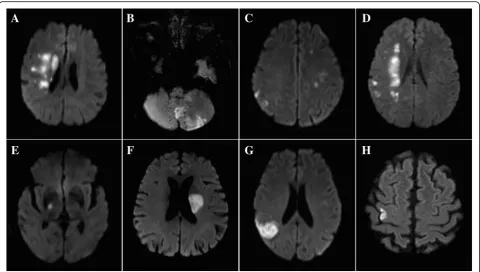
Multiple circulations; 4. Border-zone territories (Fig.
1
).
Any disagreement was decided by a third reader (L.L.).
Etiology classification
All patients were classified on the basis of The Trial of
Org 10,172 in Acute Stroke Treatment (TOAST)
classifi-cation [
20
] as previous study [
5
]. Patients with
cardi-oembolism (CE), systemic disease were excluded in
CHANCE, so there were no patients with stroke of CE
or other determined pathogenesis subtype. Finally, we
devided patients into three TOAST subtypes: large-artery
atherosclerosis (LAA), small-artery occlusion (SAO) and
stroke of undetermined pathogenesis. Subtype
classifica-tions were based on patients
’
clinical features and the
re-sults of one or more diagnostic tests, including brain MR
imaging, MRA and extracranial arteries (carotid
ultra-sound or computed tomograph angiography). All imaging
data, clinical features and diagnostic tests results
col-lected from individual centers were reviewed centrally
by two study neurologists and gave the subtype
classifications.
Follow-up and outcomes
hemorrhagic stroke as acute extravasation of blood into
the subarachnoid space or brain parenchyma with
associ-ated neurologic symptoms [
17
].
Statistical analysis
Proportions were used for categorical variables, and
me-dians with interquartile ranges were used for continuous
variables. Univariate analyses were performed to
com-pare the baseline characteristics among patients with
dif-ferent infarction patterns using one way analysis of
variance or Kruskal-Wallis test for continuous variables
and x
2test for categorical variables. Time to the event in
each imaging group illustrated using Kaplan-Meier
curve. We assessed the associations between infarction
patterns and stroke recurrence of minor stroke using
multivariable Cox regression models. Adjusted hazard
ratios (HRs) with 95% confidence intervals (CIs) were
re-ported. All the potential covariates listed in Table
1
were
included in the model. All tests were two-sided, and a
P
value < 0.05 was considered to indicate statistical
signifi-cance. All statistical analyses were performed with SAS
9.4 (SAS Institute Inc., Cary, NC).
Results
Among the 5170 patients, 1089 patients undergoing all
the MR sequences as required at baseline were included
in the CHANCE imaging subgroup. After excluding 255
patients without infarction, a total of 834 patients with
minor stroke were included.
Table
1
shows the baseline characteristics of
infarc-tion
patterns
in
lacunar
infarction
of
SAI,
non-lacunar infarction of SAI and MAIs. MAIs were
more likely to be older, have a history of congestive
heart failure and be shorter time to randomization of
the trial treatment. Lacunar infarction of SAI were
more likely to be smokers. Non-lacunar infarction of
SAI were more likely to have higher NIHSS on
ad-mission. Additional file
1
: Table S1 shows the baseline
characteristics of different infarction patterns in SAI
and MAIs respectively.
Different infarction patterns in SAI (subcortical lesion
with diameter
≤
15 mm, subcortical lesion with
diam-eter > 15 mm, cortical lesion and corticosubcortical
le-sion) had different risk of stroke recurrence (7.6%,
16.3%, 5.0% and 20.0%, respectively), however, different
infarction patterns in MAIs (unilateral anterior
circula-tion, posterior circulacircula-tion, multiple circulations and
border-zone territories) had no different risk of stroke
recurrence (14.8%, 11.8%, 19.5% and 18.5%, respectively)
(Additional file
1
: Table S2). The risk of stroke
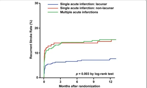
recur-rence was 7.6%, 15.1%, and 15.3% in patients with
lacu-nar infarction of SAI, non-laculacu-nar infarction of SAI and
MAIs at 1 year follow-up, respectively (Table
2
).
Com-pared with MAIs, lacunar infarction of SAI had lower
Fig. 1Infarction patterns of single acute infarction and multiple acute infarctions. Multiple acute infarctions.aUnilateral anterior circulation;
bPosterior circulation;cMultiple circulations;dBorder-zone territories. Single acute infarction.eSubcortical lesion with diameter≤15 mm;
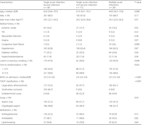
Table 1
Baseline characteristics of single acute infarction (lacunar infarction and non-lacunar infarction) and multiple acute infarctions
Characteristics Single acute infarction: lacunar infarction
n= 381
Single acute infarction: non-lacunar infarction
n= 172
Multiple acute infarctions
n= 281
Pvalue
Age,y, median (IQR) 62.6 (54.6–70.5) 61.0 (54.1–70.1) 64.8 (56.3–73.0) 0.008 Male, n (%) 261 (68.5) 105 (61.0) 193 (68.7) 0.17 Body mass index (kg/m2) 24.5 (22.7–26.2) 24.2 (22.0–26.6) 24.2 (22.0–26.2) 0.37 Medical history, n (%)
Ischemic stroke 63 (16.5) 27 (15.7) 54 (19.2) 0.55 TIA 5 (1.3) 4 (2.3) 9 (3.2) 0.25 Myocardial infarction 6 (1.6) 4 (2.3) 9 (3.2) 0.38 Angina 9 (2.4) 0 (0.0) 9 (3.2) 0.07 Congestive heart failure 2 (0.5) 2 (1.2) 10 (3.6) 0.009 Hypertension 243 (63.8) 109 (63.4) 184 (65.5) 0.87 Diabetes mellitus 77 (20.2) 35 (20.3) 69 (24.6) 0.36 Hypercholesterolaemia 45 (11.8) 20 (11.6) 32 (11.4) 0.99 Current or previous smoking, n (%) 179 (47.0) 62 (36.0) 129 (45.9) 0.046 Time to randomization, n (%) 0.025
< 12 h 160 (42.0) 88 (51.2) 145 (51.6)
≥12 h 221 (58.0) 84 (48.8) 136 (48.4)
NIHSS on admission, median(IQR) 2.0 (1.0–3.0) 2.0 (2.0–3.0) 2.0 (1.0–3.0) < 0.001 TOAST classification, n (%) < 0.001
Large-artery atherosclerosis 127 (33.3) 82 (47.7) 183 (65.1) Small-artery occlusion 254 (66.7) 0 (0.0) 0 (0.0) Undetermined cause 0 (0.0) 90 (52.3) 98 (34.9)
Group, n (%) 0.54
Aspirin only 195 (51.2) 89 (51.7) 133 (47.3) Clopidogrel+aspirin 186 (48.8) 83 (48.3) 148 (52.7) Medications, n (%)
Antihypertensive 126 (52.5) 52 (48.2) 79 (42.9) 0.15 Antidiabetic 37 (48.1) 17 (48.6) 30 (43.5) 0.82 Lipid-lowering 25 (56.8) 14 (70.0) 20 (62.5) 0.60
IQRInterquartile range,NIHSSNational Institutes of Health Stroke Scale,TOASTTrial of Org 10,172 in Acute Stroke Treatment
Table 2
Adjusted HR for stroke recurrence of different infarction patterns in single acute infarction and multiple acute infarctions at
one-year follow-up
Infarction patterns n Stroke recurrence at one year
n (n% [95%CI]) Adjusted HR (95% CI)a Pvalue Single and multiple acute infarctions 834 98 (11.8 [9.64–14.13])
Multiple acute infarctions 281 43 (15.3 [11.30–20.05]) Ref
Single acute infarction: lacunar 381 29 (7.6 [5.16–10.75]) 0.41 (0.21–0.80) 0.009 Single acute infarction: non-lacunar 172 26 (15.1[10.12–21.36]) 1.01 (0.60–1.69) 0.98
HRhazard ratio,CIconfidence interval a
risk of stroke recurrence (HR 0.41, 95% CI 0.21
–
0.80,
P
=
0.009), but not in non-lacunar infarction of SAI (HR 1.01,
95% CI 0.60
–
1.69,
P
= 0.98) (Table
2
). The Kaplan-Meier
curves shows the recurrent stroke rate of SAI (lacunar and
non-lacunar infarction) and MAIs, respectively (Fig.
2
).
Discussion
In this subgroup analysis of CHANCE, we found that
la-cunar infarction of SAI had lower risk of stroke recurrence
than MAIs, while non-lacunar infarction of SAI might
have similar risk as MAIs within one-year follow-up.
TIA
registry.org
project showed MAIs had higher
stroke recurrence than SAI in TIA or minor stroke [
5
].
However, former studies indicated there were more
kinds of infarction patterns than that showed in TIA
registry.org
project [
6
,
7
,
14
]. Traditionally, SAI were
classified according to the size and location of the
infarc-tion while MAIs were classified according to the blood
supply of different brain areas [
6
,
7
]. In our study, we
found patients with different infarction patterns had
dif-ferent risk of stroke recurrence in SAI but the difference
was not observed in MAIs. We inferred that significant
difference of etiologies and pathogenesis among distinct
infarction patterns led to the results.
Previous studies indicated that SAI with different
pat-terns were usually related to different etiologies and
pathogenesis. Lacunar infarction of SAI usually related
to SAO with pathogenesis as
‘
fibrinoid necrosis
’
or
‘
lipo-hyalinosis
’
of small perforating arteries [
22
–
24
]. SAI with
subcortical lesion with diameter > 15 mm usually related
to large-artery atherosclerosis, cryptogenic and
cardio-embolic diseases [
6
,
7
] with pathogenesis as obstruction
of the origins of penetrating arteries by parent large
intracranial artery intimal plaques or embolism [
25
–
27
].
Furthermore, SAI with corticosubcortical lesion or
cor-tical lesion were usually related to LAA, CE and
crypto-genic with pathogenesis of embolism [
8
,
9
,
14
]. In a
word, lacunar infarction was different from non-lacunar
infarction in aspect of etiologies and pathogenesis [
28
].
Traditionally, lacunar infarction usually had a favorable
outcome among different TOAST classification [
11
,
12
]
and lacunar infarction had a favorable outcome when
compared with non-lacunar infarction [
29
–
31
]. So the
above findings could explain the different risk of stroke
recurrence in different patterns of SAI for different
eti-ologies and pathogenesis.
Previous studies indicated MAIs were usually related to
LAA, CE and cryptogenic, according to the TOAST
classi-fication [
6
,
7
]. There was evidence showed that the
pathogenesis of MAIs was likely to be caused by the
em-bolism from heart or major extracranial/intracranial
ves-sels [
6
,
7
,
32
,
33
]. Hemodynamic failure and microem
bolization were the pathogenesis of border-zone
infarc-tions [
34
]. As embolism was the most common
pathogenesis of MAIs, the above findings could explain
the high and similar risk of stroke recurrence in patients
with different infarction patterns of MAIs.
Recently, imaging parameters received more attention in
order to predict recurrent stroke [
3
–
5
,
19
] and might have
better predictive value for stroke recurrence than clinical
scores in patients with TIA or minor stroke [
3
,
35
]. TIA
registry.org
project showed it was convenient and quick to
stratify the risk of stroke recurrence by infarction numbers
(NAI, SAI or MAIs) in clinical practice. However, our
study indicated that non-lacunar infarction of SAI might
have similar risk of stroke recurrence as MAIs, implying
that non-lacunar infarction of SAI could be ignored if we
simply stratified the risk of stroke recurrence by infarction
numbers. So we should not only concern about the
num-ber of infarctions, but also the size and location of
infarc-tion in order to predict the risk of stroke recurrence in
minor stroke. Improved infarction pattern classifications
of TIA and minor stroke should be established in the
fu-ture large cohort study.
Our study presented several limitations. First, since
this imaging subgroup analysis included only a small
part of patients of CHANCE, potential selection bias
might have existed. Second, potential bias might have
existed, as apparent diffusion coefficient was not
in-cluded for evaluating infarction. Third, all patients in
this imaging substudy were non-cardioembolic minor
is-chemic stroke which limited the generalizability of the
findings to cardioembolic minor ischemic stroke.
Conclusions
Lacunar infarction of SAI had lower risk of stroke
recur-rence than MAIs, while non-lacunar infarction of SAI
might have similar risk as MAIs. Except for the number
of infarctions, the size and location of the infarction
should also be considered to stratify the risk of stroke
recurrence in minor stroke.
Additional file
Additional file 1:Table S1.Baseline characteristics of different infarction patterns in single acute infarction and multiple acute infarctions respectively.Table S2.Adjusted HR for stroke recurrence of different infarction patterns in single acute infarction and multiple acute infarctions at one-year follow-up. (DOCX 31 kb)
Abbreviations
3D:3-dimensional; CE: Cardio embolism; CHANCE: Clopidogrel in High-risk Patients with Acute Nondisabling Cerebrovascular Events; CI: Confidence interval; DWI: Diffusion-weighted imaging; HR: Hazard ratio; LAA : Large-artery atherosclerosis; MAIs: Multiple acute infarctions; MR: Magnetic resonance; MRA: Magnetic resonance angiography; NAI: No acute infarction; SAI: Single acute infarction; SAO: Small-artery occlusion; TOAST: The Trial of Org 10,172 in Acute Stroke Treatment
Acknowledgements
The authors thank the participants and all who were involved in the CHANCE study.
The CHANCE Investigators
University Of Chinese Medicine, Site Investigator); Lianyuan Feng, MD, PhD (Baiqiuen International Peace Hospital Of People’s Liberation Army, Site Investigator); Lianbo Gao, MD, PhD (Fourth Affiliated Hospital Of China Medical University, Site Investigator); Bo Xiao, MD, PhD (Xiangya Hospital Central-South University, Site Investigator); Yibin Cao, MD, PhD (Tangshan Worker’s Hospital, Site Investigator); Yiping Wu, MD, PhD (The 1st Hospital In Handan, Site Investigator); Jinfeng Liu, MD, PhD (Yangquan Coal (Group) Co., Ltd. General Hospital, Site Investigator); Zhiming Zhang, MD, PhD (Tianjin Tianhe Hospital, Site Investigator); Zhengxie Dong, MD, PhD (Nantong First People’s Hospital, Site Investigator); Limin Wang, MD, PhD (The 1st Hospital Of Zhangjiakou City, Site Investigator); Li He, MD, PhD (West China Hospital, Sichuan University, Site Investigator); Xinchen Wang, MD, PhD (The Second Affiliated Hospital Of Shandong University Of TCM, Site Investigator); Xueying Guo, MD, PhD (Fenyang Hospital Of Shanxi Province, Site Investigator); Ming Wang, MD, PhD (Zhejiang Zhoushan Putuo District People’s Hospital, Site Investigator); Xiaosha Wang, MD, PhD (Xiyuan Hospital Of China Academy Of Chinese Traditional Medicine, Site Investigator); Jiandong Jiang, MD, PhD (No.2 People’s Hospital East In Lianyungang City, Site Investigator); Renliang Zhao, MD, PhD (Affiliated Hospital Of Qingdao University Medical College, Site Investigator); Shengnian Zhou, MD, PhD (Qilu Hospital Of Shandong University, Site Investigator); HaoHu, MD, PhD (Zibo Hospital Of Traditional Chinese Medicine, Site Investigator); Maolin He, MD, PhD (Beijing Shijitan Hospital, Site Investigator); Fengchun Yu, MD, PhD (Beijing Haidian Hospital, Site Investigator); Quping Ouyang, MD, PhD (Beijing Shunyi District Hospital, Site Investigator); Jingbo Zhang, MD, PhD (Dalian Third Municipal Hospital, Site Investigator); Anding Xu, MD, PhD (The First Affliated Hospital Of Jinan University, Site Investigator); Xiaokun Qi, MD, PhD (Navy Genaral Hospital Of P.L.A, Site Investigator); Lei Wang, MD, PhD (Beijing Second Artillery General Hospital, Site Investigator); Fuming Shi, MD, PhD (Beijing Daxing District Hospital, Site Investigator); Fuqiang Guo, MD, PhD (Sichuan Province People’s Hospital, Site Investigator); Jianfeng Wang, MD, PhD (Dalian Municipal Central Hospital, Site Investigator); Fengli Zhao, MD, PhD (The Second Hospital In Baoding, Site Investigator); Ronghua Dou, MD, PhD (The Hospital Combine Traditional Chinese And Western Medicine In Cang zhou, Site Investigator); Dongning Wei, MD, PhD (The 309th Hospital Of P.L.A, Site Investigator); Qingwei Meng, MD, PhD (Liangxiang Hospital Of Fangshan District, Beijing, Site Investigator); Yilu Xia, MD, PhD (HuaXin Hospital First Hospital Of Tsinghua University, Site Investigator); Shimin Wang, MD, PhD (Tianjin Huanhu Hospital, Site Investigator); Zhangcang Xue, MD, PhD (Shijiazhuang First Hospital, Site Investigator); Yuming Xu, MD, PhD (The First Affiliated Hospital Of Zhengzhou University, Site
Investigator); Liping Ma, MD, PhD (Xinzhou City People’s Hospital, Site Investigator); Chun Wang, MD, PhD (Sichuan Province People’s Hospital Of Deyang City, Site Investigator); Jiang Wu, MD, PhD (First Hospital, Jilin University, Site Investigator); Yifeng Du, MD, PhD (Shandong Provincial Hospital, Site Investigator); Yinzhou Wang, MD, PhD (Fujian Province Hospital, Site Investigator); Lijun Xiao, MD, PhD (Liaoyang City Third People’s Hospital, Site Investigator); Fucong Song, MD, PhD (Handan City Center Hospital, Site Investigator); Wenli Hu, MD, PhD (Beijing Chaoyang Hospital, Capital Medical University, Site Investigator); Zhigang Chen, MD, PhD (Beijing University Of Chinese Medicine East Hospital, Site Investigator); Qingrui Liu, MD, PhD (Hebei Medical University Fourth Hospital, Site Investigator); Jiemin Zhang, MD, PhD (The Fourth Affiliated Hospital Of Soochow University, Site Investigator); Mei Chen, MD, PhD (Zhejiang University Of Chinese Medicine Affiliated First Hospital, Site Investigator); Xiaodong Yuan, MD, PhD (Affiliated Hospital Of Kailuan Company Ltd., Site Investigator); Zhihui Liu, MD, PhD (Affiliated Hospital Of Weifang Medical University, Site Investigator); Guozhong Li, MD, PhD (The First Hospital Of Harbin Medical University, Site Investigator); Xiaohong Li, MD, PhD (Dalian Friendship Hospital, Site Investigator); Tingchen Tian, MD, PhD (Tianjin Dagang Hospital, Site Investigator).
Funding
This work was supported by the Ministry of Science and Technology of the People’s Republic of China (grant numbers 2015BAI12B04, 2015BAI12B02, 2016YFC0901000, 2016YFC0901001, 2017YFC1307900); and the Beijing Municipal Science and Technology Commission (grant numbers D151100002015001, D151100002015003, Z151100003915117) . The fund body took no part in the design of the study and collection, analysis, and interpretation of data and in writing the manuscript.
Availability of data and materials
The datasets during and/or analysed during the current study available from the corresponding author on reasonable request.
Authors’contributions
GYW study concept and design, analysis and interpretation of data, drafting of the manuscript. JJ acquisition of data, analysis and interpretation of data, drafting of the manuscript. YSP and HL acquisition of data, analysis and interpretation of data and revision of the drafting of the manuscript. XM, XQZ and LPL acquisition of data and revision of the manuscript. DW study concept and design, revising the manuscript.
YJW obtaining funding, study concept and design, study supervision or coordination, revision of the drafting of the manuscript. YLW obtaining funding, study concept and design, acquisition of data, analysis and interpretation of data, revision of the drafting of the manuscript. All authors read and approved the final manuscript.
Ethics approval and consent to participate
Name of the ethics committee is IRB of Beijing Tiantan Hospital, Capital Medical University. Ethics approval number of this study is ky2009–002. The trial was approved by the Ethics Committee of Beijing Tiantan Hospital and all the participating hospitals. Written informed consent was obtained from all participants or their legal proxies.
Consent for publication
In the patient informed consent were stated that all collected data will be analyzed and prepared for publications. All patients gave signed informed consent prior to inclusion in the study.
Competing interests
The author(s) declared no potential conflicts of interest with respect to the research, authorship, and/or publication of this article.
Publisher
’
s Note
Springer Nature remains neutral with regard to jurisdictional claims in published maps and institutional affiliations.
Author details
1Department of Neurology, Beijing Tiantan Hospital, Capital Medical
University, Beijing, China.2China National Clinical Research Center for Neurological Diseases, Beijing, China.3Center of Stroke, Beijing Institute for
Brain Disorders, Beijing, China.4Beijing Key Laboratory of Translational Medicine for Cerebrovascular Disease, Beijing, China.5Illinois Neurological
Institute Stroke Network, Sisters of the Third Order of St. Francis Healthcare System, University of Illinois College of Medicine, Peoria, USA.
Received: 18 April 2018 Accepted: 4 December 2018
References
1. von Weitzel-Mudersbach P, Andersen G, Hundborg HH, Johnsen SP. Transient ischemic attack and minor stroke are the most common manifestations of acute cerebrovascular disease: a prospective, population-based study--the Aarhus TIA study. Neuroepidemiology. 2013;40:50–5. 2. Coull AJ, Lovett JK, Rothwell PM, Oxford Vascular S. Population based study
of early risk of stroke after transient ischaemic attack or minor stroke: implications for public education and organisation of services. BMJ. 2004; 328:326.
3. Yaghi S, Rostanski SK, Boehme AK, Martin-Schild S, Samai A, Silver B, et al. Imaging parameters and recurrent cerebrovascular events in patients with minor stroke or transient ischemic attack. JAMA Neurol. 2016;73:572–8. 4. Pan Y, Meng X, Jing J, Li H, Zhao X, Liu L, et al. Association of multiple infarctions and ICAS with outcomes of minor stroke and TIA. Neurology. 2017;88:1081–8.
5. Amarenco P, Lavallee PC, Labreuche J, Albers GW, Bornstein NM, Canhao P, et al. One-year risk of stroke after transient ischemic attack or minor stroke. N Engl J Med. 2016;374:1533–42.
7. Kang DW, Chalela JA, Ezzeddine MA, Warach S. Association of ischemic lesion patterns on early diffusion-weighted imaging with TOAST stroke subtypes. Arch Neurol. 2003;60:1730–4.
8. Wong KS, Gao S, Chan YL, Hansberg T, Lam WW, Droste DW, et al. Mechanisms of acute cerebral infarctions in patients with middle cerebral artery stenosis: a diffusion-weighted imaging and microemboli monitoring study. Ann Neurol. 2002;52:74–81.
9. Lee DK, Kim JS, Kwon SU, Yoo SH, Kang DW. Lesion patterns and stroke mechanism in atherosclerotic middle cerebral artery disease: early diffusion-weighted imaging study. Stroke. 2005;36:2583–8.
10. Arsava EM, Helenius J, Avery R, Sorgun MH, Kim GM, Pontes-Neto OM, et al. Assessment of the predictive validity of etiologic stroke classification. JAMA Neurol. 2017;74:419–26.
11. Lovett JK, Coull AJ, Rothwell PM. Early risk of recurrence by subtype of ischemic stroke in population-based incidence studies. Neurology. 2004;62: 569–73.
12. Petty GW, Brown RD Jr, Whisnant JP, Sicks JD, O'Fallon WM, Wiebers DO. Ischemic stroke subtypes : a population-based study of functional outcome, survival, and recurrence.Stroke. 2000;31:1062–8.
13. Purroy F, Montaner J, Molina CA, Delgado P, Ribo M, Alvarez-Sabin J. Patterns and predictors of early risk of recurrence after transient ischemic attack with respect to etiologic subtypes. Stroke. 2007;38:3225–9. 14. Bang OY, Lee PH, Heo KG, Joo US, Yoon SR, Kim SY. Specific DWI lesion
patterns predict prognosis after acute ischaemic stroke within the MCA territory. J Neurol Neurosurg Psychiatry. 2005;76:1222–8.
15. Ko Y, Lee S, Chung JW, Han MK, Park JM, Kang K, et al. MRI-based algorithm for acute ischemic stroke subtype classification. J Stroke. 2014;16:161–72. 16. Wang Y, Johnston SC, Investigators C. Rationale and design of a
randomized, double-blind trial comparing the effects of a 3-month clopidogrel-aspirin regimen versus aspirin alone for the treatment of high-risk patients with acute nondisabling cerebrovascular event. Am Heart J. 2010;160:380–386.e381.
17. Wang Y, Wang Y, Zhao X, Liu L, Wang D, Wang C, et al. Clopidogrel with aspirin in acute minor stroke or transient ischemic attack. N Engl J Med. 2013;369:11–9.
18. Liu L, Wong KS, Leng X, Pu Y, Wang Y, Jing J, et al. Dual antiplatelet therapy in stroke and ICAS: subgroup analysis of CHANCE. Neurology. 2015;85:1154–62.
19. Wen HM, Lam WW, Rainer T, Fan YH, Leung TW, Chan YL, et al. Multiple acute cerebral infarcts on diffusion-weighted imaging and risk of recurrent stroke. Neurology. 2004;63:1317–9.
20. Adams HP Jr, Bendixen BH, Kappelle LJ, Biller J, Love BB, Gordon DL, et al. Classification of subtype of acute ischemic stroke. Definitions for use in a multicenter clinical trial. TOAST. Trial of org 10172 in acute stroke treatment. Stroke. 1993;24:35–41.
21. Wang Y, Pan Y, Zhao X, Li H, Wang D, Johnston SC, et al. Clopidogrel with aspirin in acute minor stroke or transient ischemic attack (CHANCE) trial: one-year outcomes. Circulation. 2015;132:40–6.
22. Miller Fisher C. Lacunar infarcts–a review. Cerebrovasc Dis. 1991;1:311–20. 23. Lacunes FCM. Small, deep cerebral infarcts. Neurology. 1965;15:774–84. 24. Caplan LR. Lacunar infarction and small vessel disease: pathology and
pathophysiology. J Stroke. 2015;17:2–6.
25. Jung S, Hwang SH, Lee BC. Distinct clinical expressions of striatocapsular infarction according to cortical manifestations. Eur J Neurol. 2004;11:627–33. 26. Lee KB, Oh HG, Roh H, Ahn MY. Can we discriminate stroke mechanisms by analyzing the infarct patterns in the striatocapsular region? Eur Neurol. 2008; 60:79–84.
27. Nicolai A, Lazzarino LG, Biasutti E. Large striatocapsular infarcts: clinical features and risk factors. J Neurol. 1996;243:44–50.
28. Hart RG, Diener HC, Coutts SB, Easton JD, Granger CB, O'Donnell MJ, et al. Embolic strokes of undetermined source: the case for a new clinical construct. Lancet Neurol. 2014;13:429–38.
29. Sacco S, Marini C, Totaro R, Russo T, Cerone D, Carolei A. a population-based study of the incidence and prognosis of lacunar stroke. Neurology. 2006;66:1335–8.
30. Bejot Y, Catteau A, Caillier M, Rouaud O, Durier J, Marie C, et al. Trends in incidence, risk factors, and survival in symptomatic lacunar stroke in Dijon, France, from 1989 to 2006: a population-based study. Stroke. 2008;39:1945–51. 31. Fang XH, Wang WH, Zhang XQ, Liu HJ, Zhang HM, Qin XM, et al. Incidence and survival of symptomatic lacunar infarction in a Beijing population: a 6-year prospective study. Eur J Neurol. 2012;19:1114–20.
32. Takahashi K, Kobayashi S, Matui R, Yamaguchi S, Yamashita K. The differences of clinical parameters between small multiple ischemic lesions and single lesion detected by diffusion-weighted MRI. Acta Neurol Scand. 2002;106:24–9.
33. Ferro JM. Patterns of ischaemic cerebral diseases. J Neurol. 2004;251:1–10. 34. Joinlambert C, Saliou G, Flamand-Roze C, Masnou P, Sarov M, Souillard R, et al. Cortical border-zone infarcts: clinical features, causes and outcome. J Neurol Neurosurg Psychiatry. 2012;83:771–5.