Available Online at www.ijpret.com 391
APPLIED RESEARCH IN ENGINEERING AND
TECHNOLOGY
A PATH FOR HORIZING YOUR INNOVATIVE WORK
STUDY AND ANALYSIS OF TMJ IMPLANT IN HUMAN BODY
GAJANAN D. MANDAVGADE
Assistant Professor, of Mechanical Engineering, IBSS COE-Amravati.
Accepted Date: 05/03/2015; Published Date: 01/05/2015
\
Abstract: Objective of this work is to study & investigation of TMJ (Temporomandibular
Joint) implant in human body. In this study initially measure the shape and size of artificial prosthesis which are commercially available in market, by using different tools. Then by using measurement data drafting of the implant is done by means of CAD (Computer Aided Design) software. After that analysing the behaviour of CAD model by applying various boundary condition and different loading condition in Finite Element Analysis (FEA) software.
Keywords:Human Body, TMJ Implant
Corresponding Author: MR. GAJANAN D. MANDAVGADE
Access Online On:
www.ijpret.com
How to Cite This Article:
Available Online at www.ijpret.com 392 INTRODUCTION
1.0 Temporomandible Joint Anantomy
TMJ is a distinctive joint as it having permutation of hinge and sliding motions which make these joint most complicated in human body. TMJ connects the lower jaw called mandible to the temporal bone. To keep the motion smooth a disc lies between condyle (rounded end of lower jaw) and temporal bone. This disc absorb shock from movement such as chewing, talking etc and the muscle which surrounding the joint control its position. Following are main component in skull.
A) Teeth
The human skull is composed of an upper jaw, lower jaw, and teeth. There are thirty-two teeth, sixteen top and sixteen bottoms, in the jaw. There are two main tooth sections, the crown and root. The crown is the part of the tooth that can be seen above the gum line while the root is hidden in the jaw.
B) Bones
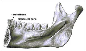
The bones act as the structural support to the body. The three main structural components in the skull associated with mastication are the lower jaw (mandible), the upper jaw (maxilla), and the lateral side of the skull (temporal) as shown in Figure.1
C) Blood Vessels And Nerves
Like any other portion of the body the jaw is surrounded by blood vessels and nerves. The blood vessels carry blood to the muscles and bones, supplying them with necessary nutrients such as oxygen. The nerves work as the communication system between the brain and all parts of the body.
Available Online at www.ijpret.com 393 A ligament is a band of fibrous tissue that attaches bone to bone or bone to cartilage. The purpose for ligaments in The TMJ is to guide and prohibit excessive movements of the mandible while also protecting sensitive tissues such as nerves and blood vessels. Disorders of the jaw joint and chewing muscles are as fallows [1]
Myofascial pain, the most common temporomandibular disorder, involves discomfort or pain in the muscles that control jaw function.
Internal derangement of the joint involves a displaced disc, dislocated jaw, or injury to the condyle.
Arthritis refers a group of degenerative / inflammatory joint disorders that can affect the temporomandibular joint.
Fig 2. The bone structure of the human mandible
3.0 MATERIAL AND METHOD.
3.1 Metallic Biomaterials
Available Online at www.ijpret.com 394 3.1.1 Stainless Steel
Stainless steel is a very strong alloy. There are many different types of stainless steel, but austenitic stainless steel especially 316L most widely used for implant fabrication, because it has following advantage. [3] Stainless steel has been used for wide range of application due to easy availability, lower cost, excellent fabrication properties, fatigue resistance, accepted biocompatibility and great strength [4].
SOLID MODEL
A solid model is a digital representation of the geometry of an existing or envisioned physical object [5]. A solid model of TMJ implant is developed by using computer aided designing (CAD) software. Various CAD software are available such as CATIA, Hypermesh, PRO/E, Solid edge, etc. A 3D TMJ model is build up from the existing real implant, during developing a implant the measurements of the individual components were measured using the different ergonomic tools, and a solid model was created by using CAD software as Pro-E 4.0. The PRO/E 4.0 software offers several different approaches to develop a solid model of prosthetics like part design, surface design, assembly etc. A 3D model was developed from the existing the real implant [6]. A TMJ solid model developed by Pro/E software is shown in fig. 4 command used for developing solid model is extrude for material add and material remove, pattern, round, fillet etc.
Available Online at www.ijpret.com 395 Table1. Analysis Results for TMJ Implants (SS 316L)
6.3 ANALYSIS OF TMJ IMPLANT
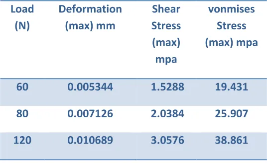
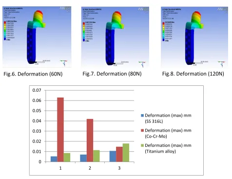
In TMJ implant analysis first import a TMJ model in ANSYS software in .sat file format then material is selected for TMJ implant, after selecting material an implant divided into a small number of solid finite elements, this processes known as meshing. A load (60N, 80N, 120N) is applied on implant by fixing its one end and then behaviour of each element is analyzed. Table No. 1 shows TMJ Implant (SS 316L) result after analysis. Above procedure is repeated for TMJ Implant (Co-Cr-Mo) and (Titanium alloy) its result shows in Table No. 2 and 3 resp.
Table 2. Analysis Results for TMJ Implants (Co-Cr-Mo) Load
(N)
Deformation (max) mm
Shear Stress (max) mpa
vonmises Stress (max) mpa
60 0.005344 1.5288 19.431
80 0.007126 2.0384 25.907
120 0.010689 3.0576 38.861
Load (N) Deformation (max) mm Shear Stress (max) mpa Vonmises Stress (max) mpa
60 0.062955 6.0113 83.859
80 0.04197 4.0075 55.906
Available Online at www.ijpret.com 396 0
0.01 0.02 0.03 0.04 0.05 0.06 0.07
1 2 3
Deformation (max) mm (SS 316L)
Deformation (max) mm (Co-Cr-Mo)
Deformation (max) mm (Titanium alloy) Load
(N)
Deformation (max) mm
Shear Stress (max) mpa
vonmises Stress (max) mpa
60 0.008596 1.4651 16.754
80 0.011461 1.9534 22.346
120 0.01792 2.9301 33.519
Fig.6. Deformation (60N) Fig.7. Deformation (80N) Fig.8. Deformation (120N) Table 3. Analysis Results for TMJ Implants (Titanium
alloy)
Available Online at www.ijpret.com 397 0
1 2 3 4 5 6 7
1 2 3
Shear Stress (max) mpa (SS 316L)
Shear Stress (max) mpa (Co-Cr-Mo)
Shear Stress (max) mpa (Titanium alloy)
Available Online at www.ijpret.com 398 0
20 40 60 80 100
1 2 3
vonmises Stress (max) mpa (SS 316L)
vonmises Stress (max) mpa (Co-Cr-Mo)
vonmises Stress (max) mpa (Titanium alloy)
CONCLUSION
SS 316L has excellent Bio-compatible properties along with physical properties which makes it an ideal implant material for fractures, when compared to other materials.
SS 316L alloy being medium light with 7.9gm /c3 density does not have any adverse effect on
the patient’s movement i.e while lifting the leg, running, jumping, walking etc
The maximum values of von mises stresses for TMJ implants of SS 316L ranges from 19.431 to 38.861 MPa is much lower when compared to the yield strength of SS 316L (690 MPa).
Stress shielding is comparatively reduced in stainless steel, as the modulus of elasticity is lower when compared to Cobalt Chromium Molybdenum.
REFERENCE
1. U.S Department of Health and Human Services. WWW.nidcr.nih.gov
2. Nitesh R. Patel, Piyush P. Gohil, “A Review on Biomaterials: Scope, Applications& Human Anatomy Significance”
3. C.N. Elias, J.H.C. Lima, R. Valiev, and M.A. Meyers Biomedical Applications of Titanium and its Alloys
4. Hench LL. Bioceramics: from concept
5. Jarek R. Rossignac, Aristides A. G. Requicha “ Solid Modelling”