CrossMark Published by DiscoverSys
ABSTRACT
Background: Malignant ovarian tumor is one of the most common types of cancer in women worldwide, and most patients come with advanced stage. Until now, a pre-operative diagnostic method to determine malignant ovarian tumor was considered unsatisfying. Neutrophil gelatinase-associated lipocalin (NGAL) allegedly plays a role in proliferation and metastasis of the cancer cells, so it is important to explore the difference of serum NGAL levels between benign and malignant epithelial ovarian tumor.
Aim: to determine the characteristics of patients with benign and malignant epithelial ovarian tumor, histologic types, and serum NGAL levels difference in benign and malignant epithelial ovarian tumor. Method: This was a comparative analytic study with a cross-sectional design. Forty patients were chosen with consecutive sampling; each group consists of 20 female patients who will undergo primary
surgery for suspicious of benign and malignant epithelial ovarian tumor. A blood sample was collected one day prior to the surgery and confirmed with histopathologic examination after surgery.
Results: most of the patients aged 20-50 years old, had ≥1 child and the most common histopathology type of malignant epithelial ovarian tumor was mucinous adenocarcinoma while the majority of the benign epithelial ovarian tumor was endometriotic cyst. The median rate of NGAL in the malignant epithelial ovarian tumor was 219.5 ng/ml, higher than median rate of NGAL in benign epithelial ovarian tumor with 132.5 ng/ml, the difference was statistically significant (p < 0.001). The median rate of NGAL in malignant epithelial ovarian tumor according to the stage of disease showed no significant difference. Conclusion: A new diagnostic criteria of epithelial ovarian tumor needs to be considered which include NGAL as one of the screening components.
Keywords: benign epithelial ovarian tumor, malignant epithelial ovarian tumor, NGAL
Cite This Article: Gafur, A., Edianto, D., Simanjuntak, R.Y., Pasaribu, H.P., Ardiansyah, E., Siregar, H.S. 2018. Serum Neutrophil Gelatinase-Associated Lipocalin (NGAL) level difference in benign and malignant epithelial ovarian tumor. Bali Medical Journal 7(1): 132-136. DOI:10.15562/ bmj.v7i1.809
Serum Neutrophil Gelatinase-Associated Lipocalin
(NGAL) level difference in benign and malignant
epithelial ovarian tumor
Abdul Gafur,* Deri Edianto, Roy Yustin Simanjuntak, Hotma Partogi Pasaribu,
Edy Ardiansyah, Henry Salim Siregar
INTRODUCTION
Ovarian cancer is a malignancy derived from the ovarian tissue. Ovarian cancer consists of approxi-mately 30 different types, in which its classification is differentiated according to the type of cancer cells originated.
Ovarian cancer accounts for 3% of all cancers in women and is the fifth leading cause of cancer deaths in women in the United States (US). In 2014, it was estimated that approximately 22,000 women diagnosed with ovarian cancer in the US and about 14,000 would die because of the disease. The incidence of ovarian cancer decrease almost 1 percent annually from 1987 to 2011, the mortality rate dropped from an average of 1.6 percent each year from 2001 to 2010. White women have a higher incidence and mortality rate than women with other race or ethnicity.1 Whereas the incidence of ovarian
cancer at the National General Hospital Dr. Cipto Mangunkusumo Jakarta during 2002-2006 also showed the highest proportion among gynecologic cancers, and mortality rate due to ovarian cancer also showed a fairly high rate of 34.1% from 327 cases
of gynecologic cancer deaths from 2002-2006. Research from Health Research and Development Agency of Indonesia in 2007 on 218 ovarian cancer patients reported that the 5-year survival rate was 41.25%, in which 76.3% in stage I, 66.6% in stage II, 24.6% in stage III and 8.1% in stage IV.2,3,4
Ovarian cancer patients are mostly diagnosed late since there is still no accurate early detection method for this cancer, so only 25-30% people are diagnosed at an early stage. Accurate screening for a malignant ovarian tumor will certainly reduce the mortality of the disease, however no effec-tive screening method has been found until now, followed by the fact that most of the early stage of malignant ovarian tumors showed no symptoms, 70% of cases are found in the advanced stage of which the tumor has spread beyond the ovaries, or stages III and IV. This has led to the need for high-effectiveness tumor markers to be used as a screening examination of malignant ovarian tumors so that it might reduce the morbidity and mortality rate of the disease.4,5
Obstetrics and Gynecology Department Medical Faculty of Sumatera Utara University Adam Malik General Hospital Jl. Bunga Lau No. 17, Medan Tuntungan, Medan
*Correspondence to: Abdul Gafur,
Obstetrics and Gynecology Department Medical Faculty of Sumatera Utara University Adam Malik General Hospital Jl. Bunga Lau No. 17, Medan Tuntungan, Medan dr.gafur.agf@gmail.com
Received: 2017-08-15 Accepted: 2017-10-22 Published: 2017-10-24
Volume No.: 7
Issue: 1
First page No.: 132
P-ISSN.2089-1180
E-ISSN.2302-2914
Recently, the use of various potential tumor markers has been identified and evaluated whether in combination with CA-125 or other markers, or as a single test. The use of microarray technol-ogy has enabled the analysis of the expression of thousands of genes and has been used extensively to find new tumor markers for cancer detection. Several studies have reported that lipocalin-2 (LCN2) or neutrophil gelatinase-associated lipo-calin (NGAL) showed increased expression in ovarian cancer.6
We saw a close relationship between NGAL with malignant ovarian tumors from several existing literature. In addition, there is the fact that malig-nant ovarian tumor is a disease that is relatively difficult to detect, so it often found in an advanced stage. Research on NGAL has not been done in Indonesia, so these encourage authors to conduct research on NGAL.
METHODS
This study was a comparative analytic study in 2 unpaired populations with cross-sectional study design, conducted in H. Adam Malik General Hospital, dr. Pirngadi Medan Hospital, the affil-iated hospitals of the Department of Obstetrics and Gynecology Faculty of Medicine University of North Sumatra and Prodia Clinical Laboratory Medan from March to May 2017.
The sample size was calculated statistically based on the formula. From the calculation, we obtained n1 = n2 = 19.77 rounded to 20 subjects (number of samples for each case and control group), N = 40 subjects. The study samples were part of the study population that meets the inclusion and exclusion criteria with consecutive sampling.
Data analysis performed with descriptive and analytical analysis. Descriptive analysis was conducted to explore the frequency of charac-teristic distribution of research subjects. The statistical test was performed with a computer program appropriate for univariate analysis to see the characteristics of the sample and bivar-iate analysis performed with t-test for normally distributed data or with Mann Whitney U-test for non-normally distributed data, to see the compar-ison of NGAL levels in patients with benign or malignant epithelial ovarian tumors. The p-value considered significant if p < 0.05 with 95% confi-dence interval.
This study has received approval from the research subject voluntarily and by the Ethics Committee of the Faculty of Medicine, University of North Sumatra who has conducted a feasibility
RESULTS
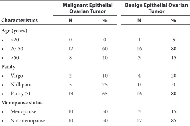
As depicted in table 1, within the malignant epithe-lial ovarian tumors group, the subjects were mostly in the 20-50 years of age, counting for 12 subjects (60%), while eight subjects (40%) were >50 years. In the benign epithelial tumor group, most of the subjects were in 20-50 years old age group, count-ing for 18 subjects (80%), and only one subject aged under 20 years old (5%) and three subjects with aged over 50 years (15%).
Most subjects in malignant epithelial ovarian tumor group have ≥1 children, counting for 13 people (65%), only two subjects (10%) were unmarried (virgo) and five people (25%) with nulliparous. In the benign epithelial ovarian tumor group, most of the subjects have ≥1 children, counting for 16 subjects (80%) and four subjects (20%) were unmarried (virgo).
As for menopausal status, the malignant epithe-lial ovarian tumor group has the same number of menopausal and nonmenopausal subjects. In the benign epithelial ovarian tumor group, the major-ity of the subjects were not menopause, which accounted for 17 subjects (85%) and three (15%) subjects have experienced menopause.
Table 2 showed the histopathological distribu-tion of epithelial ovarian tumor. The most common histopathological distribution of malignant epithe-lial ovarian tumors was mucinous adenocarcinoma (25%), serous adenocarcinoma (20%) and endo-metrioid carcinoma (5%). While in the benign epithelial ovarian tumor group mainly with endo-metriotic cyst (20%) then mucinous cystadenoma (15%) and serous cystadenoma (15%).
Normality test for NGAL level showed the data were not normally distributed, so Mann Whitney test was used to determine the mean difference of NGAL level in malignant and benign epithelial ovarian tumor. The median NGAL level of malig-nant epithelial ovarian tumor was 219.5 ng/ml with a minimum-maximum value of 112-583 ng/ml, which higher compared to NGAL level of benign epithelial ovarian tumor, which was 132.5 ng/ml (median) with a minimum-maximum value of 80-312 ng/ml. Mann Whitney test obtained signifi-cant difference (p < 0.001)
of 116-544 ng/ml. Analysis with Kruskal Wallis test showed no significant difference (p = 0.555).
DISCUSSION
This study was conducted on patients with benign and malignant ovarian tumors by examining serum NGAL as a predictor of malignancy of epithelial ovarian tumors and involved 40 people as study sample, with 25% of the sample were mucinous adenocarcinoma.
The results of current study obtained most of the study subjects were in 20-50 years old age group, with malignant epithelial ovarian tumors counted for 60% and benign epithelial ovarian tumor counted for 80%. Only a small percentage of subjects with age under 20 years old or above 50 years old.
The risk of ovarian cancer increases with age. About two-thirds of women are diagnosed with ovarian cancer at age 55 or older. The mean age for the onset of ovarian cancer is 63 years of age. However it may occur at various ages.7,8,9,10
Most of the research subjects have ≥ one children (65% of subjects and 80% of subjects in malignant and benign epithelial ovarian tumor, respectively). However, there were also study subjects with virgo and nullipara status, although only a few.
In terms of menopausal status, subjects who have experienced menopause in malignant epithelial ovarian tumors group were 50%. While in benign epithelial ovarian tumor group, most subjects were not menopause (85%). Women have a risk of ovar-ian cancer if they do not have children, have chil-dren after 30 years of age, or have early menopause (before age 50).7,8,9,10
The increased risks that have also been associ-ated with ovarian cancer are early menarche and late menopause. Risk factors which associated with undisturbed ovulation cycle for years also give rise to the hypothesis that repeated stimulation of the ovarian epithelial surface may lead to malignancy. This pathogenesis also called the “incessant ovula-tion” hypothesis. The process of ovarian epithelial tissue repair due to repeated and cyclic periods of ovulation leads to frequent cellular proliferation. This will lead to the mutation of the p53 gene in the DNA phase. Thus these events are considered to contribute toward carcinogenesis process of ovar-ian tumors.11,12
The most common histopathologic distribution in malignant epithelial ovarian tumor was muci-nous adenocarcinoma (25%), followed by serous adenocarcinoma and endometrioid carcinoma, with 20% and 5%, respectively. In benign ovarian Table 1 Characteristics of Research Subjects
Characteristics
Malignant Epithelial
Ovarian Tumor Benign Epithelial Ovarian Tumor
N % N %
Age (years)
• <20 0 0 1 5
• 20-50 12 60 16 80
• >50 8 40 3 15
Parity
• Virgo 2 10 4 20
• Nullipara 5 25 0 0
• Parity ≥1 13 65 16 80
Menopause status
• Menopause 10 50 3 15
• Not menopause 10 50 17 85
Tabel 2 Histopathological Distribution of Epithelial Ovarian Tumor
Histopathology Total
N %
Malignant
• Mucinous adenocarcinoma 10 25
• Serous adenocarcinoma 8 20
• Endometrioid carcinoma 2 5
Benign
• Endometriotic cyst 8 20
• Mucinous cystadenoma 6 15
• Serous cystadenoma 6 15
Table 3 Differences of NGAL Level Between Malignant and Benign Epithelial Ovarian Tumors
Epithelial Ovarian Tumor
NGAL Level (ng/ml)
p*
n Median Min-Max
Malignant 20 219.5 112-583 < 0.001
Benign 20 132.5 80-312
* Mann Whitney test
Table 4 Differences in NGAL Level Based on Stage of Ovarian Carcinoma
Stage of Epithelial Ovarian Cancer
NGAL level (ng/ml)
p*
N Median Min-Max
I 6 201 112-258 0.555
II 3 266 212-432
III 9 223 127-583
IV 2 330 116-544
tumors, most subjects with endometriotic cysts (20%) then mucinous cystadenoma (15%) and serous cystadenoma (15%). Approximately 90% of primary malignant ovarian tumors derived from epithelial cells, and are thought to arise from surface epithelial of ovaries or derived from surface epithelial of inclusion cysts. Epithelial tumors have various cell types, indicating the potential for meta-plasia from differentiated mesothelial tissues. Cell types in descending order are as follows: (1) serous; (2) mucinous; (3) endometrioid; resemble endo-metrium; (4) clear cells, and (5) Brenner tumors, resembling urothelial metaplasia.13 However, in
this study the most common type of tumor was mucinous. Researchers suspect this happens due to consecutive sampling in a short time (3 months) so it can not describe the whole incidence of ovarian epithelial tumors.
The median NGAL levels of malignant epithelial ovarian tumors in this study were 219.5 ng/ml, higher than NGAL levels of benign epithelial ovarian tumor with 132.5 ng/ml (median), p < 0.001. NGAL may be found although in low levels in healthy human plasma, with a mean level of 72 ng/ml (range 40-109 ng/ml). In this study, the average number of NGAL levels of benign epithe-lial ovarian tumors was higher than healthy human, indicates the involvement of NGAL in the develop-ment of epithelial ovarian tumors. In endometriotic cysts, the NGAL level was increased, although it was not statistically tested in this study, researchers assumed that it is due to the inflammatory process and the role of pro-inflammatory cytokines in endo-metriosis. NGAL can form dimeric complexes with MMP-9 which plays a role in extracellular matrix degradation in the pathogenesis of endometriosis. This is similar to Triebel et al. who identified a link between NGAL and gelatinolytic enzyme MMP-9 (matrix metalloproteinase 9 or gelatinase B) that is known to degrade some of the basement membrane components including type I gelatin and collagen type I, IV, V, XI, which is part of ovarian cancer carcinogenesis.6,14
In the tumor microenvironment, NGAL is thought to be involved in the cell cycle. Iron is essen-tial in the main process of DNA synthesis by ribo-nucleotide reductase. Iron ions (Fe++) are useful for cells to continue the cell cycle process from G1 to S phase. Tumor cells have a high requirement for iron and show elevation of transferrin receptor-1 levels. NGAL may accumulate in the cytoplasm and affect or suppress the iron-responsive gene. Iron discharge depends on the NGAL cycle through the acidic endosom.15
The result of the current study showed that there was an association between serum NGAL
levels with malignant epithelial ovarian tumors (p < 0.001). So it can be concluded that serum NGAL can be considered as one of the screening compo-nents that are good enough for ovarian tumors. However, there was no significant difference in serum NGAL levels in various stages of cancer (p = 0.555). This study is in accordance with some previous studies. A study by Jian Wu et al. (2016) which concluded that the increase of NGAL and MMP-9 expression in epithelial ovarian cancer associated with the initiation and progression of epithelial ovarian cancer. These data suggest that NGAL may be a good marker for monitoring the changes in benign lesions, premalignant and malig-nant ovarian tumors and this molecule may be involved in the development of epithelial ovarian cancer. Similarly, a study by Cho (2009) reported an increase in NGAL expression and its association with tumor differentiation in ovarian cancer.15,16,17
Lim et al. (2007) analyzed NGAL expression in various cell strains. In various strains of cancer cells, only SKOV3 and OVCA433 were found to express NGAL, no NGAL expression was found in cancer cell strains such as mesenchymes in HEY, OVHS1, and PEO. NGAL also not found in IOSE29 cancer cell strain. These results suggest that NGAL expres-sion associated with ovarian tumor phenotype.18,19
In clinical practice, CA-125 is the most commonly used tumor marker to detect ovarian cancer, but CA-125 has poor sensitivity and speci-ficity. Positive predictive value of CA-125 in women with adnexal masses was 35-91%, and the negative predictive value ranged from 67-90%. The sensitiv-ity of CA-125 to differentiate benign or malignant masses was varied with the range of 61-90%, and specificity between 35% and 91%. The low positive predictive value of CA-125 makes this examination less than ideal as screening examination since it may be found in other diseases. The US Preventive Services Task Force assigns a low rating (recom-mendation D) to CA-125 as screening for ovarian cancer.20,21
The expression of NGAL was proven to be elevated in malignant ovarian tumors, so serum NGAL should be further studied and developed as a diagnostic tool for ovarian tumors, especially malig-nant epithelial ovarian tumors, in order to achieve higher survival rate in patients with ovarian cancer.
REFERENCES
1. National Cancer Institute. A Snapshot of Ovarian Cancer.
November 2014. https://www.cancer.gov
3. Oemiati R. Rahajeng E. Kristanto AY. Prevalensi Tumor dan Beberapa Faktor yang Mempengaruhinya di Indonesia. Bul. Penelit. Kesehat, Vol. 39, No.4, 2011: 190 – 204 4. The American Cancer Society. Early Detection, Diagnosis,
and Staging. February 2016. https://www.cancer.org
5. Busmar B. Kanker Ovarium. Dalam: Aziz MF, Andrijono, Saifuddin AB (Ed) Buku Acuan Nasional Onkologi Ginekologi. Yayasan Bina Pustaka Sarwono Prawirohardjo. Jakarta. 2006. p468-527
6. Cho HB, Kim JH. Lipocalin2 Expressions Correlate Significantly With Tumor Differentiation in Epithelial Ovarian Cancer. Journal of Histochemistry & Cytochemistry Volume 57(5): 513–521, 2009
7. Simon H. In: Zieve D (Ed). Ovarian cancer risk factor and prevention. The New York Times Health Guide. February 2017.
8. Bodelon C et al. Ovarian cancer risk factors by parity sta-tus. British Journal of Cancer (2013) 109, 769–776. doi:
10.1038/bjc.2013.344
9. Gajjar K, Ogden G, Mujahid MI, Razvi K. Symptoms and Risk Factors of Ovarian Cancer: A Survey in Primary Care. International Scholarly Research Network ISRN Obstetrics and Gynecology Volume 2012, Article ID 754197, 6 pages
doi:10.5402/2012/754197
10. Salehi F, Dunfield L, Phillips KP, Krewski D, Vanderhyden BC. Risk Factors for Ovarian Cancer: An Overview with Emphasis on Hormonal Factors. Journal of Toxicology and Environmental Health Part B 11:3. (2008) p301 – 321 11. Cytogenetics in Oncology and Haematology.
2004;8(2):115-133.
12. National Cancer Centre Singapore. Ovarian Cancer. 2015.
https://www.nccs.com.sg
13. National Ovarian Cancer Coalition. Types & Stages of
Ovarian Cancer. http://ovarian.org
14. Axelsson L, Bergenfeldt M, Ohlsson K. Studies of the release and turnover of a human neutrophil lipocalin. Scand J Clin Lab Invest. 1995 Nov;55(7):577-88.
15. S. Candido et al. Roles of NGAL and MMP-9 in the tumor microenvironment and sensitivity to targeted therapy. Biochimica et Biophysica Acta 1863 (2016) 438–448 16. Cho HB, Kim JH. Lipocalin2 Expressions Correlate
Significantly With Tumor Differentiation in Epithelial Ovarian Cancer. Journal of Histochemistry & Cytochemistry Volume 57(5): 513–521, 2009
17. MH Wu et al. Endometriosis and possible inflammation markers. Gynecology and Minimally Invasive Therapy 4 (2015) 61-67
18. Wu J, Shang AQ, Lu WY. Clinical significance of NGAL and MMP-9 protein expression in epithelial ovarian can-cers. Int J Clin Exp Med 2016;9(2):3069-3075
19. Lim R et al. Neutrophil gelatinase-associated lipocalin (NGAL) an early-screening biomarker for ovarian cancer: NGAL is associated with epidermal growth factor-induced epithelio-mesenchymal transition. Int. J. Cancer: 120, 2426–2434 (2007)
20. Hussain F, Hussain AN. In: Huh WK (Ed) Gynecologic
Tumor Marker. January 2015.http://emedicine.medscape.
com
21. Ferraro S, Braga F, Lanzoni M, Boracchi P, Biganzoli EM, Panteghini M. Serum human epididymis protein 4 vs carbohydrate antigen 125 for ovarian cancer diag-nosis: a systematic review. Journal of clinical pathology. 2013;66(4):273-81.