ISSN:2278 7496
AJPER April-June. 2019, Vol 8, Issue 2 (16-27)
HEPATOPROTECTIVE AND ANTIOXIDANT ACTIVITY OF CNIDOSCOLUS
CHAYAMANSA AGAINST D-GALACTOSAMINE INDUCED LIVER DAMAGE IN RATS
Supyar Singh, Salaj Khare, B. K. Dubey, Amit Joshi, Amit Jain, Grijesh Pandey
Technocrats Institute of Technology-Pharmacy Education and Research, Bhopal (M.P.)
*Corresponding Author’s E mail: supyarsingh12@gmail.com
Received 03/01/2019; Revised 12/01/2019; Accepted 04/02/2019, Available online 15/04/2019.
ABSTRACT
In the deficiency of dependable liver-protective drugs in contemporary medicine, a large number of medicinal preparations are recommended for treatment of liver disorders. Cnidoscolus chayamansa Mc Vaugh (Euphorbiaceae) is a medicinal and edible plant known as Chaya, is commonly used as an antiinflammatory, antiprotozoal, antibacterial agent and as a remedy for respiratory illness, gastrointestinal disorders and vaginal infections related with the inflammation process. Although C. chayamansa is one of most used and valued medicinal plants, only few studies on documenting its pharmacological properties can be found. The antioxidant and hepatoprotective activities of ethanolic extracts of C. chayamansa leaves are evaluated here. Ethanolic extract of C. chayamansa (EECP, 200and400mg/kg,p.o.) was evaluated for its hepatoprotective and antioxidant activity in d-galactosamine (d-GalN)-induced hepatotoxicity in rats. Biochemical and histopathological studies were performed to assess hepatoprotective activity. d-GalN administration induced hepatotoxicity in rats which was manifested by increased levels of serum alanine aminotransferase (ALT), aspartate aminotransferase (AST), alkaline phosphatase (ALP), total bilirubin (TB), Gamma-glutamyl transpeptidase(GGTP) and decrease levels of Total protein(TP) and Total albumin(TA).In liver homogenate, there was significant decrease in SOD, CAT and GPx levels and increase in LPO levels were observed in animals treated with galactosamine as compared to normal control group. Pretreatment with EECP significantly protected the liver in d-GalN administered rats. EECP significantly elevated antioxidant enzymes like superoxide dismutase, catalase, glutathione peroxidase and decreased lipid peroxidation levels in liver. Histological studies showed that EECP at 400 mg/kg attenuated the hepatocellular necrosis in d-GalN intoxicated rats. From this study, it can be concluded that the ethanolic extract of C. chayamansa is not only an effective hepatoprotective agent, but also possesses significant antioxidant activity.
Keywords: Cnidoscolus chayamansa, Hepatoprotective, Antioxidant activity, D-galactosamine, Biochemical and histopathological studies.
INTRODUCTION:
Impairment of vital organs like liver leads to serious consequences for the health of an individual and, in the majority of cases, is life threatening. Management of liver diseases is still a challenge to the modern medicine 1. Modern medicines have little role to alleviation of hepatic diseases and the plant-based preparations which are chiefly available medicines employed for the treatment of liver disorders (Raju et al., 2008). The effectiveness of these plant products must be proved so as to identify newer medicaments acting against hepatic injury. In the absence of a reliable liver protective drug in the modern system of medicine, a number of medicinal plants in Ayurveda are recommended for the treatment of
liver disorders. Natural treatments from medicinal plants are considered to be effective and safe medicaments for hepatotoxicity2. Liver injury can be caused by different agents, such as viruses, chemicals, alcohol and auto-immune disease3. D-Galactosamine (D-GalN) is a well-established hepatotoxicant, it induces a diffuse type of liver injury closely resembling human viral hepatitis 4 and acute self-limiting hepatitis with necrosis, inflammation and regeneration, resembling drug-induced diseases in humans 5. The toxicity of D-GalN is mainly related to the depletion of uridine pools that are associated with limited ribonucleic acid (RNA) and protein synthesis, thus altering hepatocellular function 6. Cnidoscolus chayamansa Mc Vaugh, (Euphorbiaceae), called Chaya in South Texas, is popular in Mexico and Central America and has been introduced into the United States and now presently available in and around Southern part of India, for potential uses as a leafy vegetable and or as a medicinal plant. The edible parts of C. chayamansa plant which taste such as spinach when cooked, provide important nutritional sources for proteins, vitamins (A and C), minerals (calcium, iron, phosphorus), niacin, riboflavin, and thiamine. Among populations that cannot afford expensive foods rich in these nutrients 7. C. chayamansa traditionally has been recommended for a number of ailments including diabetes, obesity, kidney stones, hemorrhoids, acne and eye problems 8. C. chayamansa shoots and leaves have been used as a laxative, diuretic, circulation stimulant to improve digestion to stimulate lactation and to harden the finger nails 9. The leaves contain mineral constituents such as K, Ca, Mg, Na, Fe, Mn,
Zn, and Cu, flavonoids such as amentoflavone, Astragatin, kaempferol-3o-ruttinoside and
dihydromyricetin. Leaves also contain hydrocyanic glycosides, a toxic compound easily destroyed by cooking, even though some people tend to eat raw C. chayamansa leaves, it is unwise to do so while the
nutritional value of C. chayamansa has been demonstrated 10. But still, no scientific investigation has so far been reported in the literature regarding its action on the liver. Therefore, the present study was aimed at evaluating the hepatoprotect.
Materials and Methods
Chemicals
All chemicals were of analytical grade and purchased from Himedia Lab Limited, India. D-Galactosamine (d-GalN) was purchased from Merck India Ltd.,Mumbai, India. Biochemical estimations were carried out using kits purchased from Ecoline Merck Limited, India.
Plant collection and extraction
ether, chloroform and ethanol at 75-79°C for 72hrs. The extract obtained was evaporated at 45°C, then dried and stored in airtight container. Finally the percentage yields were calculated of the dried extracts. The yield of the petroleum ether, chloroform and ethanol extracts was found to be 3.6, 5.2 and 8.4% (w/w), respectively.
Acute toxicity study
This study was carried out as per OECD test guideline 423 (OECD, 2001) in Wistar albino rats. The Animal Ethics Committee of the institution approved the study protocol. The extract fell under class 4 (LD50 > 2000 mg/kg). One-tenth and one-fifth of this dose was selected as the therapeutic dose for the evaluation of hepatoprotective activity 11.
Experimental animals
Male/female albino Wistar rats weighing 200 ± 20 gm used in this study were obtained from the Animal House of PBRI, Bhopal (). The animals were maintained under standard laboratory conditions of constant temperature (24 ± 20C), relative humidity (50% ± 15%), 12 h light: 12 h dark cycle, and allowed free access to food and water. Animal care and handling was done according to the guidelines set by the World Health Organization, Geneva, Switzerland and approved by the Committee for Animal Care at the National Center for Radiation Research and Technology (NCRRT), Atomic Energy Authority (AEA).
Experimental design
In the present study animals were divided into five main groups with six rats in each group:
Group 1: Served as vehicle control.
Group 2: Toxic control received 25mg/kg of D-galactosamine through I.P for 21 days
Group 3: Standard control received 25mg/kg of vitamin E orally for 21 days
Group 4: The treatment control received 200mg/kg of EECP for 21 days
Group 5: The treatment control received 400mg/kg of EECP for 21 days
ive and antioxidant activity of C. chayamansa leaves on rat liver damage induced by d-GalN.
Statistical analysis
Values are expressed in mean ± SD for six rats in each group. P value was calculated using one way ANOVA followed by Newmann Keul’s multiple range tests. Values of p<0.01 were considered
significant in all cases.
Results
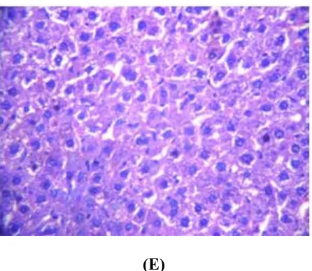
In the acute toxicity studies, EECP did not show any toxic signs or mortality at 2000 mg/kg dose. The elevated levels of AST, ALT, ALP, GGPT and TB in d-GalN intoxication were significantly reduced in the rats pre-treated with EECP and increased levels of TP and TA significantly (Table 1). Pre-treatment with EECP (400 mg/kg) exhibited significant hepatoprotective activity which was comparable with the standard drug Vitamin E. Table 2 also depicts there is a decrease in the levels of SOD, CAT, GPx and increase in LPO in d-GalN treated rats liver in comparison to vehicle treated group. EECP pre-treatment dose dependently caused a significant increase in the levels of antioxidants and decreased the LPO levels in comparison to d-GalN treated rats. Table 3 shows the levels of non-enzymatic antioxidants such as reduced glutathione, Vitamin C and Vitamin E in the tissues (liver) of D-galactosamine hepatotoxic and control rats. The levels of non-enzymatic antioxidants in D-galactosamine hepatotoxic rats significantly decreased. EECP both doses administered rats showed significantly increased levels of these non-enzymic antioxidants as compared with untreated hepatotoxic rats. Histology of liver sections of normal
control animals (Group I) showed normal liver architecture with were brought out central vein, were preserved cytoplasm and prominent nucleus and nucleolus. The liver sections of galactosamine treated
Table.1Effect of EECP and Vitamin Epre-treatment on biochemical parameters of the rats intoxicated with D-Galactosamine
Group. No.
Treatment AST
(IU/ml) ALT (IU/ml) ALP (IU/ml) TP (gm/dl) TB (mg/dl) GGTP (mg/dl) TA (g/dl)
I Normal control 10ml/kg
44.40±1.52 30.09±1.49 23.68±1.30 5.15±0.08 1.92±0.08 96.90±2.75 3.80±0.16
II *a Toxic control
D-galactosamine 25mg/kg
105.90± 2.40
94.49±1.05 144.10±2.35 3.16±0.22 4.40±0.26 173.42±2.90 2.20±0.07
III *b Standard control Vitamin E
25mg/kg
60.10±1.20 40.56±1.06 56.4±1.70 3.90±0.08 2.8±0.15 122.20±1.95 2.90±0.05
IV *b Treatment control EECP
200mg/kg
68.65±1.46 54.82±2.72 65.86±2.30 4.60±0.25 3.30±0.20 136.30±3.04 2.54±0.04
V *b Treatment control EECP
400mg/kg
62.45±1.15 47.94±0.97 58.50±1.95 4.05±0.26 2.95±0.18 130.94±1.23 2.30±0.09
Values are given as mean ± SD from six rats in each group; Values are found out by using one way ANOVA followed by Newmannkeul’s multiple range tests; *a-values are significantly different from Normal control at P< 0.01;*b-values are significantly different from Toxic control (G2) at p< 0.01.
Table2 Effect of EECP and Vitamin E pre-treatment on biochemical liver parameter in D-Galactosamine induced hepatotoxicity.
Group. No.
Treatment SOD
(U/mg protein) CAT (U/mg protein) GPX (U/mg protein) LPO (nmol of NDA/mg protein) I
Normal control 10ml/kg Normal saline
132.25±2.40 290.40±2.40 1.10±0.05 3.90±0.17
II
*aToxic control
D-galactosamine 25mg/kg
68.20±1.65 190.75±2.70 0.40±0.02 7.40±0.12
III
*bStandard control
Vitamin E 25mg/kg
118.05±2.80 260.45±1.92 0.85±0.02 4.50±0.14
IV
*b Treatment control
EECP 200mg/kg
96.50±1.60 230.05±1.80 0.55±0.02 5.60±0.28
V
*b Treatment control EECP
400mg/kg
105.65±2.62 240.75±2.65 0.74±0.02 4.80±0.08
Table 3 Effect of EECP on non enzymatic antioxidants in the liver tissue of
d-galactosamine-hepatotoxic and control rats
Groups Glutathione
mg/100g Tissue
Vitamin-C mg/100g Tissue
Vitamin-E mg/100g Tissue
Normal control 10ml/kg
132.60±3.45 0.82±0.08 5.92±0.60
*a Toxic control
D-galactosamine 25mg/kg
73.55±1.70 0.30±0.02 2.40±0.30
*b Standard control
Vitamin E 25mg/kg
110.32±2.70 0.74±0.07 5.60±0.55
*b Treatment control
EECP 200mg/kg
98.05±2.16 0.60±0.04 4.92±0.50
*b Treatment control
EECP 400mg/kg
91.90±1.95 0.69±0.06 5.02±0.48
Values are given as mean ± SD from six rats in each group; Values are found out by using one way ANOVA followed by Newmannkeul’s multiple range tests; *a-values are significantly different from Normal control at P< 0.01;*b-values are significantly different from Toxic control (G2) at p< 0.01.
(A) (B)
(E)
Fig. 1(A): Magnified view of liver from normal control group (B): Toxic Control group (C): Standard Drug
control (D): (C. Chayamansa 200 mg/kg/rat) (E) (C. Chayamansa 400 mg/kg/rat) Data were presented as
mean±SD, No. of animals (n) =6
DISCUSSION
Elevated levels of ALT, AST, ALP enzymes are indicative of cellular leakage and loss of functional integrity of cell membrane in liver [18]. Pre-treatment with EECP decreased the level of hepatic enzymes
towards their respective normal value is an indication of stabilization of plasma membrane as well as repair of hepatic tissue damage caused by d-GalN. The 400mg/kg dose had a better effect than the low
dose of EECP (200mg/kg). The higher concentration might have resulted in the production of more by
products that would have interfered with the activity. Treatment with EECP significantly decreased these
enzyme activities, indicating that EECP has a hepatoprotective effect against a D-galactosamine- induced
liver injury. An increase in TB and ALP reflects the pathological alteration in biliary flow 19. EECP mediated suppression of the increased TB level suggests the possibility of the extract being able to stabilize biliary dysfunction. These biochemical findings were further substantiated by histopathological studies. D-galactosamine-induced oxidative damage is generally attributed to the formation of the highly reactive hydroxyl radical (OH·), the stimulator of lipid peroxidation and the source of destruction and
damage to the cell membrane 20. D-galactosamine toxicity enhanced lipid peroxidation and reduced
antioxidants were reported in the kidney 21. The previous studies show that D-galactosamine-induced
rats significantly increased thiobarbituric acid reactive substances, lipid hydroperoxides and conjugated
dienes in liver and kidney 22,23. In the present study, we observed an increase in the levels of
thiobarbituric acid reactive substances, lipid hydroperoxides and conjugated dienes in the tissues of
cause functional degradation; thus, the degradation of vital tissue leading to complications may be
indirectly due to increased oxidative stress. Treatment with EECP and Vitamin-E showed a significant
reduction which might be due to the antioxidant ability of these compounds and the consequent reduction
in lipid peroxidation. EECP possesses antioxidative and free-radical scavenging effects. Oxidative stress
is an imbalance between reactive oxygen species and the antioxidant defense mechanisms of a cell or
tissue, which leads to lipid peroxidation, DNA damage and the inactivation of many enzymes 24. The
enzymatic antioxidant defense system is the natural protector against lipid peroxidation that includes
superoxide dismutase, catalase and glutathione peroxidase. Reduced activities of these enzymes in the
tissue of D-galactosamine- hepatotoxic rats were observed in our study. Superoxide dismutase protects
against the superoxide radical (O2·−), which damages the membrane and its biological structure. Catalase
primarily decomposes hydrogen peroxide to H2O at a much faster rate, sharing this function with
glutathione peroxidase. Glutathione peroxidase may play an important role in the removal of lipid
hydroperoxides. The balance between these enzymes is important for the efficient removal of oxygen
radicals from tissues 25. Therefore, reduction in the activity of these enzymes may result in a number of
deleterious effects due to the accumulation of superoxide radicals and H2O2. Significant increases in the
activities of these enzymes were observed on EECP administration. The second line of defense consists
of the non-enzymic scavenger’s glutathione, ascorbic acid, and α-tocopherol, which scavenge residual
free radicals escaping from decomposition by the antioxidant enzymes. Moreover, enzymic antioxidants
are inactivated by the excessive levels of free radicals and hence the presence of non-enzymic
antioxidants is presumably essential for the removal of these radicals 26. Glutathione a major non-protein
thiol in living organism’s plays a central role in coordinating the antioxidant defense process. Glutathione
reacts directly with reactive oxygen species and electrophilic metabolites, protects the essential thiol
group from oxidation, and serves as a substrate for several enzymes including glutathione peroxidase 27.
The lowered glutathione in D-galactosamine induced rats represents the increased utilization of
glutathione as a result of oxidative stress. Perturbation in the redox status of glutathione not only impairs
cellular defense against toxic compounds but also results in enhanced oxidative stress and oxidative
injury 28. Apart from glutathione, α-tocopherol and ascorbic acids are important free-radical scavengers
which protect cell membrane against toxic agents. Both vitamins C and E have a synergistic action in
scavenging oxygen-derived free radicals 29. Vitamin C functions as a free-radicals cavenger of oxygen
radicals and successfully prevents detectable oxidative damage under all types of oxidative stress.
Ascorbic acid appears to trap the peroxyl radical in the aqueous phase with a rate large enough to lipids
ascorbate. The thiol cycle consists of a GSSG/GSH couple 30. Thus glutathione in blood keeps up the
cellular levels of the active form of vitamin C. When there is a reduction in glutathione, the cellular level
of ascorbic acid is also lowered. The observed decrease in the levels of α-btocopherol and ascorbic acid
in the D-galactosamine rats might be due to an antioxidant defense against increased ROS or due to a
decrease in glutathione levels in D-galactosamine-hepatotoxic rats. In this respect, reported that ascorbic
acid and α-tocopherol decreased in liver diseases, particularly in D- galactosamine- hepatotoxic rats. Our
study observed increase the levels of these antioxidants in EECP and Vitamin-E administered rats. The
ability of EECP to enhance the levels of antioxidants along with its anti lipid peroxidative activity
suggests that this compound might be potentially useful in counteracting free-radical-mediated tissue
damage caused by hepatotoxicity. Studies on the antioxidative potency of various flavonoids have
confirmed the importance of the distribution and quantity of the hydroxyl groups. In general, the
antioxidative properties of polyphenols depend on hydroxylation of ring B. The present results
corroborate the protective action of EECP in D- galactosamine intoxication of rats, particularly
noticeable with the high dose used by us (400 mg/kg body weight). Since C. chayamansa is rich in flavonoids and phenolics, the possibility of the mechanism of hepatoprotection of C. chayamansa extract may be due to its antioxidant action.
Conclusion
Our findings demonstrated that EECP at both doses possesses hepatoprotective and antioxidant activity,
which is evidenced by lowered serum hepatic marker enzyme activities. Among the two dosages tested,
400mg/kg/body weight showed more promising hepatoprotective and antioxidant activity, and is
comparable to the standard drug Vitamin-E. The hepatoprotective effect is documented by the biochemical and histopathological data obtained. It may be speculated that the constituents of C.
chayamansa leaf are to be responsible, at least in part, for the observed hepatoprotective effect. A
possible mechanism of C. chayamansa extract as hepatoprotective may be due to its antioxidant effect
which impairs the activation of D-galactosamine into the reactive form, it may be speculated that the
high flavonoid content of C. chayamansa, was responsible for the observed protective effects. Further
studies need to be done on C. chayamansa extract to identify the active constituent(s) responsible for the
hepatoprotective activity and elucidate the mechanism of action.
References
2. Raju RW, Radhika SS, Kunal MT, Kalpana SP and Sunil SJ. Screening of roots of Baliospermum montanum for hepatoprotective activity against paracetamol induced liver damage in albino rats.
International Journal of Green Pharmacy 2008; 2: 220-223.
3. Sugiyama K, He P, Wada S and Saeki S. Teas and other beverages suppress D-galactosamine induced liver injury in rats. J Nutr 1999;129:1361-7.
4. Wills PJ and Asha VV. Protective effect of Lygodium flexuosum (L.) Sw. (Lygodiaceae) against D-galactosamine induced liver injury in rats. J Ethnopharmacol 2006; 108:116-23.
5. Jonker AM, Dijkhuis FW, Boes A, Hardonk MJ and Grond J. Immunohistochemical study of extracellular matrix in acute D-Galactosamine hepatitis in rats. Hepatology 1992; 15:423-31. 6. Keppler DO, Pausch J and Decker K. Selective uridine triphosphate deficiency induced by
D-galactosamine in liver and reversed by pyrimidine nucleotide precursors. Effect on ribonucleic acid synthesis. J Biol Chem. 1974; 246:211-6.
7. Yang YH. Tropical home gardens as a nutritional intervention. In: Inglett GE, Charalambous G, editors. Tropical Food Chemistry and Nutrition. New York: Academic Press; 1979. p. 417-36. 8. Bolio JD. Chaya (Cnidoscolus chayamansa, Euphorbiaceae), a marvellous food. Tierra. 1975;
30:407- 8, 427-8.
9. Rowe L. Plant guards secret of good health. Vol. 4. Harlingen, Texas: Valley Morning Star; 1994. p. 1-12.
10.Martin FW and Chaya RR. Cnidoscolus chayamansa includes composition and nutritional value, culture in Puerto Rico. In: Vegetables of Hot Humid Tropics. New Orleans, LA: USDA, ARS;
1978.
11.Roy S, Niranjan C, Jyothi T, Shankrayya M, Vishawanath K, Prabhu K, Gouda V and Setty R. Antiulcer and anti-inflammatory activity of aerial parts Enicostemma littorale Blume. J Young Pharm 2010; 2: 369–373.
12.Najmi AK, Pillai KK, Pal SN and Aqil M. Free radical scavenging and hepatoprotective activity of jigrine against galactosamine induced hepatopathy in rats. J Ethnopharmacol. 2005; 97, 521– 525.
13.Lowry OH, Rosebrough NJ, Farr AL and Randall RJ. Protein measurement with the Folin phenol reagent. J Biol Chem. 1951; 193, 265–275.
14.Kakkar P, Das B and Viswanathan PN. A modified spectrophotometric assay of superoxide dismutase. Indian J Biochem Biophys. 1984; 21, 130–132.
hydrogen peroxide by catalase. J Biol Chem. 1952; 195, 133–140.
16.Paglia DE and Valentine WN. Studies on the quantitative and qualitative characterization of erythrocyte glutathione peroxidase. J Lab Clin Med. 1967; 70, 158–169.
17.Ohkawa H, Ohishi N and Yagi K. Assay for lipid peroxides in animal tissues by thiobarbituric acid reaction. Anal Biochem. 1979; 95, 351–358.
18.
Jadon A, Bhadauria M and Shukla S. Protective effect of Terminalia belerica Roxb. and gallicacid against carbon tetrachloride induced damage in albino rats. J Ethnopharmacol. 2007; 109: 214–218.
19.Ravikumar V, Shivashangari KS and Devaki T. Hepatoprotective activity of Tridax procumbens against d-galactosamine/ lipopolysaccharide-induced hepatitis in rats. J Ethnopharmacol. 2005; 101, 55–60
20.Ryan CJ, Aslam M and Courtney JM. Transference of hepatic coma to normal rats from galactosamine treated donors by reverse plasma exchange. Biomat. Artif Cells Artif Organs.1990,18: 477–482.
21.Barry H and Gutteridge JMC. Free radicals in biology and medicine. Barry, H.(Ed.), Oxford Clarendon Press. 1989: 254–255.
22.Chikhi N, Holic N, Guellaen G, Laperche Y. Gamma-glutamyl transpeptidase gene organization and expression: a comparative analysis in rat, mouse, pig and human species. Biochem. Physiol.1999, 122: 367–380.
23.Sakaguchi S and Yokota K. Role of Ca2+on endotoxin-sensitivity by galactosamine challenge: lipid peroxide formation and hepatotoxicity in zymosan primed mice. Pharmacol. Toxicol.1995,
77: 81–86.
24.Mourella M and Meza MA. Colchicine prevents D-galactosamine-induced hepatitis. J. Hepatol. 1989; 8: 165–172.
25. Speaker SD, Fabris M, Ferrari V and Dalle MA. Quercetin protects cutaneous tissue associated
types including sensory neurons from oxidative stress induced by - glutathione depletion: cooperative effects of ascorbic acid. Free Rad. Biol. Med. 1997;22: 669–678.
27.Allen RG. Oxygen-reactive species and antioxidant responses during development: the metabolic paradox by cellular differentiation. Proc. Soc. Exp. Biol. Med. 1991, 196:117–129.
28.Reed DJ. Glutathione: toxicological implications. Ann. Rev. Pharmacol. Toxicol. 1990,30: 603– 631.
29.Wojacki JL, Samachowiec B, Gonet S, Juzwiak E and Dabrowska Zamojen M. Effect of buckwheat extract on free radical generation in rabbits administered high fat diet. Phytother. Res.1995; 9: 323–326.