Utilizing Cell-SELEX, as a Promising Strategy to Isolate ssDNA Aptamer Probes for
Detection of
Staphylococcus aureus
Mitra Yazdi Yahyaabadi1, Ghambar Sultan Dorraj1, Mohammad Heiat1*, Ali Mohammad Latifi1**
Abstract
Introduction
Staphylococcus aureus is a pathogenic gram-positive,
facultative anaerobe bacterium that can cause infection in humans and animals, ranging from simple skin infections to life-threatening disease. S. aureus is considered as one of the five common causes of nosocomial infections [1, 2]. Many people annually are infected with S. aureus in hos-pitals. Currently traditional methods which are often time-consuming and cost-intensive, applied to diagnose S.
aureus. For instance LCR (ligase chain reaction) and PCR
(polymerase chain reaction) account as highly sensitive diagnostic systems [3] that are able to track a small num-ber of bacteria within a few hours but they require some obstacles such as isolation of bacterial DNA, post amplifi-cation procedure and expensive instruments to amplify nucleic acid. Such handicapping barriers limit the wide-spread use of these technologies for clinical diagnosis. Latex Immuno-agglutination Assay (LIA) and ELONA are also employed which are insensitive or time-consuming methods [4, 5]. Accordingly it is essential to develop a fast, inexpensive and nevertheless accurate method to identify S. aureus contaminated samples. Aptamer is a single stranded oligonucleotide of DNA or RNA which have an equal or higher sensitivity than antibodies. Apta-mers are able to bind specifically to the targets in quanti-ties of pico to nano molar ranges [6, 7]. Unlike antibodies, aptameric probes are not immunogenic, and have long half-life and stability under different conditions. Aptamers are smaller in size, which leadsthem to create more surface
density on the receptors and easier binding to the analytes [8]. These probes are isolated via a systematic evolution procedure so-called SELEX (systematic evolution of ligands by exponential enrichment). Indeed SELEX is an in vitro method to isolate target-specific single-stranded oligonucleotides among a random sequence library. SELEX conditions can be massively altered to enrich ap-tameric sequences with desired characteristics. Developing different SELEX procedures have led to the isolation of large variety of aptamers dedicated to many targets, rang-ing from small organic molecules to a protein complex or even whole cells. Cell-SELEX is a term that explains the pathway in which whole live cells are employed as target to isolate aptamers (Fig. 1). Thus, it is useful enrichment strategy for lots of applications, including bacterial detec-tion [9]. Aptamers bind to their targets with a specific three-dimensional structure [10].
In the dimensions of research, aptamers are broadly utilized for manufacturing various biosensors so-called apta-sensors. In the past few recent years, aptamers have been exhibited to be worthy substitute for antibodies in serological diagnostic techniques with higher merits. Ap-tamer probes are appropriate alternative tools to detect interested targets in multifarious formats such as [11], Sur-face plasmon resonance (SPR), enzyme-linked oligonuc-leotide assay (ELONA) [12-14], aptamer-linked immobi-lized sorbent assay (ALISA), lateral flow assay (LFA) [15], fluorescent labeled aptamers and electrochemical and other biosensors [11, 16, 17].
Staphylococcus aureus is one of the most important pathogens in hospital-acquired infections. Annually, many people are infected with S. aureus in hospitals. Rapid detection of this bacterium is extremely helpful in preventing and managing this bacterium mediated diseases. Aptamers are powerful probes, which can be used as a target explorer in a wide range of diagnostic systems.To isolate a specific aptamer against S. aureus, a library of single-stranded DNA molecules was designed, and enriched through Cell-SELEX procedure. In the Cell-SELEX, the DNA library was exposed to the S. aureus bacterium in 8 reiterative quadruple rounds including: binding, separation, elution and amplification. After 8 rounds, the PCR product was cloned and sequenced. Cloned aptameric sequences were evaluated through enzyme-linked oligonucleotide assay (ELONA), and a sequence with the best outcomes was selected as ideal aptamer. Eight rounds of Cell-SELEX procedure led to isolation of a specific ssDNA aptamer against S. aureus and named as “STAPT” (conflation of STAphylococcus and APTamer). Using ELONA technique, the detection limit of this aptamer was determined as 4 × 10³ CFU/ml. The aptamer “STAPT” showed the promising and potent abilities and features to be utilized as a bio-detection element likely in advanced detection systems. Although more extended researches are needed for this purpose.
Keywords: Staphylococcus aureus, Detection, Aptamer, Cell-SELEX, ELONA
1. Applied Biotechnology Research Center, Ba-qiyatallah University of Medical Science, Tehran, Iran
* Corresponding Authors Mohammad Heiat
Applied Biotechnology Research Center, Baqiya-tallah University of Medical Science, Tehran, Iran E-mail: [email protected]
Ali Mohammad Latifi
E-mail: [email protected]
Mitra Yazdi Yahyaabadi, et al. Utilizing Cell-SELEX, as a Promising Strategy to Isolate ssDNA Aptamer
178 Journal of Applied Biotechnology Reports, Volume 4, Issue 3, Summer 2017
However, the prerequisite for manufacturing an efficient biosensor is to have a capable aptamer. Therefore, in this study we tried to design a Cell-SELEX procedure to isolate a specific aptamer against S. aureus bacterium.
Materials and Methods
Materials
To run Cell-SELEX procedure all biological and chemical agents were prepared from reliable companies. Some of the most important agents include; PCR ready master-mix from Topaz gen Co. Lambda exonuclease from Thermo Fisher Inc. HRP-conjugated streptavidin was purchased from Invitrogen Co. PTG19-T vector and other cloning requirements were prepared from Sinaclon Co. S. aureus
strain, as target, (ATCC 25923), Streptococcus epidermi-dis, (ATCC 12228) and Streptococcus pyogenes (ATCC 19615), as counter SELEX, were prepared from reference laboratory.
DNA Library and primers
The ssDNA library (5'-GCCTGTTGTGAGCCTCCTAAC (N38) CATGCTTATTCTTGTCTCCC-3̛') and primers were designed and synthesized by Metabion Co, Germany which below is shown:
Primer 1 Forward 5′-GCCTGTTGTGAGCCTCCTACC-3′ Reverses 5′-GGGAGACAAGAATAAGCATG-3′
Primer 2 Forward 5′-biotin-GCCTGTTGTGAGCCTCCTACC-3′ Reverses 5′-phosphate GGGAGACAAGAATAAGCATG-3′
Cell-SELEX procedure
Synthetic ssDNA library (2 nM) was denatured by heating at 90°C for 5 min and immediately cooled on ice for 10 min. After running the negative SELEX step (removal of cross-reactive aptamers), the denatured ssDNA library was mixed with 1 × 10⁷ CFU/ml of S. aureus resuspended in 300 ml of screening buffer (Tris-HCl 25 mM, KCl 50 mM, Nacl 200 mM, EDTA 0.2 mM, Glycerol %5, Dtt 0.5 mM)
Figure 1. SELEX schematic procedure. Synthesized ssDNA random pool was mixed with bacteria cells at the first round. Unbounded aptamers were discarded and bound aptamers were collected and amplified via asymmetric-PCR. PCR products were served as second-ary pool for the second round. After 8 reiterative rounds, the final pool was cloned and then analyzed by enzyme-linked oligonucleotide assay (ELONA). Finally, high binding clones were sequenced and their affinities were analyzed by SPR system.
and was incubated at 37°C for 1 h. In the following, the mixture was centrifuged and the supernatant containing unbound oligonucleotides was discarded. The pellet co taining aptamer-bacteria complex was washed 3 times with 200 ml of washing buffer (TBST buffer: PBS buffer plus Tween 0.05%), then 100 ml of distilled water was added and incubated at 80°C for 10 min. Following the centrif gation, the supernatant containing eluted
transferred into a new tube and recovered using alcohol glycogen precipitation procedure (ethanol: 2.5 fold, acetate sodium 0.1 fold and glycogen 0.05 fold of supernatant) and the ssDNA concentration was assessed using optical densitometry in the wavelength of 260 nm.
The recovered ssDNA was amplified by PCR procedure, and after conversion to the single-stranded oligonucleotide (by Lambda exonuclease enzyme), used for the next round of Cell-SELEX. Every SELEX round condition became restricted through reducing the amount of ssDNA pool at each round (2 to 0.1 nM), reducing the incubation time (from 1 h to 45 min), and increasing the number of rounds. The aptamer enrichment repetitive cycles were followed and monitored by ssDNA binding quality. To
cell-SELEX process, the binding quantity of ssDNA was determined based on the ratio of used to recovered conce tration of ssDNA in each Cell-SELEX round. According to this checkpoint assessments, 8 rounds of Cell
were continued and then the procedure was entered the next step (Fig. 1). The final ssDNA pool were amplified and cloned into E. coli DH5α, as cloning host, by means of the PTG19-T vector. In order to identify the cloned aptameric DNA sequences, several randomly selected colonies were sequenced (BioneerDNA sequencing se vice).
Evaluation of the specificity and binding properties (Quality, cross-reactivity and LOD) of isolated aptamer
Isolated aptamer was biotinylated by 5′ primers using PCR. Then the indirect ELON
measure the limit of detection (LOD) of aptamers as follows: the microplate was first covered with
consecutive dilutions from 0.5 McFarland with reducing concentration steps of 1
10 in each well), then the plate was
washed (3 times with 200 ml of PBST buffer). Afte wards, the wells were blocked using blocking buffer (commercially purchased from Roche Co). In the follo ing, 2 μl aptamer (with the concentration of 1 ng/μl PBS) was added in each well and incubated at 37°C with gentl shaking for 60 minutes. After washing, 100 μl streptav din-HRP conjugate was added (1/5000 in PBST buffer) and again incubated at 37°C with gently shaking for 60 minutes. After washing, 100 ml of TMB substrate was added to each well and the microplate was transferred to a dark place. The reaction was stopped by addition of 50 µl of 2.5 M sulfuric acid after 15 min. Finally, the optical density of appeared yellow color was read by ELISA reader at the wavelength of 450 nm. A well without bact ria was considered as negative control. As the final EL NA evaluation, the aptamer cross reactivity with similar bacteria was also assessed against S. epidermidis
Streptococcus through the same procedure. All exper ments for evaluation of binding quality, cross
and was incubated at 37°C for 1 h. In the following, the mixture was centrifuged and the supernatant containing unbound oligonucleotides was discarded. The pellet
con-bacteria complex was washed 3 times with 200 ml of washing buffer (TBST buffer: PBS buffer plus Tween 0.05%), then 100 ml of distilled water was added and incubated at 80°C for 10 min. Following the centrifu-gation, the supernatant containing eluted ssDNA was transferred into a new tube and recovered using alcohol-glycogen precipitation procedure (ethanol: 2.5 fold, acetate sodium 0.1 fold and glycogen 0.05 fold of supernatant) and the ssDNA concentration was assessed using optical
e wavelength of 260 nm.
The recovered ssDNA was amplified by PCR procedure, stranded oligonucleotide (by Lambda exonuclease enzyme), used for the next round SELEX. Every SELEX round condition became rough reducing the amount of ssDNA pool at ), reducing the incubation time (from 1 h to 45 min), and increasing the number of rounds. The aptamer enrichment repetitive cycles were followed and monitored by ssDNA binding quality. To evaluate the SELEX process, the binding quantity of ssDNA was determined based on the ratio of used to recovered
concen-SELEX round. According to this checkpoint assessments, 8 rounds of Cell-SELEX the procedure was entered the next step (Fig. 1). The final ssDNA pool were amplified DH5α, as cloning host, by means of T vector. In order to identify the cloned aptameric DNA sequences, several randomly selected es were sequenced (BioneerDNA sequencing
ser-Evaluation of the specificity and binding properties reactivity and LOD) of isolated aptamer
′-Biotin-forward primers using PCR. Then the indirect ELONA was used to measure the limit of detection (LOD) of aptamers as follows: the microplate was first covered with S. aureus (9 consecutive dilutions from 0.5 McFarland with reducing in each well), then the plate was es with 200 ml of PBST buffer). After-wards, the wells were blocked using blocking buffer (commercially purchased from Roche Co). In the follow-ing, 2 μl aptamer (with the concentration of 1 ng/μl PBS) was added in each well and incubated at 37°C with gently shaking for 60 minutes. After washing, 100 μl
streptavi-HRP conjugate was added (1/5000 in PBST buffer) and again incubated at 37°C with gently shaking for 60 minutes. After washing, 100 ml of TMB substrate was s transferred to a dark place. The reaction was stopped by addition of 50 µl of 2.5 M sulfuric acid after 15 min. Finally, the optical density of appeared yellow color was read by ELISA reader at the wavelength of 450 nm. A well without
bacte-dered as negative control. As the final ELO-NA evaluation, the aptamer cross reactivity with similar
S. epidermidis and
Streptococcus through the same procedure. All experi-ments for evaluation of binding quality, cross-reactivity
and LOD calculation were repeated at least 3 times and the average values were used as final outputs.
Determination of the secondary structure of aptamers
Secondary structure of the isolated sequence was dete mined by using of mfold online softwar (http://mfold.RNA.albany.edu/q=mfold) at the folding temperature of 37°C. The concentration of Na
were also adjusted in the base of screening buffer.
Results
Cell-SELEX and PCR
Aptamers obtained at each round of Cell
be amplified for the next round. One of the difficulties in this case was the optimization of PCR conditions at every round. In a general conclusion, reducing the number of PCR cycles, increasing the a
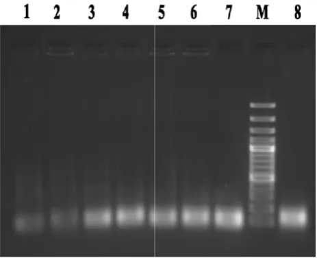
diluting the template oligonucleotide pool, resulted in the elimination of smear and unwanted products. By conside ing these key items, the PCR procedure with the most optimized condition was performed for amplification of recovered ssDNA at the each round (Fig. 2).
Figure 2. Lane 1 to 8: amplification of recovered ssDNA pool at the end of each round (round 1 to 8), M: DNA marker.
Investigation of SELEX performance
In order to ensure about how the SELEX works and to confirm that the SELEX procedure enriched the oligonu leotide fragments with the higher levels of affinity to ba terial cells, the binding quantity of the all oligonucleotide pools were measured based on the ratio of used to reco ered amount of ssDNA for each roun
(Fig. 3). According to the chart illustrated at Figure 3, the ssDNA pool of round five (R5) showed the highest bin ing quantity compared to other rounds even rounds of six (R6), seven (R7) and eight (R8). It could be due to the maximum tendency of oligonucleotides to the target. These findings somehow indicated the end of SELEX in round eight (R8).
Evaluation of binding quality of isolated aptamers
The pool of ssDNA obtained from round eight were cloned and sequenced. Randomly sel
sized and their binding qualities were independently and LOD calculation were repeated at least 3 times and the average values were used as final outputs.
Determination of the secondary structure of aptamers
Secondary structure of the isolated sequence was deter-mined by using of mfold online software (http://mfold.RNA.albany.edu/q=mfold) at the folding temperature of 37°C. The concentration of Na+ and Mg2+ were also adjusted in the base of screening buffer.
Aptamers obtained at each round of Cell-SELEX need to be amplified for the next round. One of the difficulties in this case was the optimization of PCR conditions at every round. In a general conclusion, reducing the number of PCR cycles, increasing the annealing temperature and diluting the template oligonucleotide pool, resulted in the elimination of smear and unwanted products. By consider-ing these key items, the PCR procedure with the most optimized condition was performed for amplification of
red ssDNA at the each round (Fig. 2).
. Lane 1 to 8: amplification of recovered ssDNA pool at the end of each round (round 1 to 8), M: DNA marker.
Investigation of SELEX performance
In order to ensure about how the SELEX works and to that the SELEX procedure enriched the oligonuc-leotide fragments with the higher levels of affinity to bac-terial cells, the binding quantity of the all oligonucleotide pools were measured based on the ratio of used to recov-ered amount of ssDNA for each round of the Cell-SELEX (Fig. 3). According to the chart illustrated at Figure 3, the ssDNA pool of round five (R5) showed the highest bind-ing quantity compared to other rounds even rounds of six (R6), seven (R7) and eight (R8). It could be due to the tendency of oligonucleotides to the target. These findings somehow indicated the end of SELEX in
Evaluation of binding quality of isolated aptamers
Mitra Yazdi Yahyaabadi, et al. Utilizing Cell-SELEX, as a Promising Strategy to Isolate ssDNA Aptamer
178 Journal of Applied Biotechnology Reports, Volume 4, Issue 3, Summer 2017
evaluated by ELONA procedure. Analytical results dem-onstrated that more aptamers could significantly raise the optical density in ELONA (Fig. 4). Meanwhile, the apta-mer with the highest binding properties was selected to continue research procedure. The selected aptamer was named “STAPT” which is a logical conflation of STAphy-lococcus and APTamer.
Figure 3. Binding quantity of ssDNA pool in the SELEX rounds (ratio of used amount to recovered amount of ssDNA) from pri-mary library to round 8.
Figure 4. Binding quality of sequences obtained from final pool of Cell-SELEX (Round 8).
STAPT cross-reactivity analysis
In order to study the cross reactivity of selected aptamer, it was employed against some other bacteria such as S.
epi-dermidis and Streptococcus in ELONA. Results revealed
that the STAPT is definitely dedicated to S. aureus with a powerful feature (Fig. 5).
Calculation of ATAPT aptamer LOD
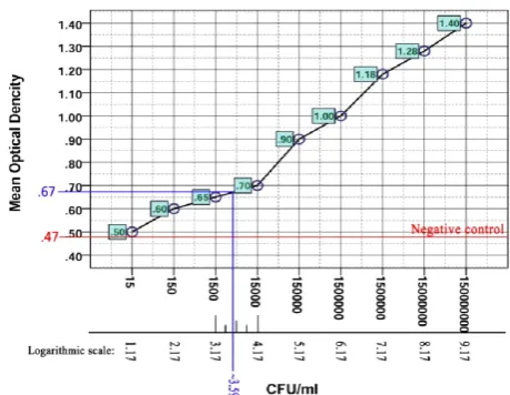
At the first step, according to the formulas 1 and 1ʹ, the limit of blank (LOB) (the highest optical density, expected to be recorded in replications of a blank sample without analyte) was calculated. Based on LOB, and by use of formulas 2 and 2ʹ, the limit of detection (LOD) ( the lowest number of bacteria at which detection is reliably
feasi-ble)was estimated [18]. Calculations revealed that the ap-tamer detection limit was approximately at the OD = 0.67. The OD corresponding point on the logarithmic scale and subsequently bacterial number (CFU/ml) revealed that the LOD of STAPT was ~ 4 × 10³ CFU/ml. it means that the minimum number of bacteria that can determine by STAPT
is ~ 4 × 10³ CFU/ml (Fig. 6).
Figure 5. STAPT aptamer cross-reactivity test. S. aureus used as specific and S. epidermidis and Streptococcus as non-specific targets.
Figure 6. The graphical illustration for calculation of STAPT LOD.
Secondary structure of STAPT aptamer
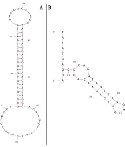
The secondary structures of selected aptamer (STAPT) were determined by using mfold online software. This software predicted the secondary structure of STAPT with
and without constant regions. Illustration of ATAPT sec-ondary structure revealed that both core region of STAPT
(aptamer without constant regions) and full length STAPT
(aptamer with constant regions) have two functional loops (Fig. 7).
Figure 7. Secondary structure of aptamers predicted by mfold. A) Full length STAPT with the minimum energy of -27.65 Kcal/mol. B) Core region of STAPT with the minimum energy of -1.50 Kcal/mol.
Discussion
Staphylococci can be normally isolated from skin,
naso-pharynx and also public places specially hospitals [19]. Even when have no symptoms, they can show the potential to cause disease. Three major pathogens of this gram-positive genus of bacteria include S. aureus, S. epidermitis,
and S. saprophyticus. Today, S. aureus is known as a
lead-ing cause of disease in hospital [1]. S. aureus causes a wide range of diseases which can be classified into two categories including: 1) Diseases caused by exotoxin re-lease such as gastroenteritis (food poisoning) and toxic shock syndrome, 2) Diseases caused by direct bacterial onset such as dermal infection and bacteremia/sepsis [20]. Accordingly, the diagnosis of S. aureus can pave the way for disease prevention and management. How to detect
Staphylococci from Streptococci is very important because
most Staphylococci are resistant to penicillin G. There are three methods for their diagnosis: Gram-positive test, cata-lase test, and direct culture. It is more important that we can identify S. aureus as the most pathogenic species from other species of Staphylococci. Aptamer, known as chemi-cal antibody [21], is a potent probe which can be served as a nimble bio-detector element and a replacement for anti-body. SELEX with all its derivatives is a high throughput strategy to isolate aptamer through a nucleic acid pool with a large diversity. In this study, the Cell-SELEX method
was employed for the isolation of specific aptamers against
S. aureus whole cell. Performing consecutive Cell-SELEX
rounds is aimed to increase the affinity and proprietary performance of aptamer. Rising trend in the affinity of aptamers during the SELEX process will confirm and veri-fy the accuracy of SELEX for aptamers isolation. For this purpose, different methods have been developed, such as capillary electrophoresis [22], flow cytometry [23] affinity matrix [24] surface plasmon resonance (SPR) etc [25]. In the present study, the Cell-SELEX accuracy was moni-tored by measuring round binding quantity, whereby the ratio between the used and recovered ssDNA was calcu-lated at the end of each round. Here we also used ELONA to investigate binding quality, cross-reactivity and LOD of isolated aptamer. Finding proved that our isolated aptamer-ic probe named “STAPT” has acceptable binding quality and LOD (~ 4 × 10³ CFU/ml) and also a promising speci-ficity for S. aureus. A large number of studies entitled bac-terial detection technique based on aptamer have been pub-lished, and readers will be involved in a confusing diversi-ty of methods, findings and conclusions. However, we tried to simplify some of them and evaluate our STAPT
aptamer. Lavu and colleagues [26], isolated ssDNA apta-mers against Salmonella enterica serovar Typhimurium
using Cell-SELEX. They reported that their beat isolated aptamer (SAL 26) had sensitivity limit of 102 CFU/ml. Wang et al. [27], used two DNA aptamers against S.
typhimurium to manufacture a label-free detection system
by SPR. They immobilized aptamers on the gold chips in SPR experiment and operated the detection procedure. Finally, they determined that the LOD of their aptamer-based sensor was as 3 × 10⁴ CFU/ml. Hu and colleagues [28] isolated, a single-stranded DNA aptamer against Bifidobacterium breve through 12 rounds of whole-cell-SELEX. Their findings showed that the LOD of their aptamer with the highest affinity (BB16-11f) was 103 CFU/ml. Duan et al., [29] isolated and characterized some aptamers against S. typhimurium using Cell-SELEX. By a superficial looking at the structure of their best apta-mer (ST2P) its complexity is revealed. Of course, this ap-tamer with a quad-blade structure and with five complete loops, establishes themore complicated and stronger bind-ing. As expected, this aptamer was very desirable and its LOD was equal to 25 CFU/ml. Savory and colleagues [30] developed a colorimetric apta-sensor for detection of
Streptococcus mutans. They demonstrated that their
im-mobilized aptamer on gold colloids, in the form of a flow-through diagnostic system, could detect S. mutans with a LOD of 1 × 10⁵ CFU/ml. There are many examples with various outcomes, however, in comparison with other sim-ilar studies, STAPT aptamer is placed in the middle class position with satisfactory properties. The relatively large discrepancies between the aptamers are often rooted in the quality of the implementation of SELEX, its own inherent characteristics and binding assessment techniques.
Conclusions
Mitra Yazdi Yahyaabadi, et al. Utilizing Cell-SELEX, as a Promising Strategy to Isolate ssDNA Aptamer
178 Journal of Applied Biotechnology Reports, Volume 4, Issue 3, Summer 2017
what shows itself is no exception and can play a decisive role in manufacturing of biosensors.
Acknowledgements
This study is extracted from a MSc thesis which was ap-proved by the Applied Biotechnology Research Centers, Baqiyatallah University of Medical Sciences, Tehran, Iran. The authors would like to thank colleagues in this centre for their kind and generous assistance.
References
1. Shittu, A.O., Okon, K., Adesida, S., Oyedara, O., Witte, W., Strommenger, B., Layer, F., Nübel, U., Antibiotic resistance and molecular epidemiology of Staphylococcus aureus in Nigeria. BMC Microbiol, 2011, Vol. 11, pp. 92-99.
2. Chang, Y.C., Yang, C.Y., Sun, R.L., Cheng, Y.F., Kao, W.C., Yang, P.C., Rapid single cell detection of Staphylococcus aureus by aptamer-conjugated gold nanoparticles.Sci Rep, 2013, Vol. 3, pp. 1863-1869.
3. Mothershed, E.A., Whitney, A.M., Nucleic acid-based methods for the detection of bacterial pathogens: present and future considerations for the clinical laboratory.Clin Chim Acta, 2006, Vol. 363, pp. 206-220.
4. Chapin, K., Musgnug, M., Evaluation of three rapid methods for the direct identification of Staphylococcus aureus from positive blood cultures. J Clin Microbiol, 2003, Vol. 41, pp. 4324-4327.
5. Tan, T.Y., Corden, S., Barnes, R., Cookson, B., Rapid identification of methicillin-resistant Staphylococcus aureus from positive blood cultures by real-time fluorescence PCR. J Clin Microbiol, 2001, Vol. 39, pp. 4529-4531.
6. Jenison, R.D., Gill, S.C., Pardi, A., Polisky, B., High-resolution molecular discrimination by RNA.Science, 1994, Vol. 263, pp. 1425-1429.
7. Dorraj, G.S., Rassaee, M.J., Latifi, A.M., Pishgoo, B., Taval-laei, M., Selection of DNA aptamers against Human Cardiac Troponin I for colorimetric sensor based dot blot application.J Biotechnol, 2015, Vol. 208, pp. 80-86.
8. Tabarzad, M., Kazemi, B., Vahidi, H., Aboofazeli, R., Shah-hosseini, S., Nafissi-Varcheh, N., Challenges to Design and De-velop of DNA Aptamers for Protein Targets. I. Optimization of asymmetric PCR for generation of a single stranded DNA library. Iran J Pharm Res, 2014, Vol. 13, pp. 133-141.
9. Sefah, K., Shangguan, D., Xiong, X., O'Donoghue, M.B., Tan, W., Development of DNA aptamers using Cell-SELEX. Nat Protoc, 2010, Vol. 5, pp. 1169-1185.
10. Heiat, M., Najafi, A., Ranjbar, R., Latifi, A.M., Rasaee, M.J., Computational approach to analyze isolated ssDNA aptamers against angiotensin II.J Biotechnol, 2016, Vol. 230, pp. 34-39. 11. Hwang, K.S., Lee, S.M., Eom, K., Lee, J.H., Lee, Y.S., Park, J.H., Yoon, D.S., Kim, T.S., Nanomechanical microcantilever operated in vibration modes with use of RNA aptamer as receptor molecules for label-free detection of HCV helicase.Biosens Bio-electron, 2007, Vol. 23, pp. 459-465.
12. Mir, M., Lozano-Sánchez, P., Katakis, I., Towards a target label-free suboptimum oligonucleotide displacement-based de-tection system. Anal Bioanal Chem, 2008, Vol. 391, pp. 2145-2152.
13. Balogh, Z., Lautner, G., Bardóczy, V., Komorowska, B., Gyurcsányi, R.E., Mészáros, T., Selection and versatile applica-tion of virus-specific aptamers. FASEB J, 2010, Vol. 24, pp. 4187-4195.
14. Chen, F., Hu, Y., Li, D., Chen, H., Zhang, X.-L., CS-SELEX generates high-affinity ssDNA aptamers as molecular probes for hepatitis C virus envelope glycoprotein E2.PloS one, 2009, Vol. 4, pp. e8142.
15. Wang, C., Zhang, L., Shen, X., Development of a nucleic acid lateral flow strip for detection of hepatitis C virus (HCV) core antigen.Nucleosides Nucleotides Nucleic Acids, 2013, Vol. 32, pp. 59-68.
16. Balogh, Z., Lautner, G., Bardoczy, V., Komorowska, B., Gyurcsanyi, R.E., Meszaros, T., Selection and versatile applica-tion of virus-specific aptamers. FASEB J, 2010, Vol. 24, pp. 4187-4195.
17. Vallian, S., Khazaei, M., Medical applications of aptamers. Research in Pharmaceutical Sciences, 2009, vol. 2, pp. 59-66. 18. Armbruster, D.A., Pry, T., Limit of Blank, Limit of Detection and Limit of Quantitation.Clin Biochem Rev, 2008, Vol. 29, pp. S49-S52.
19. Moghaddam, M.M., Abolhassani, F., Babavalian, H., Mirne-jad, R., Barjini, K.A., Amani, J., Comparison of in vitro antibac-terial activities of two cationic peptides CM15 and CM11 against five pathogenic bacteria: Pseudomonas aeruginosa, Staphylococ-cus aureus, Vibrio cholerae, Acinetobacter baumannii, and Escherichia coli.Probiotics and antimicrob proteins, 2012, Vol. 4, pp. 133-139.
20. Lin, Y.-C., Peterson, M.L., New insights into the prevention of staphylococcal infections and toxic shock syndrome. Expert Rev Clin Pharmacol, 2010, Vol. 3, pp. 753-767.
21. Zhou, G., Wilson, G., Hebbard, L., Duan, W., Liddle, C., George, J., Qiao, L., Aptamers: A promising chemical antibody for cancer therapy.Oncotarget, 2016, Vol. 7, pp. 13446-63. 22. Mosing, R.K., Bowser, M.T., Isolating aptamers using capil-lary electrophoresis-SELEX (CE-SELEX). Methods Mol Biol, 2009, Vol. 535, pp. 33-43.
23. Nabavinia, M.S., Charbgoo, F., Alibolandi, M., Mosaffa, F., Gholoobi, A., Ramezani, M., Abnous, K., Comparison of flow cytometry and ELASA for screening of proper candidate aptamer in Cell-SELEX pool.Appl Biochem Biotechnol, 2017, Vol. pp. 1-9.
24. Setlem, K., Mondal, B., Ramlal, S., Kingston, J., Immuno affinity SELEX for simple, rapid, and cost-effective aptamer enrichment and identification against Aflatoxin B1.Frontiers in Microbiology, 2016, Vol. 7, pp. 1909-1914.
25. Dausse, E., Barre, A., Aime, A., Groppi, A., Rico, A., Ainali, C., Salgado, G., Palau, W., Daguerre, E., Nikolski, M., Toulme, J.J., Di Primo, C., Aptamer selection by direct microfluidic re-covery and surface plasmon resonance evaluation. Biosens Bio-electron, 2016, Vol. 80, pp. 418-425.
26. Lavu, P.S.R., Mondal, B., Ramlal, S., Murali, H.S., Batra, H.V., Selection and characterization of aptamers using a mod-ified whole cell bacterium SELEX for the detection of Salmonel-la enterica Serovar Typhimurium.ACS Comb Sci, 2016, Vol. 18, pp. 292-301.
27. Wang, B., Park, B., Xu, B., Kwon, Y., Label-free biosensing of Salmonella enterica serovars at single-cell level. J Nanobio-technol, 2017, Vol. 15, pp. 40-46.
28. Hu, L., Wang, L., Lu, W., Zhai, Q., Fan, D., Liu, X., Zhao, J., Zhang, H., Chen, W., Selection, identification and application of DNA aptamers for the detection of Bifidobacterium breve.RSC Adv, 2017, Vol. 7, pp. 11672-11679.
29. Duan, N., Wu, S., Chen, X., Huang, Y., Xia, Y., Ma, X., Wang, Z., Selection and characterization of aptamers against Salmonella typhimurium using whole-bacterium systemic evolu-tion of ligands by exponential enrichment (SELEX). J of Agric Food Chem, 2013, Vol. 61, pp. 3229-3234.
30. Savory, N., Takahashi, Y., Tsukakoshi, K., Hasegawa, H., Takase, M., Abe, K., Yoshida, W., Ferri, S., Kumazawa, S., Sode, K., Ikebukuro, K., Simultaneous improvement of specifici-ty and affinispecifici-ty of aptamers against Streptococcus mutans by in silico maturation for biosensor development. Biotechnol Bioeng, 2014, Vol. 111, pp. 454-461.