Original Article
Prevalence of Toxoplasma gondii Infection among Healthy
Blood Donors in Northeast of Iran
Saeed SADOOGHIAN 1, Hossein MAHMOUDVAND 2, Mohammad Ali MOHAMMADI 1, Naser NAZARI SARCHESHMEH 3, Amir TAVAKOLI KARESHK 1, Hossein KAMIABI 1,
*Naser ZIA-ALI 1
1. Dept. of Medical Parasitology and Mycology, Kerman University of Medical Sciences, Kerman, Iran 2. Dept. of Medical Parasitology and Mycology, Lorestan University of Medical Sciences, Khorramabad, Iran
3. Blood Transfusion Organization of Khorasan Razavi, Mashhad, Iran
Received 16 Nov 2016
Accepted 22 Apr 2017 Abstract Background: This cross-sectional investigation aimed to evaluate the prevalence of IgM and IgG anti- Toxoplasma gondii antibodies and the associated risk factors among healthy blood donors in Khorasan Razavi Province, northeast of Iran from Nov 2014 to May 2015.
Methods: Overall, 491 serum samples from apparently healthy blood donors re-ferred the six biggest blood centers in Razavi Khorasan, Iran, were screened for IgG and IgM anti-T. gondii antibodies by enzyme-linked immunosorbent assay (ELISA). A structured questionnaire was used to obtain information on risk factors for T. gondii infection. Nested PCR was also used to detect DNA of T. gondii in the IgM-positive samples by using of B1 and RE (Repetitive Element) as marker for amplifying fragment size of 531 bp and 164 bp in PCR method.
Results: Totally, 200 (40.7%) samples were seropositive for anti-T. gondii
antibod-ies; 184 (37.5%) donors tested seropositive for only IgG antibody, 8 (1.6%) tested seropositive for both IgM and IgG and 8 (1.6%) were positive for IgM antibody alone. Several risk factors significantly related to T. gondii seropositivity in the uni-variate analysis at P<0.05 included age (P<0.001), and raw/half-cocked meat con-sumption (P=0.015). T. gondii DNA was found in all sixteen IgM-positive samples.
Conclusion: T. gondii infection was present among healthy blood donors in
northeast of Iran. Thus, it is suggested to design screening programs for preventing transfusion-transmitted toxoplasmosis.
Keywords:
Toxoplasmosis, Blood transfusion, IgG antibody, IgM antibody, Nested-PCR, Iran
*Correspondence
Email:
naserzia@yahoo.com
Iranian Society of Parasitology http://isp.tums.ac.ir
Iran J Parasitol
Open access Journal at http://ijpa.tums.ac.ir Tehran University of Medical
Introduction
oxoplasma gondii, an obligate intra-cellular parasite, is a generally suc-cessful microorganism that infects around 30% of the human population globally (1). The transmission of infection occurs by ingestion of water, vegetables and/or soil con-taminated with oocysts from cat feces; or raw/undercooked meat containing tissue cysts, and congenitally (2, 3). Toxoplasma infection may be transmitted via the whole blood or white blood cell transfusions or organ trans-plantation to vulnerable recipients (3, 4).
Among immunocompetent people, toxo-plasmosis is generally asymptomatic, whereas, the infection in the immunocompromised in-dividuals such as patients with acquired im-munodeficiency syndrome, and neonates can be fatal for their living (5-8). Considering a number of factors for example community and cultural behavior, climate, and form of transmission, the prevalence of toxoplasmosis is varying from 10% to 80% (9-12). In Iran, the prevalence of this infection varies depend-ing on geographical regions was approximately 18% to 70% (13). At present time, there are numerous serological investigations on the prevalence of T. gondii antibodies between blood donors in various regions of the world (14-24); however, there are a small number of studies on seroprevalence of toxoplasmosis in healthy blood donors of Iran (25-30).
The present cross-sectional investigation aimed to determine the prevalence of IgM and IgG anti-T. gondii antibodies and the associat-ed risk factors among healthy blood donors in Razavi Khorasan Province, northeastern Iran. Moreover, as a second objective, to confirm the presence of T. gondii DNA and parasitemia in blood donors, all IgM-positive analyzed us-ing molecular tests with diagnostic markers.
Materials and Methods
Study design
This cross-sectional study was carried out in the six biggest blood centers of Razavi
Khora-san Blood Transfusion Organization (RKBTO) in Razavi Khorasan, Iran. This province covers an area of 144681 km2 with the population of nearly 6000000 and is locat-ed in the northeast of Iran.
Sample collection and participants
A total of, 491 serum samples were collected from apparently healthy blood donors referred the six biggest blood centers of RKBTO in Razavi Khorasan, Iran, during the period from Nov 2014 to May 2015.
Questionnaire
The applied questionnaire anchored in de-mographic data including age, gender, educa-tion, residence, and blood group was prepared before collection of blood samples. Moreover, possible risk factors, such as animal contacts (cats), raw/half-cooked meat consumption (lamb and beef), consumption of raw vegeta-bles and raw egg/milk, gardening or agricul-ture activity, and blood transfusion were eval-uated.
ELISA test
To evaluate the anti-T. gondii antibodies, all the serum samples were tested using the com-mercially available ELISA kit (Dia.Pro, Italy). Analyses were performed following the manu-facturer's instructions. Based on the ELISA kit, positive results for IgG and IgM were defined as values of ≥500 international units (IU)/ml and index values of ≥0.6, respectively. Range of equivocal results was from 250 to 500 IU/ml and index values of 0.5 to 0.6 were as-sumed for IgG and IgM, respectively. In addi-tion, negative results were defined as < 250 IU/ml and index values of < 0.5 were consid-ered for IgG and IgM, respectively.
Molecular study using Nested PCR
Nested PCR was used to detect DNA of T.
gondii in the IgM-positive samples. DNA was
extracted from the buffy coat of all of the IgM positive samples according to the method
scribed elsewhere (27). DNA of each sample was extracted using the DNeasy blood and tissue kit (Bioneer, South Korea) according to manufacturer’s instructions. DNA was stored at -20 °C until further use in PCR analysis. Specific primers related to both regions of B1 and RE (Repetitive Element) were used. PCR primers used for B1 gene amplification are as follows: Pml/S1, 5-TGTTCTGTCCTATCGCAACG
(positions 128 to 147); Pml/S2,
5-TCTTCCCAGACGTGGATTTC (positions 152 to
171); Pml/AS1, 5-ACGGATGCAGTTCCTTT-CTG (positions 707 to 688); and Pml/AS2, 5- CTCGACAATACGCTGCTTGA (positions 682 to 663). PCR primers used for RE gene amplifica-tion are as follows: RE nested PCR1,
5′-TGACTCGGGCCCAGCTGCGT (positions 71 to
90); RE nested PCR1, 5′-CTCCTCCCTTCGTCC-AAGCCTCC (positions 490 to 468); RE nested PCR2, 5′-AGGGACAGAAGTCGAAGGGG (po-sitions 187 to 206); and RE nested PCR2,
5′-GCAGCCAAGCCGGAAACATC (positions 350 to
331) (31-33).
Each reaction was carried out in a final vol-ume of 25 µl, containing 1µl of each primer, 10-µl water, 8-µl of master (Ampliqon™) and 5µl of DNA for both B1 and RE gene.
The target B1 gene amplified using the fol-lowing conditions: one cycle of 3 min initial denaturation at 94 °C followed by 35 cycles of 94 °C for 30 sec, 60 °C for 1 min,72 °C for 2 min and was ended by one cycle of final extension at 30 °C for 1 min. Reaction for RE were started with an initial denaturation at 94 C for 3 min, and then cycled 30 times with dena-turation at 94 °C for 30 sec, follow by anneal-ing at 60 °C for 30 sec, and finally an extension step at 72 °C for 2 min follow by 1 min final extension at 30 sec. PCR products of second round of the PCR were loaded onto a 1.5% agarose gel and the results were compared with standard band markers of T. gondii, 531 bp.
Ethics
Ethics Committee of Kerman University of Medical Sciences approved this study. In addi-tion, a written informed consent was obtained
from all the participants before blood sam-pling.
Statistical analyses
Statistical analysis was carried out using SPSS 17.0 software (Inc., Chicago, IL, USA). Logistic regression models were used to eval-uate univariate between T. gondii seropositivity and the potential risk factors. Multivariate lo-gistic analysis was performed with the full model, including all potential risk factors in the analyses. In this survey, P<0.05 was con-sidered to be statistically significant.
Results
Participants
Overall, 491 blood donors were included in the present study; the mean age of the partici-pants was 36.29±4.16 yr old (ranging from 18 to 57 yr old). Most participants were male (93.9%), aged 30-40 yr old, living in urban are-as, which had college education or above.
Seroprevalence of anti-T. gondii antibodies
From 491 blood donors, 200 (40.7%) sam-ples were seropositive for anti-T. gondii anti-bodies; 184 (37.5%) donors tested seropositive for only IgG antibody, 8 (1.6%) tested sero-positive for both IgM and IgG and 8 (1.6%) were positive for IgM antibody alone; indicat-ing the seroprevalence of IgG and IgM anti-T.
gondii antibodies were 37.5% and 3.2%,
(P≤0.001) and IgM (P=0.95) anti-T. gondii an-tibodies among different age groups (Table 1).
Risk factors for being T gondii anti-bodies
Several risk factors significantly related to T.
gondii seropositivity in the univariate analysis at
P<0.05 included age (P<0.001), and
raw/half-cocked meat consumption (P<0.01). However, other demographic and risk factors of the blood donors did not show any correlation with T. gondii seropositivity(Table 2).The cor-relation between risk factors and status of an-ti-T. gondii IgG antibodies in the univariate analysis (crude OR) are shown in Table 2.
Table 1: Demographic characteristics and T. gondii sero-prevalence among healthy blood donors in Razavi
Khorasan Province, Iran
Variables No. (%) IgG positive IgM positive
Gender
Male
Female 461 (93.9)30 (6.1) (37.3) (40) (2.4) 16.7
Age group(yr)
18-30 30-40 40-50 50-60 >60yrs
161 (32.8) 170 (34.6) 116 (23.6) 42(8.6)
2 (4)
36 39.4 34.5 42.8 0.5
0 0.58
4.3 23.8
0
Residential place
Urban
Rural 421 (85.5) 70 (14.3) 38.2 32.9 2.9 5.7
Education
Less than diploma
Diploma and above 200 (40.7) 291 (59.3) 29.5 43 4.5 2.4
Blood type
A B AB O
150 (30.5) 150 (30.5) 46 (9.4) 145 (29.5)
35.3 33.3 39.1 43.4
4 3.3 2.2 2.8
Being in contact with cat
No
Yes 478 (97.4) 13 (2.6) 37.7 308 3.1 7.7
Raw/half-cooked meat consumption
No
Yes 443 (90.2) 48 (9.8) 37.7 35.4 2.9 6.3
Gardening or agriculture
No
Yes 346 (70.5) 145 (29.5) 38.2 35.9 3.5 2.8
Blood transfusion
No 478 (97.4) 37.4 3.3
Yes 13 (2.6) 38.5 0
In multiple logistic regression, age (P<0.001),
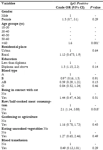
sero-Table 2: Univariate (crude OR) logistic regression analysis of the potential factors associated with T. gondii
IgG seroprevalence among healthy blood donors in Razavi Khorasan Province, Iran
Variables IgG Positive
Crude OR (95% CI) P value
Gender
Male
Female 1.5 (0.7, 3.1) 1 0.29-
Age groups (yr)
18-30 30-40 40-50 50-60 >60
- - - - 1.6
- - - - 0.001*
Residential place
Urban
Rural 1.13 (0.675, 1.9) 1 0.64
Education
Less than diploma
Diploma and above 1.5 (1.15, 2.3) 1 0.14 -
Blood type
A B AB O
1 0.97 (0.16, 1.5) 0.59 (0.29, 1.21) 0.84 (0.52, 1.34)
- 0.91 0.15 0.46
Being in contact with cat
No
Yes 1.44 (0.47, 4.36) 1 0.51 -
Raw/half-cooked meat consump-tion
No Yes
1
2.1 (1.14, 3.88) 0.018- *
Gardening or agriculture
No
Yes 1.16 (0.78, 1.73) 1 0.45 -
Eating uncooked vegetables No
No
Yes 1.27 (0.65, 2.46) 1 0.49 -
Blood transfusion
No 1 -
Yes 0.49 (0.13,1.81) 0.29
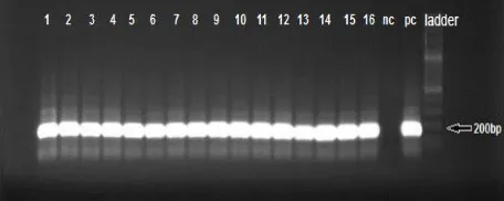
Nested-PCR
To confirm the presence of T. gondii DNA in IgM-positive blood donors, all IgM-positive were analyzed using B1 and RE primers (Fig. 1 and 2). The findings revealed that T. gondii DNA was found in all sixteen IgM-positive samples. For all of samples, fragment of about
Fig. 1: B1- nested PCR analysis of T. gondii DNA (531 bp). Lane 1-16:IgM- positive blood samples; C-: negative control (D.W); C+: positive control (T.gondii RH strain); ntc: negative test control (T.gondii negative sample); (ladder (100 bp)
Fig. 2: RE- nested PCR analysis of T. gondii DNA
(164bp). Lane 1-16:IgM -positive blood samples; nc: negative control (D.W); pc: positive control (T.gondii RH strain); ladder (100 bp)
Discussion
Toxoplasmosis in immunocompromised persons could cause severe manifestations such as central nervous system involvements as encephalitis and some brain disorders. This infection in blood donors potentially indicates a threat of parasite transmission to immuno-compromised or immunosuppressed blood transfusion recipients (5-8). Because of in-progress treatments are not efficient and there is no successful vaccine, it is obligatory to per-form labors for preventing of Toxoplasma transmission to blood recipients for likely in-cidence of reactivated toxoplasmosis specifi-cally in immunodeficient persons. In this in-vestigation, totally 491 blood samples were collected from blood donors of Razavi Khorasan, Iran. Overall, 200 (40.7%) samples were seropositive for anti-T. gondii antibodies;
so that 37.5% and 3.2% were positive for IgG and IgM anti-T.gondii antibodies, respectively.
Many studies have reported similar T. gondii seropositivity in the blood donors of Czech Republic (19), Mexico (22), and southeastern Iran (25, 29, 30); whereas this T. gondii sero-positivity was more than those reported in India (14), northeast of Thailand (23), and Taiwan (17), and some regions of Iran (26, 28, 30). In contrast, T. gondii seropositivity in the blood donors of Razavi Khorasan, Iran was less than the one reported among blood do-nors in central Iran (26), northeast of Brazil (30), north of India (16), and Egypt (15), where the seroprevalence of T. gondii has var-ied from 50% to 75%. This difference in T.
gondii seropositivity among the blood donors
around the world could be associated to some factors such as geographical and environmen-tal factors, sociocultural habits, transmission routes, sample size in the studied population (9-12). Here, consumption of raw/half-cocked meat is the main risk factor for T. gondii sero-positivity, indicating that among the blood donors in this study, the consumption of tis-sue cysts in meat (food-borne transmission) is one of the main routs of infection. Similarly, several studies have reported the food-borne transmission as main infection route in blood donors (26, 34, 35).
By gender, although seroprevalence of
anti-T. gondii antibodies in female donors were
Similar to the other studies we found that T. gondii seropositivity increased with age, indi-cated age because of increased opportunity for exposure; such a finding was in agreement with those observed in other studies (15, 24, 36).
There was no significant association be-tween education, residence, blood group, raw-milk/egg consumption, and blood transfusion, agriculture activity, and contact with cat and seroprevalence of anti-T. gondii antibodies in blood donors.
By Nested-PCR analysis, T. gondii DNA was found in all sixteen IgM-positive samples. The presence of parasitemia in IgM-positive healthy blood donors, whereas ensures the likelihood of transmission of Toxoplasma through blood transfusion. Parasitemia in two (1.9%) of the IgM-positive subjects were demonstrated from blood donors in south-ern Iran (27). Moreover, T. gondii DNA was reported in one (9.0%) of IgM-positive sam-ples healthy blood donors from Kerman Province, southeast of Iran (25). These varia-tions could be related to some factors such as the short duration of parasitemia and the low numbers of trophozoites circulating in periph-eral blood, which caused false negative results in such cases (37).
Conclusion
T. gondii infection was prevalent among
healthy blood donors of Razavi Khorasan Province, in the northeastern of Iran with the overall seroprevalence rate of 40.7% and pres-ence of parasite DNA of T. gondii in IgM-positive samples. Ingestion of undercooked meat is associated with increase of seropositiv-ity in the blood donors in the northeast of Iran. The results can be a warning for blood transfusion organizations in order to pay spe-cial attention to toxoplasmosis among blood donors and design screening programs as pre-ventive affairs for any probable transmission of toxoplasmosis.
Acknowledgements
The authors would like to thank the staff of Razavi Khorasan Blood Transfusion Organi-zation for sample collection.
Conflict of Interests
The authors declare that there is no conflict of interest in this study.
References
1. Hill D, Dubey J. Toxoplasma gondii: transmission, diagnosis and prevention. Clin Microb Infect. 2002; 8(10): 634-640.
2. Robert-Gangneux F, Darde ML. Epidemiology of and diagnostic strategies for toxoplasmosis. Clin Microbiol Rev. 2012; 25: 264–296. 3. Sukthana Y. Toxoplasmosis: beyond animals to
humans. Trends Parasitol. 2006; 22: 137–142. 4. Mahmoudvand H, Ziaali N,Ghazvini H,
Shojaee S, Keshavarz H, Esmaeeilpour K, Sheibani V. Toxoplasma gondii infection promotes neuroinflamation through cytokine networks and induced hyperalgesia in BALB/c mice. Inflamation. 2016; 39(1): 405-412. 5. Derouin F, Pelloux H. Prevention of
toxoplasmosis in transplant patients. Clin Microbiol Infect. 2008; 14: 1089–1101.
6. Biesiada G, Kalinowska-Nowak A, Czepiel J, et al. Toxoplasmosis—epidemiology, clinical manifestation and infection in pregnant women. Przegl Lek. 2006; 63: 97–99.
7. Signorini L, Gulletta M, Coppini D, et al. Fatal disseminated toxoplasmosis during primary HIV infection. Curr HIV Res. 2007; 5: 273– 274
8. Mahmoudvand H , Sheibani B, Keshavarz H, Shojaee S, EsmaeelpourKh, Ziaali N. Acetyl-cholinesterase inhibitor improves learning and memory impairment induced by Toxoplasma gondii infection. Iran J Parasitol. 2016; 11 (2): 177-185.
9. Mahmoudvand H, Ziaali N, Aghaei I, Sheibani V, Shojaee S, Keshavarz H, Shabani M. The possible association between Toxoplasma gondii
disorders in BALB/c mice. Pathogen Glob Health. 2016;109(8):369-76.
10. Siegel SE, Lunde MN, Gelderman AH, et al. Transmission of toxoplasmosis by leukocyte transfusion. Blood. 1971; 37: 388–394.
11. Shulman IA. Parasitic infections and their impact on blood donor selection and testing. Arch Pathol Lab Med. 1994; 118: 366–370. 12. Mahmoudvand H, Sheibani V, Esmaeelpour K
et al. Toxoplasma gondii infection potentiates cognitive impairments of Alzheimer's disease in the BALB/c mice. J Parasitol. 2016; 102(6): 629-635.
13. Mostafavi SN, Jalali Monfared L. Toxoplasmosis epidemiology in Iran: a systematic review. J Isfahan Med Sch. 2010; 30(176): 74-87.
14. Sundar P, Mahadevan A, Jayshree R, et al.
Toxoplasma seroprevalence in healthy voluntary blood donors from urban Karnataka.Indian J Med Res. 2007;126(1): 50-56.
15. Elsheikha HM, Azab MS, Abousamra NK, et al. Seroprevalence of and risk factors for
Toxoplasma gondii antibodies among asymptomatic blood donors in Egypt. Parasitol Res. 2009; 104(6): 1471-1476.
16. Yazar S, Eser B, Yay M. Prevalence of
anti-Toxoplasma gondii antibodies in Turkish blood donors. Ethiop Med J. 2006;44(3): 257-261. 17. Chiang TY, Hsieh HH, Kuo MC, et al.
Seroepidemiology of Toxoplasma gondii infection among healthy blood donors in Taiwan. PloS One. 2012; 7(10): :e48139.
18. Elhence P, Agarwal P, Prasad KN, et al. Seroprevalence of Toxoplasma gondii antibodies in North Indian blood donors: Implications for transfusion transmissible toxoplasmosis. Trans Apher Sci. 2010; 43(1): 37-40.
19. Svobodova V, Literak I. Prevalence of IgM and IgG antibodies to Toxoplasma gondii in blood donors in Czech Republic. Eur J Epidemiol. 1998; 14 : 803-805.
20. Makki SM, Abdel-Tawab AH. Anti-Toxoplasma gondii antibodies among volunteer blood donors in eastern Saudi Arabia. J Egypt Soc Parasitol. 2010; 40(2): 401-12.
21. Yazar S, Eser B, Yay M. Prevalence of
anti-Toxoplasma gondii antibodies in Turkish blood donors. Ethiop Med J.2006; 44(3): 257-261. 22. Galvan Ramirez ML, Covarrubias X,
Rodriguez R, et al. Toxoplasma gondii antibodies
in Mexican blood donors. Transfusion. 2005; 45: 281–282.
23. Pinlaor S, Ieamviteevanich K, Pinlaor P, et al. Seroprevalence of specific total immunoglobulin (Ig), IgG and IgM antibodies to Toxoplasma gondii in blood donors from Loei province, Northeast Thailand. Southeast Asian J Trop MedPublic Health. 2000; 31: 123-127. 24. Alvarado-Esquivel C, Mercado-Suarez MF,
Rodríguez-Briones A, et al. Seroepidemiology of infection with Toxoplasma gondii in healthy blood donors of Durango, Mexico. BMC Infect Dis. 2007; 7:75-81.
25. Mahmoudvand H, Saedi Dezaki E, Soleimani S, Baneshi MR, Kheirandish F, Ezatpour B, Zia-ali N. Seroprevalence and risk factors of
Toxoplasma gondii infection among healthy blood donors in southeast of Iran. Parasite Immunol. 2015, 37(7), 362-367.
26. Ormazdi H, Sanikhani N, Hadighi R, et al. Investigation of antibodies (IgG and IgM) against Toxoplasma gondii in blood donors referred to Tehran blood transfusion organization by ELISA. Urmia Med J. 2010; 21 (2): 212-216.
27. Sarkari B, Shafiei R, Zare M, Sohrabpour S, Kasraian L. Seroprevalence and molecular diagnosis of Toxoplasma gondii infection among blood donors in southern Iran. J Infect Dev Ctries. 2014;15; 8(4):543-7.
28. Ferdowsi S, Farsi L, Tajalli SM, Soltani H. Seroprevalence Anti-Toxoplasma gondii
antibodies and anti- Epstein-Barr virus (EBV) antibody among volunteer blood donors referred Gonabad blood transfusion. J Zabol Uni Med Scie Health. 2013; 5( 2): 60– 9. 29. Modrek MJ, Mousavi M, Saravani R.
Toxoplasma gondii seroprevalence among blood donors in Zahedan, southeastern Iran. Int J Infect. 2014; 2(1): e21111.
30. Zainodini N, Zare-Bidaki M, Abdollahi S, Afrooz M, Ziaali N, Ebrahimian M, Kazemi Arababadi M. Molecular and serological detection of acute and latent toxoplasmosis using Real-Time PCR and ELISA techniques in blood donors of Rafsanjan city, Iran. Iran J Parasitol. 2014; 9(3): 336– 41.
fragment length polymorphism analysis at the B1Gene. J Clin Microbial. 2001;398–400. 32. Chiabchalard R, Wiengcharoen JT, Sukthana
Y. Sensitivity and specificity of PCR for the detection of Toxoplasma gondii and added to laboratory samples. Southeast Asian J Trop Med Public Health 2005; 36: 408–411.
33. Kong QM, Lu SH, Tong QB et al. Loop-mediated isothermal amplification (LAMP): early detection of Toxoplasma gondii infection in mice. Parasites Vectors. 2012; 5. 2:1-7.
34. Baril L, Ancelle T, Goulet V, et al. Risk factors for Toxoplasma infection in pregnancy: a case-control study in France. Scand J Infect Dis. 1999; 31: 305–309.
35. Jones JL, Dargelas V, Roberts J, et al. Risk factors for Toxoplasma gondii infection in the United States. Clin Infect Dis. 2009; 49: 878– 84.
36. Coelho RA, Kobayashi M, Carvalho LB jr. Prevalence of IgG antibodies specific to
Toxoplasma gondii among blood donors in Recife, Northeast Brazil. Rev Inst Med Trop Sao Paulo. 2003; 45: 229-231.
37. Nimri L, Pelloux H, Elkhatib L. Detection of