Makuch Marcelina, Szmygin-Milanowska Katarzyna, Michnar Marek, Palonka Michał, Krusiński Adam, Grzywa-Celińska Anna. Tracheopbronchopatia osteochondroplastica – a case presentation. Journal of Education, Health and Sport. 2018;8(11):222-228. eISNN 2391-8306. DOI http://dx.doi.org/10.5281/zenodo.1482819
http://ojs.ukw.edu.pl/index.phpohs/article/view/6284
The journal has had 7 points in Ministry of Science and Higher Education parametric evaluation. Part B item 1223 (26/01/2017). 1223 Journal of Education, Health and Sport eISSN 2391-8306 7
© The Authors 2018;
This article is published with open access at Licensee Open Journal Systems of Kazimierz Wielki University in Bydgoszcz, Poland
Open Access. This article is distributed under the terms of the Creative Commons Attribution Noncommercial License which permits any noncommercial use, distribution, and reproduction in any medium, provided the original author (s) and source are credited. This is an open access article licensed under the terms of the Creative Commons Attribution Non commercial license Share alike.
(http://creativecommons.org/licenses/by-nc-sa/4.0/) which permits unrestricted, non commercial use, distribution and reproduction in any medium, provided the work is properly cited. The authors declare that there is no conflict of interests regarding the publication of this paper.
Received: 25.10.2018. Revised: 25.10.2018. Accepted: 11.11.2018.
Tracheopbronchopatia osteochondroplastica – a case presentation
Marcelina Makuch
Chair and Department of Pneumonology, Oncology and Allergology, Medical University of Lublin, Poland
Katarzyna Szmygin-Milanowska
Chair and Department of Pneumonology, Oncology and Allergology, Medical University of Lublin, Poland
Marek Michnar
Chair and Department of Pneumonology, Oncology and Allergology, Medical University of Lublin, Poland
Michał Palonka
Chair and Department of Pneumonology, Oncology and Allergology, Medical University of Lublin, Poland
Adam Krusiński
Chair and Department of Pneumonology, Oncology and Allergology, Medical University of Lublin, Poland
Anna Grzywa-Celińska
Abstract
Narrowing of the lower respiratory tract is a rare pathology. It may be associated with pathologies of the primary respiratory system or connective tissue diseases (systemic scleroderma, granulomatosis with polyangiitis, relapsing polychondritis). It can lead to non-specific clinical symptoms. We present an interesting case of a 52-year old patient with a history of several months’ dyspnea. Imaging tests revealed significant stenosis of the trachea and the left main bronchus. Tracheobronchopatia osteochondroplastica was diagnosed. Moreover, we carry out the differential diagnosis of the above pathology.
Key words: airway diseases, tracheobronchopatia osteochondroplastica, tracheal stenosis
CASE REPORT
A fifty-two-year old woman was admitted to the Department of Pulmonology because of dyspnea on exertion and rest. She had a six months’ history of hoarseness, recurrent infections of the upper and lower respiratory tract, chest pain and joint pain. In addition, she suffered from skin psoriasis and psoriatic arthritis. 14 months ago computed tomography of the chest showed no significant pathologies.
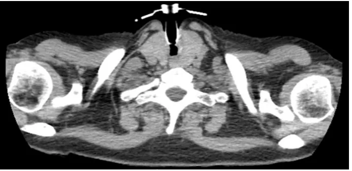
Fig. 1 Imaging findings revealed by chest computed tomography: generalised thickening of the cartilagous portion of the trachea
Bronchofibroscopy revealed thickened wall of the trachea along the entire length, without focal changes of mucosa in the upper 1/3 narrowed tracheal lumen. Stenosis of the left main bronchus was shutting the left upper lobe bronchus. Bronchial lavage was negative for acid fast bacilli or any other pathogen. Histopathological examination from the bronchial biopsy was unremarkable, including fragments of the mucous membrane covered with snapshot epithelium, quite intense inflammation in the stroma, inflammatory infiltrates around the bronchial glands, erythrocytes and fibrinous effusion. We recommended inhaled bronchodilators (ipratropium), oral steroids (methylprednisolone) and antifungal medications (nystatin) in outpatient treatment
The patient was admitted to the Department of Otolaryngology a month later and classified to tracheal biopsy. It brought complications - inflammatory spastic reaction requiring urgent tracheotomy. The collected sample gave the following histological description: inflammatory infiltrates of lymphocytes and plasma cells in the bronchial mucosa with the features of stromal fibrosis, hyperplasia in the tracheal scar tissue and metaplasia.
Histopathological slides have been consulted with the Institute of Tuberculosis and Lung Diseases in Warsaw - the microscopic features were consistent with tracheobronchopatia osteochondroplastica, but also systemic disease or toxicity of the drugs could not be excluded. Basing on the above consultation, the patient was diagnosed with tracheobronchopatia osteochondroplastica.
Two years later because of persisting tracheal stenosis, silicone stent was implanted into the trachea. In the chest CT scans - no progression of airway narrowing, no interstitial changes (fig. 2). The Institute of Tuberculosis and Lung Diseases offered establishment of a T prosthesis in the tracheal stenosis, but the patient did not agree to that procedure. Because of regression of changes in the imaging studies and the lack of patient’s consent we resigned form the mechanical removal of stenosis. Decision was made to continue conservative treatment.
Fig. 2 The control chest computed tomography: silicone stent implanted into the trachea
DISCUSSION
Apart from tracheobronchopatia osteochondroplastica the differential diagnosis of tracheal stenosis in our patient included:
relapsing polychondritis - a rare systemic connective tissue disease of unknown etiology. Inflammatory infiltration destroys the cartilage tissue, especially cartilage related with the ears, nose, trachea and bronchi. Disease is usually associated with other connective tissue diseases (rheumatoid arthritis, ankylosing spondylitis, systemic vasculitis). According to MacAdam criteria cartilage inflammation of the respiratory tract is observed, which can lead to severe pneumonia, respiratory failure and even death.
amyloidosis - amyloid deposition in the bronchial tree causes disturbance of flow through the bronchi. We observe plurality of gray-white discs located submucosally, mucosal thickening and infiltration of tumor-like changes in bronchoscopy. Amyloidosis does not spare the posterior wall of the trachea, which sets it apart from tracheobronchopatia osteochondroplastica. In our patient we might exclude amyloidosis also due to the lack of a distinctive coloring of the histological preparation – no amyloid deposits were identified by histochemical staining (Congo red, Thioflavin T).
Immunological studies have revealed high concentration of cANCA, typical for this disease. But we observed no changes in paranasal nose, lung and kidney parenchyma. scleroderma - characterized by progressive fibrosis of the skin and internal organs. In the
described case, it was excluded on the basis of lack of Raynaud's symptom. We have not observed the characteristic hardening of the skin, esophagus and lung parenchyma.
calcification in the respiratory tract related to age - usually the study of the mucosa in bronchoscopy appears as physiological.
We also excluded malignant transformation.
Tracheobronchopatia osteochondroplastica (TBO) is a rare disease of unknown etiology
characterized by the presence of hard, calcified nodules in the airways. Changes locate on the walls of the trachea and main bronchi, causing their narrowing. The most frequent clinical signs of TBO are chronic cough, extertional dyspnea, bleeding, and even airway obstruction. The therapy involves the use of bronchodilators and mechanical removal of lesions during bronchofiberoscopy [1].
For the first time, changes typical for TBO were described in the first half of the nineteenth century by three independent scientists: Rokitansky, Luschka and Wilks. Then the diagnosis was limited to post-mortem examination, until 1970 when TBO was identified during bronchoscopy [2, 3, 4]. Nowadays, because of the increasingly widespread use of bronchofiberoscopy techniques in clinical practice, the disease usually is diagnosed intravitally [5].
TBO occurs usually in over 50-year-old patients, but there are reports of the disease occuring in younger patients, even 9-year-old girls. TBO etiology has not been known so far. Some authors proposed several theories describing the formation of the lesions in the trachea and bronchi. Virchow suggested echondrosis concept, according to which echondromas are the initial lesions and eventually undergo ossification and take the form of a bony spur. Aschoff came to the conclusion that the lesions arise from the cartilaginous and osseous metaplasia of undifferentiated connective tissue cells. In contrast, Dalgaard suggested that changes typical for TBO occur as a result of the conversion of elastic fibers of the fiber membrane into the trachea cartilage which undergoes calcification [6].
Morphological and biochemical study of peripheral blood are not useful for the diagnosis of TBO. Pulmonary function tests may be normal. Only a small percentage of patients presents features of airway obstruction, which correlates with disease progression.
Radiological picture of TBO is multifaceted. Initially correct, but the advanced form of the disease (as in the case of our patient) is visible as significant stenosis of the trachea. The chest CT may show as follows: thickening of the tracheobronchial tree and a number of calcified nodules. Changes can extend anywhere in the airways, even to the peripheral bronchi, but usually they are present in 2/3 of the distal portions of the trachea and proximal bronchi. Due to the fact that the nodules are formed from cartilage, the rear wall of the trachea (membranous) remains free of the disesae, which distinguishes TBO from amyloidosis or granulomatosis with polyangitis [8].
The most important test to confirm the diagnosis of TBO is bronchofiberoscopy. The most representative endoscopic image is the presence of hard, nodular formations associated with the cartilages or between cartilage. These nodules are hard on touch and give gritty sensation while passing the scope through the lumen. Nodules can imitate the image of crazy-paving pattern. TBO is localised usually in anterior and posterior walls of the airways. In the early stages of the disease desribed changes appear as a small eminence, then hardening nodules tending to confluence. As the disease progresses the airway walls stiffen and further narrowing is observed [9].
It is believed that the appearance of the airways in the bronchofiberoscopy is so distinctive that the histopathological examination is not required. Therefore, the biopsy of the nodules is not done routinely - it is technically difficult because of their hard consistency. The histopathological study that allows to rule out other diseases (such as cancer or amyloidosis) points out lumps of submucosal cartilage. In the nodules might be islands of bone marrow, even with hematopoiesis, and inflammatory infiltrates suggesting chronic inflammation [10]. TBO prognosis is favorable in most cases. The disease is mild, so the treatment is used only in case of persistent symptoms. Treatment consists of humidifying the respiratory tract, avoidance of disease triggers and effective treatment of respiratory tract infections. The administration of a bronchodilator helps for some people (as in our patient). Only when substantial narrowing of the airways is carried out, trials of mechanical removal of the lesions are done during laryngoscopy or bronchoscopy. Other methods include, among others, laser ablation, implanting the stent at the site of stenosis, cryotherapy or radiation theraphy. The described methods usually alleviate the symptoms and improve the results of pulmonary function tests, but complications may also happen e.g. secondary airflow obstruction, as those observed in our patient during laryngological surgery. In extreme cases, a surgical treatment is needed: resection of the larynx, trachea segmental resection or even removal of the diseased portions of the lung parenchyma [11, 12].
REFERENCES:
2. Chroneou A Zias N, Gonzalez AV Beamis JF. Jr. Tracheobronchopathia osteochondroplastica. An entity underrecognized? Monaldi Arch Chest Dis 2008; 69: 65-69.
3. Prakash UB. Tracheobronchopathia osteochondroplastica. Semin Respir Crit Care Med 2002; 23: 167-175.
4. CJ Martin. Tracheobronchopathia osteochondroplastica. Arch Otolaryngol 1974; 100: 290-293
5. Porzezińska M.A Janowicz.Janowiak P.Cynowska B.A Sternau.R PęksaEt al. Tracheobronchopathia osteochondroplastica-case report and literature review.Pneumonol Alergol Pol. 2015; 83 (2): 135-9.
6. S. RizzoTracheobronchopathia osteochondroplastica associated with calcification of falx cerebri and rhinobronchial aith nasal polyposis syndrome. J. Bronchol. 1998, 5, 128-131
7. Prakash UB. Tracheobronchopathia osteochondroplastica. Semin Respir Crit Care Med 2002; 23: 167-175.
8. Nieves-Nieves J, Gonzalez-Viridiana S, Fernandez-Gonzalez R, Fernandez-Medero R. Tracheobronchopatia osteochondroplastica: an underdiagnosed central airway disease. Int J Med Med Sci 2012; 4: 211-213
9. Pirożyński M. bronchofiberoscopy Alfa Medica Press, Warsaw; , 2011: 86-87
10. Luo S, Wu L, Zhou J, Xu S, Yang Q, Li Y, H Shen, et al.Tracheobronchopathia osteochondroplastica: two cases and a review of the literature.International Journal of Clinical and Experimental Pathology2015; 8 (7):8585-8590.
11. Doshi H, R Thankachen, Philip MA, Kurien S, V Shukla, Korula RJ. Tracheobronchopathia osteochondroplastica presenting as an isolated nodule in the right upper lobe bronchus with upper lobe collapse. J Thorac Cardiovasc Surg 2005; 130: 901-902.