Engida Abebe, Minewor Abdlhadi, Ayelign Tsehay, Befekadu Lemmu, Mahteme Bekele. Ethiop Med J, 2019, Vol. 57, No. 2
ORIGINAL ARTICLE
ANALYSIS OF THE ETIOLOGICAL SPECTRUM AND CLINICAL PROFILE OF
EXTRA- HEPATIC BILIARY TREE OBSTRUCTION AT A TERTIARY HOSPITAL
IN ADDIS ABABA
Engida Abebe, MD1*, Minewor Abdlhadi, MD1, Ayelign Tsehay, MD1 , Befekadu Lemmu, MD1, Mahteme Bekele , MD1
ABSTRACT
Background: Extra-hepatic biliary tree obstruction is a common problem in surgical gastroenterological practice.
The condition can be caused by the obstruction of the bile duct with gallstones, strictures and hepato-biliary malignancy.
Objective: To assess and report the etiological spectrum, clinical presentation, treatment and outcome of patients
with extra-hepatic biliary tree obstruction.
Methods: A retrospectives analysis of all patients admitted with a diagnosis of extra-hepatic biliary tree
obstruc-tion and operated at St. Paul Hospital Millennium Medical College from January 1,2012 to December 30, 2015 was performed. The data were collected from patient medical records identified using the operation theater log-book. A pretested, structured data extraction format was used to collect the data, which were entered onto and analyzed using SPSS version 20.
Results: A total of 116 patients, 62 (53.4%) females, were operated for extra-hepatic biliary tree obstruction. Their
age ranged from 21 to 80 years with a mean (±SD) of 40.3(11.2) years. Abdominal pain seen in 107 (92.2%) of the patients and jaundice in 98 (84.5%) were the two most common presenting complaints. Abdominal ultrasound was the main imaging modality used to identify the etiology in 88.8% of the patients. Benign conditions accounted for 79 (68.1% of the underlying etiology, common bile duct stone being the most common, 70 (60.3%). Pancreatic head tumor was the commonest malignant cause, 19 (51.3%), followed by cholangiocarcinoma, 15 (40.5%). Cholidochoduodenostomy was performed for 50 (43.1%) of the patients and cholecystojejunostomy with Braun’s anastomosis for 22 (19%).
Conclusion: Abdominal pain and jaundice were the main presenting symptoms of extra-hepatic biliary tree
ob-struction, which was mainly caused by common bile duct stone obstruction and pancreatic head tumor. Ultra-sound was the main modality for diagnosing the underlying causes and bypass operation was commonly done
Key word: Common bile duct, Stone, Extra-hepatic biliary tree obstruction, Jaundice
1 St. Paul’s Hospital Millennium Medical College
* Corresponding Author, e-mail: engidaabebe@yahoo.com
INTRODUCTION
Extra Hepatic Biliary Obstruction (EHBO) is a con-dition in which bile exit/flow is obstructed starting from hepatic ducts up to the second part of the duo-denum and it may result in obstructive jaundice (OJ) (1). Jaundice refers to yellowish discoloration of the skin, sclera and mucous membrane due to an in-creased level of bilirubin concentration in body fluid. It is detectable when plasma bilirubin exceeds 50 mmol/L or 2-2.5 mg/dl. Jaundice is called obstructive when the cause is blockage of bile flow from the liver to the intestine (1).
EHBO is one of the most common hepatobiliary sur-gical conditions managed by general surgeons and hepatobiliary surgeons (2-4). The condition can be caused by either benign or malignant conditions.
Benign pathologies which occur relatively more in younger patients includes biliary stones (choledocolithiasis), benign biliary strictures (iatrogenic or sclerosing), parasite infestations (ascaris, liver flukes and hydatid cysts), etc. Malig-nant causes of EHBO includes pancreatic head tu-mors, tumors of the biliary tree, tumors of the sec-ond part of the duodenum, Ampula of Vater tumors and others (2,5). Pancreatic head tumor is the most common cause of malignant EHBO (1,3,4).
Differentiating causes of EHBO/OJ needs laboratory and imaging studies. Laboratory investigations such as liver enzyme, serum bilirubin level and uribilino-gen level help in defining the type of jaundice. A significant rise in Alkaline phosphatase (ALP) com-pared to a rise in Alanine Aminotransferase (ALT) or Aspartate Aminotransferase (AST) is common char-acteristics of OJ due to EHBO (4). Definitive diagno-sis is usually reached by imaging studies like ab-dominal ultrasound (AUS), Endoscopic Ultrasound (EUS), abdominal computed tomography (CT), Ma-gentic Resonant Cholangiopancreatoprahpy (MRCP), Endoscopic Retergrade Cholangiopancreatography (ERCP) and Percuatneos Cholangiography (PTC) (7,8). AUS is considered as the first line investiga-tion while MRCP the gold standard (9). CT is an important evaluation and staging study in patients with malignant obstruction (1).
The type of treatment for EHBO can be minimally invasive procedures or open surgery depending on the diagnosis and the hospital setting (10). In areas where the technology and the skill is available stones in the Hepatic Duct (HD) or Common Bile Duct (CBD) are treated by ERCP (8). In resource limited countries like Ethiopia open surgery is still the pre-ferred option of treatment for both benign and nant conditions. Because most patients with malig-nant EHBO present late, resection surgery is done only for few percent of the patients. The objective of this study was to determine the etiologic spectrum, clinical presentation, treatment and outcome of pa-tients with extra hepatic biliary tree obstruction who were admitted to St. Paul Hospital Millennium Medi-cal College (SPHMMC), surgiMedi-cal wards from Janu-ary 1, 2012 - December 30, 2015.
PATIENTS AND METHODS
A cross-sectional descriptive study was conducted at SPHMMC department of surgery, Addis Ababa, Ethiopia. SPHMMC is a tertiary level teaching hos-pital engaged in both undergraduate and postgraduate programs. During the study period department had 10 general surgeons, 04 operation theaters and 110 surgical beds. The study included all patients admit-ted and operaadmit-ted from January 1, 2012 to December 31, 2015 at the department of surgery with a diagno-sis of EHBO. EHBO was defined as obstruction to the bile exit/flow starting from hepatic ducts up to the second part of duodenum. The hospital had no ERCP or PTC service during the study period. Pa-tients were identified from operation theater log books and ward admission and discharge book. Pa-tients admitted but not operated and those with in-complete medical records were excluded from the study.
Data was collected from individual patient’s medi-cal records by trained second year surgimedi-cal residents using a pretested structured data collection format. Data collection was supervised by the investigators. Collected data was checked for completeness, cleaned, coded, entered and analyzed with SPSS version 20. Patient socio-demographic characteris-tics, presenting complaint, laboratory and imaging finding, intra-operative findings, type of surgery done and post operative complications were col-lected. A rise in ALT, AST or ALP 2-3 times above the normal values was considered significant. Chi square test was used to test the presence of associa-tion and P value <0.05 was considered as a statisti-cally significant. Ethical clearance was obtained from SPHMMC Institution Review Board (IRB).
RESULTS
Totally 120 patients were operated for EHBO in the study period and charts of 116 (96.7%) patients with complete medical records used for this study. There was a slight female preponderance, (53.3%) with male to female ration of 1:1.14. Age of patients ranged from 21 to 80 years with a mean (±SD) of 40. 3 years(11.2). The majority of patients, 90 (77.6%), were above 40 years of age. Among pa-tients under 49 years of age females (35) out num-ber males (14 ) while in those above 50 years, male (40) were more commonly affected than females (27) (Figure 1). The difference in age and sex dis-tribution of EHBO was found to be statistically sig-nificant (p=0.013, p=0.000, respectively).
Abdominal pain was the most common symptom, 107 (92.2%), followed by history of yellowish dis-coloration of the eye, 98 (84.5%), and both abdomi-nal pain and yellowish discoloration of the eye, 89 (76.7%). Abdominal pain associated with jaundice (painful jaundice) was less prevalent presentation in patients with malignant EHBO, 27 (72.9%), com-pared to benign EHBO, 62 (78.4%). On physical examination 95 (81.9%), 27 (23.3%) and 25 (21.6%) patients found to have icteric sclera, right upper quadrant abdominal tenderness and hepa-tomegaly/palpable gall bladder respectively. Table 1 Sixteen (13.8%) patients had co-morbid illness: hypertension, DM or both.
Significant rise in ALT& AST level was seen in 61 (52.6 %) and 63 (54.3%) patients respectively. Sig-nificant ALP rise was seen in 72(62.7%) of the pa-tients. Serum bilirubin was determined in 94 (81%) patients, total bilirubin raised above normal in 75 (79.8%) patients while direct bilirubin was raised in 80 (85.1%). The rise in both total and direct bilirubin level was significantly higher in malignant EHBO compared to benign causes (P=0.000)
Figure 1: Age and sex distribution patients with extra hepatic biliary tree Obstruction at SPHMMC, Addis Ababa, Ethiopia, 2015.
Clinical manifestation Number of patients Frequency (%)
Abdominal pain 107 92.2
Yellowish discoloration of the eye 98 84.8
Icteric sclera (on P/E) 95 81.9
Abdominal pain and yellowish
discol-oration of the eye 89 76.7
Dark color urine /pale color stool 59 50.9
Abdominal Tenderness 27 23.3
Hepatomegaly/palpable GB 25 21.6
Pruritis 23 19.8
Weight loss 17 14.7
Abdominal ultrasound was the main radiologic/ imaging study, which was done for all patients. US missed diagnosis in 10 (8.6%) patients. Computerized tomography was done in 33(89.2%) of the patients with malignant EHBO but missed diagnosis of 7 (21.2%) patients.
CBD stone was the most commonly miss labeled pathology 7(6.1%). Distal cholangiocarcinoma found to be more prone for under diagnosis than any other cause of EHBO .
Table 2: The degree of serum bilirubin increment among benign and malignant Causes
of EHBO at SPHMMC, Addis Ababa, Ethiopia, 2015.
Serum bilirubin benign Pathology malignant Total P-value
Total serum
bilirubin <1mg/dl 19 0 19
0.000
1-5 20 5 25
5-10 14 10 24
10-15 6 8 14
>15 2 10 12
Total 61 33 94
Direct bilirubin
<0.3mg/dl 14 0 14
0.000
0.3-1.8 12 1 13
1.8-3.3 14 1 15
3.3-4.8 9 10 19
>4.8 12 21 33
Total 61 33 94
Table 1: Clinical finding of patients with extra hepatic biliary obstruction at
The etiology of EHBO was benign in 79 (68.1%) cases. Stone diseases of the biliary tree (choledocholithiasis) were the leading cause, 70 (60.3%), followed by pancreatic head tumor 19 (16.4%). Pancreatic head tumor made 51.4% of all causes of malignant EHBO and 62.3 % of peri-ampularry tumors Table 3.
In benign EHBO Females were almost two times, 51 (64.5%), more commonly affected than males (28 (35.4%)) while in malignant conditions males were more commonly affected, 26 (70%), verses 11(30%).
The sex difference in the causes of EHBO was sta-tistically significant (P =0.000) Table 4. Among patients with malignant EHBO, 81% of them were older than 50 years of age. The difference in occur-rence of malignant EHBO in different age group was statistically significant (P < 0.013) (Table 4).
Table 3. Frequency of Benign and malignant causes of EHBO at St. Paul hospital,
Addis Ababa, Ethiopia, 2015.
Pathologic entity Diagnosis Number % (of total)
Benign CBD/CHD stone 70 60.3
Stricture 3 2.6
Choledochal cyst 1 1.4
Hydatid cyst ruptured into the biliary
tree 1 1.4
External compression by Hydatid cyst 1 1.4
Other 3 2.6
Total 79 68.1
Malignant Pancreatic head tumor (PHT) 19 16.4
Distal cholangiocarcinoma 9 7.75
Proximal cholangiocarcinoma 3 2.58
Gall bladder Ca 3 2.58
Ampula of vater tumor 2 1.7
Duodenal cancer 1 0.8
Total 37 31.9
Table 4: Age and sex distribution of benign Vs malignant causes of extra-heaptic biliary
obstruction at St. Paul’s hospital, Addis Ababa, Ethiopia, 2015.
Age group
Benign Malignant Total P value
No. % No. % No. %
20-29 8 10.1 2 5.4 10 8.6
0.013
30-39 16 20.3 - - 16 13.8
40-49 18 22.8 5 13.5 23 19.8
50-59 14 17.7 12 32.4 26 22.4
60-69 14 17.7 11 29.7 25 21.6
>70 9 11.4 7 18.9 16 13.8
Total 79 100 37 100 116 100
Sex Male Female 28 51 35.4 64.6 26 11 70.3 29.7 54 62 46.6 53.4 0.000
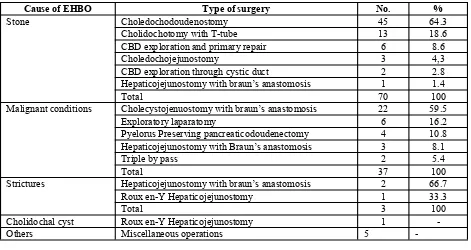
The types of surgery performed according to the cause of EHBO are shown in Table 5. For benign conditions the most common intervention was dochodoudenostomy, 45 (64.3%), followed by Choli-dochotomy with T-tube insertion, 13(18.6%). Pa-tients with stricture were managed with Hepaticoje-junostomy. In Malignant EHBO, the most common surgery was Cholecystojujunostomy with Braun’s anastomosis. Pyelorus preserving pancreaticodouden -ectomy was done only for 4 (12.9 %) of the patients who can be managed by that surgery. A good number of patients had only exploration Table 5.
There were 32 complications on 18 (15.5%) pa-tients. Surgical site infection was the most common complication, 16 (13.4%), followed by pulmonary infections 5(4.3%). Ascitis leak (2.6%) and wound dehiscence (2.6%) were seen in malignant condi-tions only. None of the patients developed anasto-motic leak. Overall complication rate was higher in patients with malignancy than benign conditions (21.6% Vs 12.6%). There was no death among pa-tients with benign causes but three (8.1%) papa-tients with malignant EHBO died.
Table 5: The type operation done for patients with extra-hepatic biliary obstruction at St. Paul Hospital, Addis
Ababa, Ethiopia, 2015.
Cause of EHBO Type of surgery No. %
Stone Choledochodoudenostomy 45 64.3
Cholidochotomy with T-tube 13 18.6
CBD exploration and primary repair 6 8.6
Choledochojejunostomy 3 4,3
CBD exploration through cystic duct 2 2.8
Hepaticojejunostomy with braun’s anastomosis 1 1.4
Total 70 100
Malignant conditions Cholecystojenuostomy with braun’s anastomosis 22 59.5
Exploratory laparatomy 6 16.2
Pyelorus Preserving pancreaticodoudenectomy 4 10.8 Hepaticojejunostomy with Braun’s anastomosis 3 8.1
Triple by pass 2 5.4
Total 37 100
Strictures Hepaticojejunostomy with braun’s anastomosis 2 66.7
Roux en-Y Hepaticojejunostomy 1 33.3
Total 3 100
Cholidochal cyst Roux en-Y Hepaticojejunostomy 1 -
Others Miscellaneous operations 5 -
DISCUSSION
The finding of this study showed females and males to be affected almost equally except when age cate-gory is considered where relatively young females are more commonly affected by benign causes. Khurram and etals also showed benign conditions to occur at younger age, mean age of 42 years (12). Older age groups are affected more by malignant causes of EHBO and are mostly males. The occur-rence of malignancy at older age is a well established fact (1).
Regarding clinical presentation most of the findings of our study are consistent with the literatures report where abdominal pain and yellowish discoloration of the eye top the list in the table (2-5).
Regarding live enzymes the finding in our study, lower rate of significant rise in liver enzyme and bilirubin levels, was due to a higher rate of choledo-cholithiasis, where obstruction is not always com-plete and persistent. Though the rise in liver enzyme level is lower, the rate of ALP is higher than that of ALT/AST which is in line with what the literature reported (4,11). Another important finding of this study was the degree of elevation of serum bilirubin (both total and direct), which was significantly dif-ferent between benign and malignant pathologies. Malignant causes of EHBO had higher values of se-rum bilirubin because they cause persistent and pro-gressive jaundice due to complete obstruction (2-5). Though MRCP is the gold standard imaging study in biliary obstruction, due to unavailability of the tech-nology in the hospital and cost at private settings it is done only in less than 7% of the patients (19). AUS was not only the first line but also the only imaging modality done especially when malignancy was not suspected. The result of AUS was reasonably good as seen in the fact that it missed diagnosed only in 12% of our patients. In developing nations like Ethiopia, given the limited resource, late presentation of pa-tients which make clinical evaluation and judgment less difficult and cancer is a less likely diagnosis, AUS alone can be used to decided diagnosis and management. Mahteme, et al. showed AUS to be diagnostic in 97% of their patients (16). Admassie D, et al. showed AUS to have reasonably good sensitiv-ity and specificsensitiv-ity in diagnosing choledochochole-lithiasis (sensitivity of 90% and specificity of 79%) and pancreatic head tumor(sensitivity of 50% and specificity of (90%) (19,20). A study done in Tanza-nia showed abdominal ultrasound was the only diag-nostic imaging which revealed CBD stones and ab-dominal masses in 58.1% and 72.4% of the cases, respectively (5). A relatively better result was seen in Akhtar et al study where abdominal ultrasound was diagnostic in 85% of the cases (18,19).
Abdominal CT was requested only in patients where cancer was suspected based on clinical and AUS findings. The overall CT request rate is comparable to study in Nigeria where CT scan was requested in 27.5% of the cases (20) probably due to the machine capacity and inexperience of the reporting radiolo-gists the diagnostic accuracy of CT in our patients was significantly lower than reports from the devel-oped world (19).
Biliary stones and pancreatic head tumors as most common cause of EHBO were shown in our study and other literatures from Ethiopia and elsewhere (13,16,17). Abutalib found an almost similar find-ing, CBD stones accounting for 57% while PHT 25% of their cases.
The result of Talib from Bagdad was also similar, stones and pancreatic head tumors accounting for 48% and 16% of causes of EHBO respectively (11). Textbook findings are also similar though pancre-atic head tumor account for 80% of malignant EHBO (1, 16). Malignant obstruction occurring in older male Ethiopians was also shown by Mahteme, et al (16). Most cases of choledocholithiasis are secondary to the passage of gallstones from the gall-bladder into the CBD (16). Stones as a cause of EHBO were seen more often in females and younger patients probably due to the fact that fe-males are affected more commonly by biliary stones and a relatively younger population with life expec-tancy less than 60 years (21,22).
Though ERCP is the standard of care for patients for CH or CBD stones, the service was not available in the hospital. All the patients had open surgery. Cholidochotomy followed by biliary-enteric by pass - mostly choledochodoudenostomy (CDS) - the main form of surgery. CDS is a preferred surgical option in patients whose CBD is dilated to at least 1.2cm (23, 24). Anastomotic leak is a potentially serious complication of CDS, but none of the pa-tients developed that. In setting where T-tubes are scarce, CBD is reasonably dilated and the surgeon is trained, CDS should be the preferred surgical treat-ment for CBD stones.
Like in the literatures, in our patients malignant conditions which caused EHBO were advanced at presentation as revealed by less than 15% PPPD rate (1-5,16). Because we didn’t have minimally invasive means to relive EHBO all patients ended up in open surgery. Though hepaticojejunostomy is the one (probably better) option of palliation of ob-struction, cholecystojejunostomy with Braun’s anas-tomosis was the preferred surgery may be it is tech-nically easier (11) . The rate of post-op complica-tions was higher in malignant condicomplica-tions most likely related to the age of the patients, the nature and stage of the disease at presentation.
Conclusion
Limitation of the study
Since the study was done on review of documented case cards and records, the usual problems of record review may not be abolished.
ACKNOWLWDGMENT
We would like to thank SPHMMC research direc-torate for assisting us in the development of this paper. We extend our appreciation also to the hospi-tal record office and all who contributed in their own ways.
REFERENCES
1. Souba Wiley W, Fink Mitchell P, Jurkovich Gregory J, Kaiser Larry R, Pearce William H, Pemberton John H, Soper Nathaniel J.ACS surgery :Principles and Practice. 6th Ed. WebMD Inc. (Professional Publishing) 2007. 2. Ali Nayyef Assi ,Alaa Jamel Hassan, Kamal Naeem Ali. The Etiological Spectrum of Obstructive Jaundice &
Role of ERCP InThi-Qar Governorate. Iosr Journal of Pharmacy. 2013; 3(3) :26-30.
3. Tariq Wahab Khanzada, AbdulSamad, WaseemMemon, Basant Kumar. Etiological spectrum of obstructive jaundice in Isra University Hospital, Hyderabad, Pakistan. JPMI. 2008;22(02):157-160.
4. Abutalib Bader Abdullah & Zeki .A. Al-Faddagh obstructive jaundice in Basrah, Iraq. Bas J Surg .2011;1:45-57.
5. P.L. chalya, E.S. Kanumba, and M. Mchembe. Etiological spectrum and treatment outcome of Obstructive jaundice at a University teaching Hospital in northwestern Tanzania: A diagnostic and therapeutic challenges. BMC Research Notes. 2011; 4:147.
Doi:10.1186/1756-0500-4-147
6. J Sun, G Liu, Y Yuan et al. Operable severe obstructive jaundice: How should we use preoperative biliary drainage? S Afr J Surg. 2013;51(4):127-130.
7. R. Materne , B. E. Van Beers , J. F. Gigot , J. Jamart , A. Geubel , J. Pringot , P. Deprez. Extrahepatic Bil-iary Obstruction: Magnetic Resonance Imaging Compared With Endoscopic Ultrasonography. Endoscopy 2000; 32(1): 3-9.
DOI: 10.1055/s-2000-86
8. H. Dancygier, C. Nattermann. The Role of Endoscopic Ultrasonography in Biliary Tract Disease: Obstructive Jaundice. Endoscopy 1994; 26(9): 800-802.
DOI: 10.1055/s-2007-1009111
9. Amandeep Singh et al., Diagnostic Accuracy of MRCP as Compared to USG/CT in Patients with Obstructive Jaundice. J Clin Diagn Res.. 2014; 8(3): 103-107. DOI: 10.7860/JCDR/2014/8149.4120.
10. Sewnath, Miguel E., Karsten, Thomas M., Prins, Martin H. MD, Rauws Erik J. A., Obertop, Huug , Gouma Dirk J.A. Meta-analysis on the Efficacy of Preoperative Biliary Drainage for Tumors Causing Obstructive Jaundice. Ann. Surg. : 2002; 236(1): 17-27.
11. Talib A. Majid, WisamKhaleelFaraj, Mohammad JawadKadem. Surgically treated obstructive jaundice in the gastroenterology and hepatology teaching hospital in Baghdad. IJGE. 2011;5(1):24-29.
12. Khurram S. Qasim A, Shrin M, Aiza J, Aisha E Sarmad L, Asif ZM; Evaluation of aetiological spectrum of obstructive jaundiuce. J of Ayub Med Coll Abottabad. 2008; 20(4):62-66.
13. Bekele Z, Yifru A. Obstructive jaundice in adult Ethiopians in a referral hospital. Ethiop Med J. 2000; 38: 267 –75.
14. Brunicari F C ,Andersen D K ,Billiar T R. Shwartz's principles of surgery. 9th Edition. Mc Graw Hill pu-plisher; 2009. 1219-25 p.
15. Umeshchandra D. G & Jayabrata Maitra. Clinical Study of Obstructive Jaundice at Basaveshwar Teaching and General Hospital, Gulbarga. SAS J. Surg. 2015;1(3):105-118.
16. Mahteme B. et al., Surgical jaundice among Ethiopian inpatients in a university hospital Ethiop Med J. 2013;51(4):261-267.
17. Admassie D, Yesus AH, Denke A; Validity of Ultrasonography in diagnosing obstructive jaundice. East Cent Afr J Surg. 2005;82(7):379-381.
18. Akhtar S, Mufti TS. Diagnostic accuracy of obstructive jaundice on Ultrasound at Ayub Hospital complex. J of Ayub Med Coll Abottabad. 1999;11: 45–6.
19. Kumar M , Prashad R , Kumar A , Sharma R , Acharya SK , Chattopadhyay TK . Relative merits of Ultra-sonography, Computed Tomography and Cholangiography in patients of surgical obstructive jaundice. Hepatogastroenterology .1998, 45(24):2027-2032.
21. Stinton LM, Shaffer EA. Epidemiology of Gallbladder Disease: Cholelithiasis and Cancer. Gut and Liver. 2012;6(2):172-187.
Doi:10.5009/gnl.2012.6.2.172.
22. Central Statistical Agency (CSA) of Ethiopia and ICF. 2016. Ethiopia Demographic and Health Survey 2016: Key Indicators Report. Addis Ababa, Ethiopia, and Rockville, Maryland, USA. CSA and ICF.
23. De Aretxabala X, Bahamondes JC.Choledochoduodenostomy for common bile duct stones. World J Surg. 1998 ; 22(11):1171-4.
24. De Almeida ACM, Dos Santos NM, Aldeia FJ. Choledochoduodenostomy in the Management of Common Duct Stones or Associated Pathology – An Obsolete Method? HPB Surgery. 1996;10(1):27-33.