R E S E A R C H
Open Access
Bone regenerative effect of recombinant
human bone morphogenetic protein-2
after cyst enucleation
Doo Yeon Hwang
†, Sung Woon On
†and Seung II Song
*Abstract
Background:The aim of this study is to quantitatively evaluate the effect of rhBMP-2 for repair of bone defects after cyst enucleation using the osteogenesis index (OI).
Methods:Under general anesthesia, 10 patients (12 lesions) underwent oral or maxillofacial surgery for cyst enucleation. Postoperatively, 12 lesions were divided into two groups: group A (six lesions) was treated with absorbable collagen sponge (ACS) in combination with rhBMP-2, and group B (six lesions) was treated with ACS alone. After 3 months, cone-beam computed tomographic scans were obtained to measure changes in the volume of the lesions. We then calculated the OI of each group at two different Hounsfield units to determine any statistically significant difference between these two groups (Mann–Whitney U test).
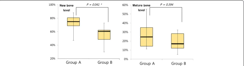
Results: As tested at the level of new bone, the mean OI was 72.37 % in group A and 55.08 % in group B—a statistically significant difference (p= 0.041). As tested at the level of mature bone, the mean OI was 27.47 % in group A and 18.88 % in group B, but the difference was not statistically significant (p= 0.394).
Conclusions:The application of rhBMP-2 after maxillofacial cyst enucleation accelerated new bone formation in the bone defects. Thus, the use of rhBMP-2 in combination with ACS may be considered an alternative to conventional bone grafting in some patients with postoperative bone defects.
Keywords:rhBMP-2, Bone regeneration, Bone defect
Background
Bone defects in the oral and maxillofacial region have many different causes, such as infection, trauma, le-sions, or invasive surgery. Because such defects require faster healing and complete reconstruction to achieve functional and esthetic recovery, numerous studies and relentless efforts have been undertaken to fulfill these requirements. As a result, a variety of bone graft mate-rials and osteogenic factors have been examined but have not led to ideal alternatives or substitutes, so rele-vant studies are ongoing.
Bone morphogenic proteins (BMPs) were discovered in 1965 by Urist [1], revealing their ability to differenti-ate undifferentidifferenti-ated osteogenic progenitor cells. Unlike
conventional bone graft materials, which are only osteoconductive, BMPs are osteoinductive and there-fore represent an optimal alternative to bone grafting for reconstruction of the oral and maxillofacial region [2]. Various applications of rhBMP-2 have been ex-plored since it was approved in 2007 as a substitute for autografts in maxillary sinus augmentation and alveolar ridge defects around an extraction socket. Previous studies have demonstrated that rhBMP-2 is effective for cleft lip and palate, alveolar bone augmentation, sinus augmentation, osteonecrosis of the jaw, and reconstruc-tion of the oral and maxillofacial region [3–5].
Intrabony cysts are common lesions of the oral and maxillofacial region and often require surgical removal. In many cases, after removal of the cyst, bone grafting is performed using a particle-type bone graft material. Re-cently, however, because bone graft materials may be-come dispersed or may migrate depending on the size or
* Correspondence:seungilsong@daum.net †Equal contributors
Department of Oral and Maxillofacial Surgery, Institute of Oral Health Science, Ajou University School of Medicine, 164, World Cup-ro, Yeongtong-gu, Suwon-si, Gyeonggi-do 16499, Republic of Korea
location of the defect and the bony housing of the lesion [6], absorbable collagen sponge (ACS) with absorbed BMPs has occasionally been used instead [7].
Although many studies have already shown the bone regenerative effects of rhBMP-2, most of these were in vitro or animal studies in which rhBMP-2 was added to other bone graft materials, and some of the clinical re-ports involved non-quantitative methods. Our study was intended to analyze the bone regenerative effects of rhBMP-2 quantitatively based on cone-beam com-puted tomographic (CBCT) images of patients treated with and without rhBMP-2.
Methods Patients
We evaluated 38 patients who visited the Department of Oral and Maxillofacial Surgery at Ajou University Hospital in South Korea from January 1, 2014, through March 31, 2015, and who underwent cyst enucleation under general anesthesia. For our study, the following selection criteria were applied:
Inclusion criteria
Both preoperative and 3-month postoperative CBCT images were available.
ACS alone or rhBMP-2 plus ACS was used for bone defect repair.
Exclusion criteria
Recipients of bone grafts (allogenic or xenogenic bone material).
Patients with diseases related to bone metabolism (e.g., osteoporosis).
Based on these criteria, we selected 10 patients for study, two of whom each had one additional, independ-ent lesion, for a total of 12 lesions. Human CBCT data were used in this study but patient consent was not
necessary. This study was reviewed and approved by the institutional review board of Ajou University Hospital (AJIRB-MED-MDB-15-203).
Methods
Study design
Two operators performed cyst enucleation for 12 lesions. After a retrospective chart review, we divided the 12 cases into two groups of six lesions each: group A was treated with rhBMP-2 plus ACS, and group B was treated with ACS alone. Preoperative and 3-month post-operative lesion volumes were measured on CBCT im-ages, and the osteogenesis index was calculated in each case, after which, a statistical analysis was performed.
Surgical procedure
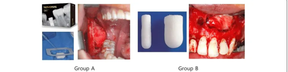
After cyst enucleation under general anesthesia, ACS with absorbed rhBMP-2 was applied on the bone de-fects in group A and primary closure was completed. In group B, ACS alone was applied after cyst enucleation, with subsequent primary closure (Fig. 1). Up to five units of ACS was used and up to 1.0 mg of rhBMP-2 (1 mg/mL) was used, depending on the size of the le-sion. The rhBMP-2 used in this study was NOVOSIS (Daewoong Pharmaceutical Company, Seoul, South Korea), and the ACS was either Ateloplug (Bioland, Cheonan, South Korea) or Rapiderm Plug (Dalim Tissen, Seoul, South Korea).
Lesion volume measurements
Each case was analyzed using Simplant software (Materi-alise Dental, Leuven, Belgium). From among the user’s tools, we selected only those pixels (Fig. 2) within the preset range of Hounsfield units (minimum =−1024 HU; maximum = 200 or 600 HU) for all the coronal or axial images believed to include lesions. The selected pixels were then remodeled into three-dimensional images, and the volumes were calculated. Hounsfield units ranged from −1024 (minimum) to 200 (new bone level)
or 600 (mature bone level) (maximum), in reference to the studies performed by Norton and Gamble [8], Sha-purian et al. [9], and Tajima et al. [10] (Fig. 3). Specific-ally, we defined two ranges: range 1 included −1024 < HU < 200 and range 2 included −1024 < HU < 600. For range 1, pixels with volumes of < 200 HU were regarded as a lesion; for range 2, pixels with volumes of < 600 HU were regarded as a lesion.
To minimize any possible visual errors during meas-urement, the patient’s head position on CBCT images was repositioned prior to volume measurement by using the line connecting the bilateral orbitales on the coronal plane, the line connecting the anterior nasal spine (ANS) and the cervical vertebra on the horizontal plane, and the line connecting the ANS to the posterior nasal spine (PNS) on the sagittal plane. To ensure accuracy, volume measurements were done twice in each case, and the mean results were used in this study.
Calculation of OI
Preoperative lesion volume was defined as V0and post-operative lesion volume asVh(HU = 200 or 600) (Fig. 4). The OI was calculated as (V0−Vh)/V0(%).
Assessment and statistical analysis
Using OI, we compared osteogenesis in groups A and B, and differences with a pvalue of < 0.05 were considered statistically significant. The Mann–Whitney U test and SPSS version 22.0 (IBM, NY, USA) were employed as the statistical technique and software program, respectively.
Results
The patients ranged in age from 16 to 50 years (mean age = 36.25), and the ratio of male-to-female patients was 9:1. Of the 12 lesions, nine were located in the man-dible and the other three in the maxilla. Histological findings for the individual lesions indicated that there were nine dentigerous cysts, two periapical cysts, and one keratocystic odontogenic tumor (KCOT) (Table 1). All the lesions healed with no remarkable complications. The measurements of preoperative lesion volume (V0) ranged from 1.24 to 14.82 cm3, with a mean of 5.25 cm3. The OI measurements at the level of new bone ranged from 29.88 to 88.21 %, with a mean of 63.72 %, and at the level of mature bone from 5.12 to 55.33 %, with a mean of 23.17 %. When postoperative lesion volume was measured, increases in the number of Hounsfield units
B
A C
Fig. 2Selection of pixels within the range of Hounsfield units desired (selection process).aOriginal CBCT image.bDrawing of lesional area.
cSelected pixels
New bone level Mature bone level
were seen throughout the lesions, especially around the margins.
When tested at the new bone level, the mean OI was 72.37 % for group A and 55.08 % for group B—a statisti-cally significant difference (p= 0.041). However, when tested at the mature bone level, the mean OI was 27.47 % for group A and 18.88 % for group B, a difference that did not reach statistical significance (p= 0.394) (Table 2 and Fig. 5).
Discussion
Human bone regenerates through patterns of maturation similar to those of bone growth in response to bone de-fects of any cause. Stable bone healing is achieved when there is an adequate blood supply and immobilization at the site of the defect. For the first 4 weeks, angiogenic and osteogenic cells originate from the surrounding bone walls and periosteum, while woven bone forms around the defect. These processes are governed by vari-ous cytokines and growth factors [11–15].
Ettl et al. suggested that although primary closure after cyst enucleation can be accomplished without bone grafts, further research regarding growth factors, osteo-blasts, stem cells, and other components is needed to understand this process more fully [16]. Bone defects up to 3 cm in diameter usually undergo complete ossifica-tion after 12 months, while larger bone defects may re-quire a longer period of ossification (24 months or more) [17, 18]. In spite of the obvious need for add-itional treatment to accelerate healing (e.g., bone graft-ing), such measures cannot always be taken when possible complications such as infection or migration are of concern. Recently, ACS with absorbed rhBMP-2 has been applied in such situations.
In his primate study, Boyne reported that rhBMP-2 alone was useful even without bone graft material for the reconstruction of facial bone defects after mandibu-lar hemisection, implant, and cleft repair [19]. After reviewing the literature on alveolar ridge augmentation, maxillary sinus augmentation, and/or extraction socket
preservation, Freitas et al. reported that ACS with absorbed rhBMP-2 appeared to function as an alterna-tive to autografting in alveolar ridge or maxillary sinus augmentation [20]. Balaji reported the use of rib graft-ing and rhBMP-2 followgraft-ing removal of an aneurysmal bone cyst [2], and in 2014, Lee et al. also reported the use of rhBMP-2 andβ-TCP/HA (tricalcium phosphate/ hydroxyapatite) in five patients with cysts [21].
Unfortunately, however, there have been some limi-tations to the use of rhBMP-2 despite the successful outcomes described above. These include the shorter half-life of BMP-2 and its rapid elimination at the ap-plication site, which requires a high dose of BMP-2 and thus expensive medical costs, overgrowth of bone, and unwanted side effects, including swelling due to im-mune reactions [7, 22, 23]. According to a recent report, excessively high doses of BMP-2 may cause oral squamous cell carcinoma [24]. However, we did not observe compli-cations in any of the patients treated at our hospital.
One can compensate for the abovementioned disad-vantages of BMP by selecting an appropriate carrier. Currently available carriers include HA, TCP, DBM,
Mature bone level New bone level
B
A C
Fig. 4Initial lesion and reduced lesion after 3 months.aPreoperative lesion.bPostoperative lesion at new bone level.cPostoperative lesion at mature bone level
Table 1Characteristics of the 12 lesions
Group Age (year) Gender Site Diagnosis V0(cm3)
A
(ACS + rhBMP-2)
16 M Mandible DC 14.82 50 F Mandible DC 1.98 21 M Mandible KCOT 6.47 45 M Mandible DC 2.27 41 M Mandible DC 3.14 20 M Mandible DC 5.02
B (ACS alone)
51 M Mandible DC 2.30 48 M Maxilla PC 6.64 37 M Mandible DC 1.24 37 M Mandible DC 2.67 45 M Maxilla DC 2.95 40 M Mandible PC 13.48
hydrogel, and ACS. Referring to the existing literature, Geiger et al. described“enhancement of osteogenic ac-tivity of BMP with a restrictive release of BMP at an effective dose during a period coincident with the ac-cumulation and proliferation of target cells” [25]. Li and Wozney reported that the releasing periods of rhBMP-2 were at least twice as long when treatment included the ACS compared with the control treat-ment without the sponge, and ACS is an appropriate carrier for BMP application [26]. In contrast, in 2008, Carter et al. mentioned that although ACS is of value for the delivery of BMP and offers good space-maintaining ability, it should be used with caution because its overcompressed use may interfere with normal bone formation [7].
Bone density can be assessed by measuring Hounsfield units and has different values depending on the type of bone. Very dense cortical bone is expressed as 600 HU or more, the dense cortical/spongy bone as 400 through 600 HU, and low-density bone as 200 HU or less [8, 9]. In 2013, Tajima et al. reported that the density of peri-implant, new bonelike tissue ranged from 185 to 713 HU (mean ± SD = 323 ± 156.2) [10].
Huh et al. found that combination therapy with bovine bone (Bio-Oss) and rhBMP-2 leads to more new bone generation than does bovine bone monotherapy and that rhBMP-2 enhanced bone regeneration [27]. In our study, the mean OI was higher in the rhBMP-2 treatment group A than in the group B, and the difference was sta-tistically significant for new bone levels with maximum
number of Hounsfield units set at 200. This result sug-gests that rhBMP-2 contributes significantly to new bone generation in the human body as well.
This study had the following limitations: difficulty in determining the margin when measuring postoperative lesion volume owing to the need for intraoperative oste-otomy to approach the lesion; several diagnoses of the lesions; preoperative secondary infections due to the le-sions; the degree of defect in the bony housing; and no consideration of the number of absorbable collagen sponges or the quantity of rhBMP-2 actually applied during the operation. Nevertheless, this study is mean-ingful in that we used a quantitative method to analyze the effect of rhBMP-2 in human subjects. Further stud-ies will be needed to perform histomorphometric ana-lyses of the effects of rhBMP-2 in the human body.
Conclusions
When rhBMP-2 was used to repair bone defects that re-main after cyst enucleation, new bone formation was in-creased. Thus, the combination of rhBMP-2 and ACS could be considered as an alternative to conventional bone grafts. We believe that rhBMP-2 is worthy of being applied to bone defects in the oral and maxillofacial re-gion in certain cases.
Ethics approval and consent
Human CBCT data were used in this study, but consent for patients was not necessary. This study was reviewed Table 2Preoperative and 3-month postoperative mean volume (±SD) and mean OI (±SD) of lesion
Group V0 New bone Mature bone
V200(cm3) OI (%) V600(cm3) OI (%)
A
(ACS+rhBMP-2)
5.62 ± 4.82 1.67 ± 1.60 72.37 ± 14.39 4.56 ± 4.57 27.47 ± 17.09 B
(ACS alone) 4.88 ± 4.60 1.79 ± 1.14 55.08 ± 15.26 3.81 ± 3.21 18.88 ± 11.15
V0preoperative volume;V200volume at new bone level (< 200 HU);V600volume at mature bone level (< 600 HU);OIosteogenesis index
and approved by the Institutional Review Board of Ajou University Hospital (AJIRB-MED-MDB-15-203).
Availability of data and materials
The dataset supporting the conclusions of this article is included in Additional file 1.
Additional file
Additional file 1:Case form and result of data. (XLSX 13 kb)
Abbreviations
ACS:absorbable collagen sponge; OI: osteogenesis index.
Competing interests
The authors declare that they have no competing interests.
Authors’contributions
S.I.S. carried out the surgical treatment of the patient and contributed to the conception of the report and critical revising. D.Y.H. and S.W.O. participated in the treatment, collection of data, and drafting of the manuscript. All authors read and approved the final manuscript
Authors’information
All of the authors have no affiliations with or involvement in any organization or entity with any financial interest or non-financial interest in this manuscript.
This manuscript represents original works and is not being considered for publication elsewhere.
Funding
We received no funding in support of this study.
Received: 29 March 2016 Accepted: 13 May 2016
References
1. Urist MR (1965) Bone: formation by autoinduction. Science 150(3698):893–899 2. Balaji S (2009) Mandibular cystic defect: a composite approach with
rhBMP-2 and rib graft. J Maxillofac Oral Surg 8(1):27–30
3. Chenard KE, Teven CM, He T-C, Reid RR (2012) Bone morphogenetic proteins in craniofacial surgery: current techniques, clinical experiences, and the future of personalized stem cell therapy. J Biomed Biotechnol Article ID 601549,14. doi:10.1155/2012/601549.
4. Gutwald R, Haberstroh J, Stricker A, Rüther E, Otto F, Xavier SP, Oshima T, Marukawa E, Seto I, Enomoto S (2010) Influence of rhBMP-2 on bone formation and osseointegration in different implant systems after sinus-floor elevation. An in vivo study on sheep. J Craniomaxillofac Surg 38(8):571–579 5. Cicciù M, Herford AS, Juodžbalys G, Stoffella E (2012) Recombinant human
bone morphogenetic protein type 2 application for a possible treatment of bisphosphonates-related osteonecrosis of the jaw. J Craniofac Surg 23(3): 784–788
6. Seok H, Lee S-K, Kim S-G, Kang T-Y, Lee M-J, Chae W-S (2014) Migration of alloplastic bone graft material in infected conditions: a case study and animal experiment. J Oral Maxillofac Surg 72(6):1093, e1091-1093. e1011 7. Carter TG, Brar PS, Tolas A, Beirne OR (2008) Off-label use of recombinant
human bone morphogenetic protein-2 (rhBMP-2) for reconstruction of mandibular bone defects in humans. J Oral Maxillofac Surg 66(7):1417–1425 8. Norton MR, Gamble C (2001) Bone classification: an objective scale of bone density using the computerized tomography scan. Clin Oral Implants Res 12(1):79–84
9. Shapurian T, Damoulis PD, Reiser GM, Griffin TJ, Rand WM (2006) Quantitative evaluation of bone density using the Hounsfield index. Int J Oral Maxillofac Implants 21(2):290–297
10. Tajima N, Ohba S, Sawase T, Asahina I (2013) Evaluation of sinus floor augmentation with simultaneous implant placement using platelet-rich fibrin as sole grafting material. Int J Oral Maxillofac Implants 28(1):77–83. doi:10.11607/jomi.2613
11. Buser D, Hoffmann B, Bernard JP, Lussi A, Mettler D, Schenk RK (1998) Evaluation of filling materials in membrane-protected bone defects. A comparative histomorphometric study in the mandible of miniature pigs. Clin Oral Implants Res 9(3):137–150
12. Ogunlewe MO, Akinwande JA, Ladeinde AL, Adeyemo WL (2006) Spontaneous regeneration of whole mandible after total mandibulectomy in a sickle cell patient. J Oral Maxillofac Surg 64(6):981–984. doi:10.1016/j. joms.2006.02.008
13. Rodeo SA, Delos D, Weber A, Ju X, Cunningham ME, Fortier L, Maher S (2010) What’s new in orthopaedic research. J Bone Joint Surg Am 92(14):2491–2501. doi:10.2106/JBJS.J.01174
14. Schenk RK, Buser D, Hardwick WR, Dahlin C (1994) Healing pattern of bone regeneration in membrane-protected defects: a histologic study in the canine mandible. Int J Oral Maxillofac Implants 9(1):13–29
15. Schilephake H (2002) Bone growth factors in maxillofacial skeletal reconstruction. Int J Oral Maxillofac Surg 31(5):469–484
16. Ettl T, Gosau M, Sader R, Reichert TE (2012) Jaw cysts—filling or no filling after enucleation? A review. J Craniomaxillofac Surg 40(6):485–493 17. Chiapasco M, Rossi A, Motta JJ, Crescentini M (2000) Spontaneous bone
regeneration after enucleation of large mandibular cysts: a radiographic computed analysis of 27 consecutive cases. J Oral Maxillofac Surg 58(9):942–948 18. Hren NI, Miljavec M (2008) Spontaneous bone healing of the large bone
defects in the mandible. Int J Oral Maxillofac Surg 37(12):1111–1116 19. Boyne PJ (2001) Application of bone morphogenetic proteins in the
treatment of clinical oral and maxillofacial osseous defects. J Bone Joint Surgery Am 83(A Suppl 1, Pt 2):S146–S150
20. Freitas RM, Spin‐Neto R, Junior EM, Pereira LAVD, Wikesjö UM, Susin C (2015) Alveolar ridge and maxillary sinus augmentation using rhBMP‐2: a systematic review. Clin Implant Dent Relat Res 17(S1):e192–e201 21. Lee JB, Ryu SH, Ryu HH, Shin YC, Hwang HS (2014) Technical reports: the
use of recombinant human bone morphogenic protein-2 (rhBMP-2) in treatment for cysts of the oral and maxillofacial regions. J Korean Maxillofac Plast Reconstr Surg 36(1):25–29
22. Shields LB, Raque GH, Glassman SD, Campbell M, Vitaz T, Harpring J, Shields CB (2006) Adverse effects associated with high-dose recombinant human bone morphogenetic protein-2 use in anterior cervical spine fusion. Spine 31(5):542–547
23. Takahashi Y, Yamamoto M, Tabata Y (2005) Enhanced osteoinduction by controlled release of bone morphogenetic protein-2 from biodegradable sponge composed of gelatin andβ-tricalcium phosphate. Biomaterials 26(23):4856–4865
24. Kim M-J, Kim K-M, Kim J, Kim K-N (2014) BMP-2 promotes oral squamous carcinoma cell invasion by inducing CCL5 release. PLoS One 9(10), e108170. doi:10.1371/journal.pone.0108170
25. Geiger M, Li R, Friess W (2003) Collagen sponges for bone regeneration with rhBMP-2. Adv Drug Deliv Rev 55(12):1613–1629
26. Li RH, Wozney JM (2001) Delivering on the promise of bone morphogenetic proteins. Trends Biotechnol 19(7):255–265
27. Huh JB, Yang JJ, Choi KH, Bae JH, Lee JY, Kim SE, Shin SW (2015) Effect of rhBMP-2 immobilized anorganic bovine bone matrix on bone regeneration. Int J Mol Sci 16(7):16034–16052. doi:10.3390/ijms160716034
Submit your manuscript to a
journal and benefi t from:
7 Convenient online submission
7 Rigorous peer review
7 Immediate publication on acceptance
7 Open access: articles freely available online
7 High visibility within the fi eld
7 Retaining the copyright to your article