S H O R T G E N O M E R E P O R T
Open Access
High-quality-draft genome sequence of the
multiple heavy metal resistant bacterium
Pseudaminobacter manganicus
JH-7
T
Xian Xia
†, Jiahong Li
†, Zijie Zhou, Dan Wang, Jing Huang and Gejiao Wang
*Abstract
Pseudaminobacter manganicus
JH-7
T(= KCTC 52258
T= CCTCC AB 2016107
T) is a Gram-staining-negative, aerobic
and non-motile strain that was isolated from a manganese mine.
The strain JH-7
Tshows multiple heavy
metal resistance and can effectively remove Mn
2+and Cd
2+. In addition, it is able to produce exopolysaccharides (EPS),
which may contribute to metal remove/adsorption. Thus, strain JH-7
Tshows a great potential in bioremediation
of heavy metal-contaminated environment. In this study, we report the draft genomic sequence of
P. manganicus
JH-7
Tand compare it to related genomes. Strain JH-7
Thas a 4,842,937 bp genome size with a G + C content of
61.2%, containing 4504 protein-coding genes and 71 RNA genes. A large number of putative genes associated
with heavy metal resistance and EPS synthesis are found in the genome.
Keywords:
Cadmium, Exopolysaccharides, Heavy metal resistance and adsorption, Manganese,Pseudaminobacter
Introduction
Genus
Pseudaminobacter
was established by Kämpfer
et al. in 1999 and contains three species represented
by
Pseudaminobacter salicylatoxidans
BN12
T(type
species) [
1
],
Pseudaminobacter defluvii
THI 051
T[
1
]
and
Pseudaminobacter manganicus
JH-7
T[
2
]. The
common characteristics of
Pseudaminobacter
strains
are Gram-staining-negative, rod-shaped and aerobic
[
1
,
2
].
P. salicylatoxidans
BN12
Tcontains a peculiar
ring-fission dioxygenase with the ability to cleave
sali-cylate in 1, 2-position to 2-oxohepta-3, 5-dienedioic
acid [
3
].
P. manganicus
JH-7
Twas isolated from a sludge
sample of a wastewater ditch in Dalong manganese
mine in 2015 [
2
]. It shows multiple heavy metal
re-sistance and can effectively remove Mn
2+and Cd
2+.
In addition, the strain produces EPS, which may
fa-cilitate heavy metal resistance and adsorption [
4
–
6
].
These features show great interests because of its
potential applications in bioremediation of heavy
metal contaminated environments. So far, only the
genome of an atypical
Pseudaminobacter
strain
Pseu-daminobacter salicylatoxidans
KCT001 has been
se-quenced [
7
]. Strain KCT001 can utilize tetrathionate
as the substrate for sulfur-oxidizing
chemolitho-trophic growth [
8
]. For better understanding the
mechanism of bacterial resistance and removal of
heavy metals, here we analyze the genome of
P.
manganicus
JH-7
T.
Organism information
Classification and features
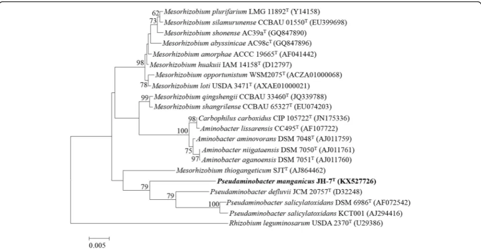
The
phylogenetic
relationship
of
P.
manganicus
JH-7
Tto the related members is shown in a 16S
rRNA gene based neighbor-joining tree. Strain JH-7
Tis closely related to
P. salicylatoxidans
BN12
T,
P.
defluvii
THI 051
Tand
P. salicylatoxidans
KCT001
(Fig.
1
). Strain JH-7
Tis Gram-staining-negative,
aer-obic, non-motile and rod-shaped (0.3
–
0.8 × 1
–
2
μ
m)
(Fig.
2
). The colonies are white, circular, entire,
slightly raised and smooth on LB agar plates. It is
positive for oxidase and catalase activities and
hy-drolysis of casein [
2
]. The major fatty acids are C
18:1ω
7
c
, C
19:0cyclo
ω
8
c
and C
16:0and the G + C content
is
61.2
mol%
[
2
].
The
major
polyamine
is
sym-homospermidine and the respiratory quinone is
* Correspondence:[email protected]
†Xian Xia and Jiahong Li contributed equally to this work.
State Key Laboratory of Agricultural Microbiology, Huazhong Agricultural University, Wuhan 430070, People’s Republic of China
ubiquinone-10. The polar lipids are
phosphatidylmo-nomethylethanolamine, diphosphatidylglycerol,
phos-phatidylglycerol, phosphatidylcholine, two aminolipids
and two lipids [
2
]. Table
1
shows the general features
of
P. manganicus
JH-7
T.
The resistant levels of
P. manganicus
JH-7
Tto
mul-tiple metal(loid)s were tested with the MIC on LB agar
plates incubated at 28 °C for 7 days. The MICs for
MnCl
2, CdCl
2,PbCl
2, CuCl
2, ZnSO
4and NiSO
4are100,
2, 10, 5, 5 and 5 mmol/L respectively. The MICs for
K
2CrO
4and Na
3AsO
3are both 0.1 mmol/L that are
lower than the above six metals. Specifically, strain
JH-7
Tcould remove nearly 60% of 5 mmol/L Mn
2+and
nearly 80% of 0.1 mmol/L Cd
2+(Fig.
3
), respectively. In
addition, strain JH-7
Tcould produce EPS based on the
aniline blue reaction incubated on LB agar in 3
–
7 days
[
9
] (data not shown). This phenomenon is consistent
with the cell image observed by TEM (Fig.
2
). A lay of
shadow around the strain was similar to the EPS
ob-served in strain
Bifidobacterium longum
35,624 [
10
].
Genome sequencing information
Genome project history
This organism was selected for sequencing particularly
due to its multiple heavy metals resistance and heavy
metal removal ability. Genome sequencing was
per-formed by Wuhan Bio-Broad Co., Ltd., Wuhan, China in
2016. The draft genome sequence of strain
P.
mangani-cus
JH-7
Thas been deposited at DDBJ/EMBL/GenBank
under accession number
MDET00000000
. The project
information is summarized in Table
2
.
Growth conditions and genomic DNA preparation
P. manganicus
JH-7
Twas grown under aerobic
condi-tions in LB medium at 28 °C for 40 h. DNA extraction
Fig. 1Phylogenetic tree highlighting the phylogenetic position ofPseudaminobacter manganicusJH-7T. The phylogenetic tree was constructed based on the 16S rRNA gene sequences. The analysis was inferred by MEGA 6.0 [41] with neighbor-joining algorithm and 1000 bootstrap repetitions were computed to estimate the reliability of the tree. Bar, 0.005 substitutions per nucleotide positionFig. 3Mn2+and Cd2+removed byP. manganicusJH-7T. Control stands for null LB medium. Strain JH-7Twas incubated until OD600reach 1.0, and then amended with 5000μmol/L MnCl2(a) and 100μmol/L CdCl2(b), respectively. The cultures were removed at 24 h intervals. After centrifuging at 12,000 rpm for 10 min, the supernatant was used to determine the residual concentration of Mn2+and Cd2+by the atomic absorption spectrometry AAS (AAS; 986A, Beijing Puxi General Instrument 197 Co., Beijing, China). Bars represent the mean ± SD of three biological replicates
Table 1
Classification and general features of
P. manganicus
JH-7
T[
42
]
MIGS ID Property Term Evidence codea
Classification DomainBacteria TAS [43]
PhylumProteobacteria TAS [44,45]
ClassAlphaproteobacteria TAS [46]
OrderRhizobiales TAS [46,47]
FamilyPhyllobacteriaceae TAS [46,47]
GenusPseudaminobacter TAS [1,2]
Speciesmanganicus TAS [2]
Type strain JH-7T(= KCTC 52258T= CCTCC AB 2016107T) TAS [2]
Gram stain negative TAS [2]
Cell shape rod-shaped TAS [2]
Motility no TAS [2]
Sporulation no TAS [2]
Temperature range 15–40 °C TAS [2]
Optimum temperature 28 °C TAS [2]
pH range; Optimum 5–9; 7 TAS [2]
Carbon source D-glucose, L-arabinose, D-fructose and D-mannose TAS [2]
MIGS-6 Habitat Mine sludge TAS [2]
MIGS-6.3 Salinity 0–6% NaCl (w/v) TAS [2]
MIGS-22 Oxygen requirement aerobic TAS [2]
MIGS-15 Biotic relationship free-living TAS [2]
MIGS-14 Pathogenicity non-pathogen NAS
MIGS-4 Geographic location Tongren city, Guizhou province, P. R. China TAS [2]
MIGS-5 Sample collection 2015 TAS [2]
MIGS-4.1 Latitude N27° 43′8" TAS [2]
MIGS-4.2 Longitude E108° 31′42" TAS [2]
MIGS-4.4 Altitude not reported
These evidence codes are from the Gene Ontology project [48]
IDAInferred from Direct Assay,TASTraceable Author Statement (i.e., a direct report exists in the literature),NASNon-traceable Author Statement (i.e., not directly observed for the living, isolated sample, but based on a generally accepted property for the species, or anecdotal evidence)
a
was performed using the QiAamp kit (Qiagen, Germany)
as the manufacturer
’
s instructions. A NanoDrop
Spec-trophotometer 2000 was used to determine the quality
and quantity of the DNA. Seven microgram of DNA was
sent to Bio-broad Technogoly Co., Ltd., Wuhan, China
for sequencing.
Genome sequencing and assembly
The genome of strain JH-7
Twas sequenced on Illumina
Hiseq2000 [
11
] and assembled by Bio-broad Technogoly
Co., Ltd., Wuhan using SOAPdenovo v2.04 [
12
]. An
Illu-mina standard shotgun library was constructed and
se-quenced, which generated 19,404,755 reads totaling
2,885,684,230 bp and average of 625 times genome
coverage. The total size of the genome is 4,842,937 bp
and a total of 60 scaffolds were obtained after arranging
68 contigs together. The part gaps of assembly were
filled and the error bases were revised using GapCloser
v1.12 [
13
].
Genome annotation
The draft genome was annotated through the NCBI
Pro-karyotic Genome Annotation Pipeline (PGAP), and
Table 2
Project information
MIGS ID Property Term
MIGS-31 Finishing quality High-quality draft
MIGS-28 Libraries used Illumina Paired-End library (300 bp in-sert size)
MIGS-29 Sequencing platforms
Illumina Miseq 2000
MIGS-31.2
Fold coverage 624.94×
MIGS-30 Assemblers SOAPdenovo v2.04
MIGS-32 Gene calling method GeneMarkS+
Locus TAG BFN67
Genbank ID MDET00000000
Genbank Date of Release
31, March, 2017
GOLD ID Gp0291525
Bioproject PRJNA338732 MIGS-13 Source material
identifier
CCTCC AB 2016107T
Project relevance Bioremediation
Table 3
Genome statistics
Attribute Value % of totala
Genome size (bp) 4,842,937 100
DNA coding (bp) 4,238,496 87.5
DNA G + C (bp) 2,963,726 61.2
DNA scaffolds 60 100
Total genesb 4685 100
Protein-coding genes 4504 96.2
RNA genes 71 1.7
Pseudo genes 110 2.3
Genes in internal clusters 1725 38.3
Genes with function prediction 3228 68.9
Genes assigned to COGs 3729 79.6
Genes with Pfam domains 3926 83.8
Genes with signal peptides 392 8.4
Genes with transmembrane helices 1119 23.9
CRISPR repeats 5
a
The total is based on either the size of the genome in base pairs or the total number of protein coding genes in the annotated genome
b
Also includes 110 pseudogenes, 54 tRNA genes, 12 rRNAs and 5 ncRNA
Table 4
Number of genes associated with the 25 general COG
functional categories
Code Value % of
totala Description
J 181 4.02 Translation
A 0 0.00 RNA processing and modification
K 299 6.64 Transcription
L 233 5.17 Replication, recombination and repair
B 3 0.07 Chromatin structure and dynamics
D 39 0.87 Cell cycle control, mitosis and meiosis
Y 0 0.00 Nuclear structure
V 46 1.02 Defense mechanisms
T 134 2.98 Signal transduction mechanisms
M 217 4.82 Cell wall/membrane biogenesis
N 35 0.78 Cell motility
Z 0 0.00 Cytoskeleton
W 0 0.00 Extracellular structures
U 106 2.35 Intracellular trafficking and secretion
O 156 3.46 Posttranslational modification, protein turnover, chaperones
C 240 5.33 Energy production and conversion
G 312 6.93 Carbohydrate transport and metabolism
E 482 10.70 Amino acid transport and metabolism
F 87 1.93 Nucleotide transport and metabolism
H 158 3.51 Coenzyme transport and metabolism
I 153 3.40 Lipid transport and metabolism
P 209 4.64 Inorganic ion transport and metabolism
Q 91 2.02 Secondary metabolites biosynthesis, transport and catabolism
R 453 10.06 General function prediction only
S 444 9.86 Function unknown
– 775 17.21 Not in COGs
a
genes were identified using the gene caller GeneMarkS
+with the similarity-based gene detection approach [
14
].
The predicted CDSs were translated and were submitted
to the Pfam protein family database [
15
] and KEGG
database [
16
]. The genes in internal clusters were
per-formed by OrthoMCL [
17
,
18
]. The protein function
classification, transmembrane helices and signal peptides
were predicted by WebMGA [
19
], TMHMM v. 2.0 [
20
]
and SignalP 4.1 [
21
], respectively. In addition, the
CRISPRfinder program [
22
] was used to predict
CRISPRs in the genome.
Genome properties
The draft genome size of strain JH-7
Tis 4,842,937 bp
with 61.2 mol% G + C content and contains 60 scaffolds.
The genome properties and statistics are shown in
Table
3
. From a total of 4685 genes, 4504 (96.2%) are
protein coding genes, 110 (2.3%) are pseudo genes and
the rest are 71 predicted RNA genes, including 54 tRNA,
12 rRNAs and 5 ncRNA. In addition, 3729 (82.8%)
pro-tein coding genes are distributed into COG functional
categories (Table
4
).
Insights from the genome sequence
Strain JH-7
Tcould tolerant multiple heavy metals (Mn
2+,
Cd
2+,Pb
2+, Cu
2+, Zn
2+and Ni
2+) and remove Mn
2+and
Cd
2+, suggesting that it has developed a number of
evo-lutionary strategies to adapt the mine environment.
Ac-cording to the genome annotation results, strain JH-7
Tharbors various putative proteins related to heavy
met-al(loid)s resistance including transporters, resistance
proteins and metal reductases (Additional file
1
: Table
S1). MntH [
23
] and metal ABC transport system [
24
]
are involved in cation uptake. Heavy metal-transporting
ATPase is responsible for the efflux of Pb
2+, Zn
2+, Cd
2+and Ni
2+[
25
–
28
]. The genome contains Cu
2+efflux
sys-tem CopABC [
29
], mercuric reductase MerA and
regula-tor MerR [
30
]. Athough the MICs for Cr
6+and As
3+are
not high, the Cr
6+efflux protein ChrA [
27
,
31
] and As
3+resistant proteins (ArsRHC and ACR3) [
32
–
34
] are
present.
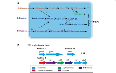
EPS are long-chain polysaccharides consisting of
branched, repeating units of sugars or sugar derivatives
[
35
]. Stain JH-7
Tcould produce EPS and all essential
proteins for EPS production are found in the genome.
Four complete nucleotide sugar synthesis (EPS
precur-sor) pathways are identified based on KEGG analysis
(Additional file
1
: Table S2) including the syntheses of
UDP-glucose,
UDP-galactose,
UDP-GlcNAc
and
GDP-D-mannose (Fig.
4a
). EPS assembly gene clusters
were also found in the genome of strain JH-7
T[
36
]
(Additional file
1
: Table S3, Fig.
4b
). Based on gene
ana-lysis, it is suggested that the EPS assembly in strain
JH-7
Tmight belong to Wzx/Wzy-dependent pathway
[
37
], e.g., repeat units are assembled by
glycosyltransfer-ases (EpsI) and translocated across the cytoplasmic
membrane to periplasm by flippase (Wzx) [
37
] and
WbaP
[
38
].
Next,
Wzy
(RfaL),
polysaccharide
co-polymerase (
Gum
C) and the outer membrane
poly-saccharide exporter (
Gum
B) transports the polymerized
repeat units to cell surface [
37
,
39
]. EPS has been
re-ported to contribute to heavy metal removal/adsorption
in bacteria [
3
–
6
]. Hence, the ability of EPS may
contrib-ute to Mn
2+and Cd
2+removal.
To gain more insight, the genomic features of strain
JH-7
Tis compared with the available genome
P.
salicyla-toxidans
KCT001 [
7
]. Strain JH-7
Thas similar genome
size (4.84 Mbp) and G + C content (61.2 mol%)
com-pared to strain KCT001 (4.61 Mbp; 62.8 mol%). A total
of 2408 core proteins are shared between the two
strains. Strain JH-7
Thas 1724 strain-specific CDSs.
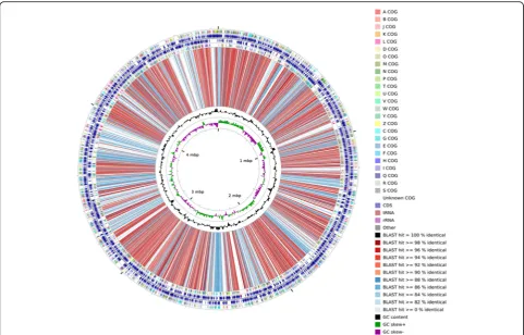
Figure
5
shows the genome comparison results of strain
JH-7
Tand strain KCT001 using CGview comparison tool
[
40
]. Comparing to
P. salicylatoxidans
KCT001, strain
JH-7
Twas unable to utilize tetrathionate for
chemo-lithoautotrophy (data not shown). However, it harbors
high quantitative and diverse heavy metal resistance
genes.
Conclusions
To the best of our knowledge, this study provides the
first typical strain genomic information of the genus
Pseudaminobacter
and revealed a consistency of
import-ant characters between genotypes and phenotypes. Strain
JH-7
Tis resistant to multiple heavy metals and capable
of removal Mn
2+/Cd
2+. Genome analysis reveal various
genes responsible for multiple heavy metal resistance,
which provides the genomic basis for this strain to adapt
the harmful environment.
Additional file
Additional file 1:Table S1.Putative heavy metal(loid)s resistance proteins.Table S2.Putative nucleotide sugars biosynthesis proteins for EPS production.Table S3.Putative proteins for EPS production. (XLSX 11 kb)
Abbreviations
EPS:Exopolysaccharides; MIC: Minimal inhibition concentration
Funding
This study was supported by National key research and development program of China (2016YFD0800702).
Authors’contributions
XX and JL performed the sequence annotation and genomic analysis and prepared the draft manuscript. ZZ, DW and JH performed the heavy metals resistance and removal tests. GW designed the study and revised the manuscript. All authors read and approved the final manuscript.
Competing interests
The authors declare that they have no competing interests.
Publisher
’
s Note
Springer Nature remains neutral with regard to jurisdictional claims in published maps and institutional affiliations.
Received: 10 January 2018 Accepted: 28 September 2018
References
1. Kämpfer P, Müller C, Mau M, Neef A, Auling G, Busse HJ, et al. Description of Pseudaminobacter gen. Nov. with two new species,Pseudaminobacter salicylatoxidanssp. nov. andPseudaminobacter defluviisp. nov. Intl J Syst Bacteriol. 1999;149:887–97.
2. Li J, Huang J, Liao S, Wang G.Pseudaminobacter manganicussp. nov., isolated from sludge of a manganese mine. Int J Syst Evol Microbiol. 2017;67(5):1589–94. 3. Hintner JP, Lechner C, Riegert U, Kuhm AE, Storm T, Reemtsma T, et al.
Direct ring fission of salicylate by a salicylate 1,2-dioxygenase activity from Pseudaminobacter salicylatoxidans. J Bacteriol. 2001;183(23):6936–42. 4. Natalia N, Bogino PC, Banchio E, Giordano W. Roles of extracellular
polysaccharides and biofilm formation in heavy metal resistance of rhizobia. Materials. 2016;9(6):418.
5. Bhunia B, Prasad Uday US, Oinam G, Mondal A, Bandyopadhyay TK, Tiwari ON. Characterization, genetic regulation and production of cyanobacterial exopolysaccharides and its applicability for heavy metal removal. Carbohydr Polym. 2018;179:228–43.
6. Nouha K, Kumar RS, Tyagi RD. Heavy metals removal from wastewater using extracellular polymeric substances produced byCloacibacterium normanense in wastewater sludge supplemented with crude glycerol and study of extracellular polymeric substances extraction by different methods. Bioresour Technol. 2016;212:120–9.
7. Alam M, Roy C, Pyne P, Agarwal A, George A, Ghosh W. Whole-genome shotgun sequence of the sulfur-oxidizing chemoautotroph
Pseudaminobacter salicylatoxidansKCT001. J Bacteriol. 2012;194(17):4743–4. 8. Deb C, Stackebrandt E, Pradella S, Saha A, Roy P. Phylogenetically diverse
new sulfur chemolithotrophs ofα-proteobacteria isolated from Indian soils. Curr Microbiol. 2004;48(6):452–8.
9. Nagaraj K, Rekha PD, Arun AB. Exopolysaccharide produced byEnterobactersp. YG4 reduces uranium induced nephrotoxicity. Int J Biol Macromol. 2016;82:557–61. 10. Altmann F, Kosma P, O’Callaghan A, Leahy S, Bottacini F, Molloy E, et al.
Genome analysis and characterisation of the exopolysaccharide produced byBifidobacterium longumsubsp.longum35624™. PLoS One. 2016;11(9): e0162983.
11. Bennett S. Solexa Ltd. Pharmacogenomics. 2004;5:433–8. 12. Luo R, Liu B, Xie Y, Li Z, Huang W, Yuan J, et al. SOAPdenovo2: an
empirically improved memory-efficient short-read de novo assembler. Gigascience. 2012;1(1):18.
13. Li R, Zhu H, Ruan J, Qian W, Fang X, Shi Z, et al.De novoassembly of human genomes with massively parallel short read sequencing. Genome Res. 2010;20(2):265–72.
14. Besemer J, Lomsadze A, Borodovsky M. GeneMarkS: a self-training method for prediction of gene starts in microbial genomes. Implications for finding sequence motifs in regulatory regions. Nucleic Acids Res. 2001;29:2607–18. 15. Finn RD, Coggill P, Eberhardt RY, Eddy SR, Mistry J, Mitchell AL, et al. The
Pfam protein families database: towards a more sustainable future. Nucleic Acids Res. 2016;44(1):279–85.
16. Kanehisa M, Goto S, Kawashima S, Okuno Y, Hattori M. The KEGG resource for deciphering the genome. Nucleic Acids Res. 2004;32:277–80. 17. Li L, Stoeckert CJ Jr, Roos DS. OrthoMCL: identification of ortholog groups
for eukaryotic genomes. Genome Res. 2003;13(9):2178.
18. Fischer S, Brunk BP, Chen F, Gao X, Harb OS, Iodice JB, et al. Using OrthoMCL to assign proteins to OrthoMCL-DB groups or to cluster proteomes into new ortholog groups. Curr Protoc Bioinformatics. 2011;36(1).
19. Wu S, Zhu Z, Fu L, Niu B, Li W. WebMGA: a customizable web server for fast metagenomic sequence analysis. BMC Genomics. 2011;12:444.
20. Krogh A, Larsson BÈ, Von Heijne G, Sonnhammer EL. Predicting
transmembrane protein topology with a hidden Markov model: application to complete genomes. J Mol Biol. 2001;305:567–80.
21. Petersen TN, Brunak S, Von HG, Nielsen H. SignalP 4.0: discriminating signal peptides from transmembrane regions. Nat Methods. 2011;8(10):785–6. 22. Grissa I, Vergnaud G, Pourcel C. CRISPRFinder: a web tool to identify
clustered regularly interspaced short palindromic repeats. Nucleic Acids Res. 2007;35:52–7.
23. Kehres DG, Zaharik ML, Finlay BB, Maguire ME. The NRAMP proteins of Salmonella typhimuriumandEscherichia coliare selective manganese transporters involved in the response to reactive oxygen. Mol Microbiol. 2000;36(5):1085–100.
24. Gabbianelli R, Scotti R, Ammendola S, Petrarca P, Nicolini L, Battistoni A. Role of ZnuABC and ZinT inEscherichia coliO157: H7 zinc acquisition and interaction with epithelial cells. BMC Microbiol. 2011;11(1):36.
25. Sharma R, Rensing C, Rosen BP, Mitra B. The ATP hydrolytic activity of purified ZntA, a Pb (II)/cd (II)/Zn (II)-translocating ATPase fromEscherichia coli. J Biol Chem. 2000;275(6):3873–8.
26. Nies DH, Silver S. Ion efflux systems involved in bacterial metal resistances. J Ind Microbiol. 1995;14(2):186–99.
27. Xia X, Li J, Liao S, Zhou G, Wang H, Li L, et al. Draft genomic sequence of a chromate-and sulfate-reducingAlishewanellastrain with the ability to bioremediate Cr and cd contamination. Stand Genomic Sci. 2016;11(1):48. 28. Xiong J, Li D, Li H, He M, Miller S, Yu L, et al. Genome analysis and
characterization of zinc efflux systems of a highly zinc-resistant bacterium, Comamonas testosteroniS44. Res Microbiol. 2011;162(7):671–9.
29. Adaikkalam V, Swarup S. Characterization ofcopABCDoperon from a copper-sensitivePseudomonas putidastrain. Can J Microbiol. 2005;51(3):209–16. 30. Nascimento AM, Chartone-Souza E. Operon mer: bacterial resistance to
mercury and potential for bioremediation of contaminated environments. Genet Mol Res. 2003;2(1):92–101.
31. Viti C, Marchi E, Decorosi F, Giovannetti L. Molecular mechanisms of Cr (VI) resistance in bacteria and fungi. FEMS Microbiol Rev. 2014;38(4):633–59. 32. Li X, Zhang L, Wang G. Genomic evidence reveals the extreme diversity and
wide distribution of the arsenic-related genes inBurkholderiales. PLoS One. 2014;9(3):e92236.
33. Kang YS, Shi Z, Bothner B, Wang G, McDermott TR. Involvement of the Acr3 and DctA anti-porters in arsenite oxidation inAgrobacterium tumefaciens5A. Environ Microbiol. 2015;17(6):1950–62.
34. Kruger MC, Bertin PN, Heipieper HJ, Arsène-Ploetze F. Bacterial metabolism of environmental arsenic–mechanisms and biotechnological applications. Appl Microbiol Biotechnol. 2013;97(9):3827–41.
35. Cui Y, Xu T, Qu X, Hu T, Jiang X, Zhao C. New insights into various production characteristics ofStreptococcus thermophilusstrains. Int J Mol Sci. 2016;17(10):1701.
36. Wu Q, Tun HM, Leung FC, Shah NP. Genomic insights into high exopolysaccharide-producing dairy starter bacteriumStreptococcus thermophilusASCC 1275. Sci Rep. 2014;4:4974.
37. Schmid J, Sieber V, Rehm B. Bacterial exopolysaccharides: biosynthesis pathways and engineering strategies. Front Microbiol. 2015;6:496. 38. Schäffer C, Wugeditsch T, Messner P, Whitfield C. Functional expression of
39. Klena JD, Pradel E, Schnaitman CA. Comparison of lipopolysaccharide biosynthesis genes rfaK, rfaL, rfaY, and rfaZ ofEscherichia coliK-12 and Salmonella typhimurium. J Bacteriol. 1992;174(14):4746–52.
40. Grant JR, Arantes AS, Stothard P. Comparing thousands of circular genomes using the CGView comparison tool. BMC Genomics. 2012;13:202. 41. Tamura K, Stecher G, Peterson D, Filipski A, Kumar S. MEGA6: molecular
evolutionary genetics analysis version 6.0. Mol Biol Evol. 2013;30(12):2725–9. 42. Field D, Garrity GM, Gray T, Morrison N, Selengut J, Sterk P, et al. The
minimum information about a genome sequence (MIGS) specification. Nat Biotechnol. 2008;26:541–7.https://doi.org/10.1038/nbt1360PMID:18464787. 43. Woese CR, Kandler O, Weelis ML. Towards a natural system of organisms:
proposal for the domains archaea, bacteria and eucarya. Proc Natl Acad Sci U S A. 1990;87:4576–9.
44. Garrity GM, Bell JA, Phylum Lilburn T. XIV.Proteobacteriaphyl nov. In: Brenner DJ, Krieg NR, Stanley JT, Garrity GM, editors. Bergey’s manual of Sytematic bacteriology, second edition. Vol. 2 (theProteobacteria), part B the Gammaproteobacteria. New York: Springer; 2005. p. 1.
45. Stackebrandt E, Murray RGE, Trüper HG.Proteobacteriaclassis nov., a name for the phylogenetic taxon that includes the“purple bacteria and their relatives”. Int J Syst Evol Microbiol. 1988;38(3):321–5.
46. Garrity GM, Bell JA, Phylum Lilburn T. XIV.Proteobacteriaphyl nov. In: Brenner DJ, Krieg NR, Stanley JT, Garrity GM, editors. Bergey’s manual of Sytematic bacteriology, second edition. Vol. 2 (theProteobacteria), part C (theAlpha-,Beta-,Delta-, andEpsilonproteobacteria). New York: Springer; 2005. p. 1.
47. List of new names and new combinations previously effectively, but not validly, published. List no. 106. Int J Syst Evol Microbiol. 2006;56:677. 48. Ashburner M, Ball CA, Blake JA, Botstein D, Butler H, Cherry JM, et al. Gene